

A combination of stromal PD-L1 and tumoral nuclear β-catenin expression as an indicator of colorectal carcinoma progression and resistance to chemoradiotherapy in locally advanced rectal carcinoma
- PMID: 35762092
- PMCID: PMC9353658
- DOI: 10.1002/cjp2.285
A combination of stromal PD-L1 and tumoral nuclear β-catenin expression as an indicator of colorectal carcinoma progression and resistance to chemoradiotherapy in locally advanced rectal carcinoma
Abstract
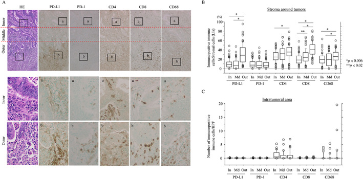
Programmed cell death-1 (PD-1) and its ligand (PD-L1) are significant mediators of immune suppression in the tumor microenvironment. We focused on the immunological impact of PD-1/PD-L1 signaling during tumor progression in colorectal carcinoma (CRC) and its association with resistance to neoadjuvant chemoradiotherapy (NCRT) in locally advanced rectal carcinoma (LAd-RC). Histopathological and immunohistochemical analyses of 100 CRC cases (including 34 RC) without NCRT and 109 NCRT-treated LAd-RC cases were performed. Membranous tumoral PD-L1 expression was identified in 9 of 100 (9%) CRC cases, including 1 of 34 (2.9%) RC cases, but PD-L1 immunopositivity was not associated with any clinicopathological factors, with the exception of deficient mismatch repair (dMMR) status. In contrast, stromal PD-L1+ immune cells, which frequently exhibited coexpression of PD-1 and CD8 markers, were significantly correlated with tumor vessel invasion, nuclear β-catenin+ tumor budding cancer stem cell (CSC)-like features, and unfavorable prognosis. In the LAd-RC cases, stromal CD8+ (but not PD-L1+) immune cell infiltration in pretreatment-biopsied samples was significantly and positively associated with therapeutic efficacy. After NCRT, tumoral PD-L1 expression was observed in only 2 of 83 (2.4%) tumors, independent of dMMR status, whereas high stromal PD-L1+ and tumoral nuclear β-catenin positivity were significantly linked to a poor response to NCRT and high tumor budding features. In addition, high stromal PD-L1 immunoreactivity was significantly associated with poorer overall survival. In conclusion, a combination of stromal PD-L1+ immune cells and nuclear β-catenin+ tumor budding may contribute to tumor progression in CRC and resistance to NCRT in LAd-RC, through formation of niche-like lesions that exhibit immune resistance and CSC properties.
Keywords: PD-1; PD-L1; cancer stem cell; colorectal carcinoma; neoadjuvant chemoradiotherapy; β-catenin.
© 2022 The Authors. The Journal of Pathology: Clinical Research published by The Pathological Society of Great Britain and Ireland and John Wiley & Sons Ltd.
Figures


Similar articles
-
S100A4 contributes to colorectal carcinoma aggressive behavior and to chemoradiotherapy resistance in locally advanced rectal carcinoma.Sci Rep. 2024 Dec 28;14(1):31338. doi: 10.1038/s41598-024-82814-9. Sci Rep. 2024. PMID: 39732925 Free PMC article.
-
Mismatch repair-deficient colorectal cancer: a model of immunogenic and immune cell-rich tumor despite nonsignificant programmed cell death ligand-1 expression in tumor cells.Hum Pathol. 2018 Feb;72:135-143. doi: 10.1016/j.humpath.2017.09.019. Epub 2017 Dec 5. Hum Pathol. 2018. PMID: 29208565
-
CMTM6 and PD-L1 coexpression is associated with an active immune microenvironment and a favorable prognosis in colorectal cancer.J Immunother Cancer. 2021 Feb;9(2):e001638. doi: 10.1136/jitc-2020-001638. J Immunother Cancer. 2021. PMID: 33579737 Free PMC article.
-
PD-1/PD-L1-dependent immune response in colorectal cancer.J Cell Physiol. 2020 Jul;235(7-8):5461-5475. doi: 10.1002/jcp.29494. Epub 2020 Jan 21. J Cell Physiol. 2020. PMID: 31960962 Review.
-
The efficacy and safety of neoadjuvant chemoradiotherapy combined with immunotherapy for locally advanced rectal cancer patients: a systematic review.Front Immunol. 2024 May 15;15:1392499. doi: 10.3389/fimmu.2024.1392499. eCollection 2024. Front Immunol. 2024. PMID: 38846948 Free PMC article.
Cited by
-
WNT/β-catenin regulatory roles on PD-(L)1 and immunotherapy responses.Clin Exp Med. 2024 Jan 27;24(1):15. doi: 10.1007/s10238-023-01274-z. Clin Exp Med. 2024. PMID: 38280119 Free PMC article. Review.
-
Total neoadjuvant treatment and PD-1/PD-L1 checkpoint inhibitor in locally advanced rectal cancer.Front Immunol. 2023 Mar 24;14:1149122. doi: 10.3389/fimmu.2023.1149122. eCollection 2023. Front Immunol. 2023. PMID: 37033988 Free PMC article. Review.
-
Therapeutic challenge for immunotherapy targeting cold colorectal cancer: A narrative review.World J Clin Oncol. 2023 Feb 24;14(2):81-88. doi: 10.5306/wjco.v14.i2.81. World J Clin Oncol. 2023. PMID: 36908678 Free PMC article. Review.
-
Contrasting Roles of Programmed Death-Ligand 1 Expression in Tumor and Stroma in Prognosis of Esophageal Squamous Cell Carcinoma.Cancers (Basel). 2024 Mar 13;16(6):1135. doi: 10.3390/cancers16061135. Cancers (Basel). 2024. PMID: 38539470 Free PMC article.
-
EBP50 Depletion and Nuclear β-Catenin Accumulation Engender Aggressive Behavior of Colorectal Carcinoma through Induction of Tumor Budding.Cancers (Basel). 2023 Dec 29;16(1):183. doi: 10.3390/cancers16010183. Cancers (Basel). 2023. PMID: 38201610 Free PMC article.
References
-
- Onyoh EF, Hsu W‐F, Chang L‐C, et al. The rise of colorectal cancer in Asia: epidemiology, screening, and management. Curr Gastroenterol Rep 2019; 21: 36. - PubMed
-
- Galon J, Costes A, Sanchez‐Cabo F, et al. Type, density, and location of immune cells within human colorectal tumors predict clinical outcome. Science 2006; 313: 1960–1964. - PubMed
Publication types
MeSH terms
Substances
LinkOut - more resources
Full Text Sources
Research Materials
