Review
. 2022 Oct 3;24(10):1613-1646.
doi: 10.1093/neuonc/noac118.

Brain metastases: A Society for Neuro-Oncology (SNO) consensus review on current management and future directions

Affiliations
Review

Brain metastases: A Society for Neuro-Oncology (SNO) consensus review on current management and future directions

Ayal A Aizer et al. Neuro Oncol. .

Abstract

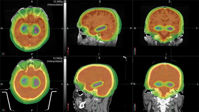
Brain metastases occur commonly in patients with advanced solid malignancies. Yet, less is known about brain metastases than cancer-related entities of similar incidence. Advances in oncologic care have heightened the importance of intracranial management. Here, in this consensus review supported by the Society for Neuro-Oncology (SNO), we review the landscape of brain metastases with particular attention to management approaches and ongoing efforts with potential to shape future paradigms of care. Each coauthor carried an area of expertise within the field of brain metastases and initially composed, edited, or reviewed their specific subsection of interest. After each subsection was accordingly written, multiple drafts of the manuscript were circulated to the entire list of authors for group discussion and feedback. The hope is that the these consensus guidelines will accelerate progress in the understanding and management of patients with brain metastases, and highlight key areas in need of further exploration that will lead to dedicated trials and other research investigations designed to advance the field.

Keywords: brain metastases; consensus; expert; guidelines; recommendations; treatment.


Figures

Fig. 1
Fig. 1
Incidence and research output as measured by ASCO abstracts (annual meeting) and active/completed prospective trials based on clinicaltrials.gov among patients with brain metastases versus other oncologic entities of similar incidence. (Abbreviations: ASCO, American Society of Clinical Oncology; CRC, colorectal cancer; US, United States).
Fig. 2
Fig. 2
Pathogenesis of brain metastases. The development of brain metastases depends on a complex interplay of factors involving tumor cells migrating from an extracranial site into the vasculature via a series of epigenetic changes, proliferation of blood vessels, and an epithelial-to-mesenchymal transition. Once the tumor cells reach the brain, they traverse the blood brain barrier, which involves upregulation of genes and proteins involved in proteolysis, extracellular matrix destruction, and mitogenesis/growth. Once inside the brain, the cells must adhere to the brain endothelia and undergo further stimulatory processes to allow for proliferation.
Fig. 3
Fig. 3
Characteristic MRI of a brain metastasis. T1-weighted postgadolinium MRI of a right frontal brain metastasis displaying characteristic rim enhancement (left) and associated T2-weighted FLAIR sequence showing extensive surrounding vasogenic edema (right). (Abbreviations: FLAIR, Fluid Attenuated Inversion Recovery; MRI, Magnetic Resonance Imaging).
Fig. 4
Fig. 4
MRI-based appearance of radiation necrosis. A left parietal metastasis is shown prior to resection (top left) and postresection, preadjuvant stereotactic radiation (top right). Five years later, the patient developed enhancement at the treated site (bottom left), which enlarged with time (bottom right). The patient was taken to the operating room for resection, and the lesion proved to be radiation necrosis. (Abbreviations: MRI, Magnetic Resonance Imaging).
Fig. 5
Fig. 5
Supportive medication utilization among patients with brain metastases. Retrospective data from a population-based study of 17 957 patients with brain metastases demonstrating the high prevalence of supportive medication use in the first 30 days following a diagnosis of brain metastases (as stratified by race and medication class). Opioids, anti-emetics, headache aids, and appetite stimulants were among the most frequently utilized medications among this patient population. Reproduced with permission from Lamba et al., Neuro-Oncology, 2020.
Fig. 6
Fig. 6
Pachymeningeal seeding. Pachymeningeal seeding after neurosurgical resection of a brain metastasis. Note the multifocal pachymeningeal recurrences (red) occurring in the absence of a cavity recurrence (green).
Fig. 7
Fig. 7
Classical leptomeningeal disease. Classical leptomeningeal disease as noted by linear enhancement along the cerebellar folia (A), supratentorial sulci (B), and cranial nerves VII/VIII bilaterally (C, red circles).
Fig. 8
Fig. 8
Laser interstitial therapy. LITT technique showing stereotactic laser fiber insertion to the target through a skull anchoring bolt (A) and MRI-based assessments before, during, and after LITT procedure (B). In Part (B), images reveal: (left) T1 and T2-weighted MRI showing regrowth of brain metastasis 12 months after radiosurgery; (left middle) intraoperative images displaying laser inserted into lesion; (right middle) 2-week post-LITT MRI showing postlaser ablation lesion increased in size but FLAIR signal improved; (right) MRI revealing resolution of lesion at 3 months post-LITT. (Abbreviations: FLAIR, Fluid Attenuated Inversion Recovery; LITT, Laser Interstitial Thermal Therapy; MRI, Magnetic Resonance Imaging).
Fig. 9
Fig. 9
Hippocampal-sparing whole brain radiation in a patient with brain metastases. The right and left hippocampi are contoured in blue and pink, respectively. The top and bottom panel shows the planning magnetic resonance imaging and computed tomography scans, respectively. The red, orange, yellow, and green dose-based shading depict the 33 Gy, 30 Gy, 27 Gy, and 16 Gy isodose lines, respectively.

Similar articles

Cited by

References

    1. Nayak L, Lee EQ, Wen PY. Epidemiology of brain metastases. Curr Oncol Rep. 2012; 14(1):48–54. - PubMed
    1. Barnholtz-Sloan JS, Sloan AE, Davis FG, et al. Incidence proportions of brain metastases in patients diagnosed (1973 to 2001) in the Metropolitan Detroit Cancer Surveillance System. J Clin Oncol. 2004; 22(14):2865–2872. - PubMed
    1. Lamba N, Wen PY, Aizer AA. Epidemiology of brain metastases and leptomeningeal disease. Neuro Oncol. 2021; 23(9):1447–1456. - PMC - PubMed
    1. Achrol AS, Rennert RC, Anders C, et al. Brain metastases. Nat Rev Dis Primers. 2019; 5(1):5. - PubMed
    1. Fabi A, Felici A, Metro G, et al. Brain metastases from solid tumors: disease outcome according to type of treatment and therapeutic resources of the treating center. J Exp Clin Cancer Res. 2011; 30(1):10. - PMC - PubMed