. 2022 Oct;55(10):e13299.
doi: 10.1111/cpr.13299. Epub 2022 Jun 28.

TGF-β1 peptide-based inhibitor P144 ameliorates renal fibrosis after ischemia-reperfusion injury by modulating alternatively activated macrophages

Affiliations

TGF-β1 peptide-based inhibitor P144 ameliorates renal fibrosis after ischemia-reperfusion injury by modulating alternatively activated macrophages

Delun Li et al. Cell Prolif. 2022 Oct.

Abstract

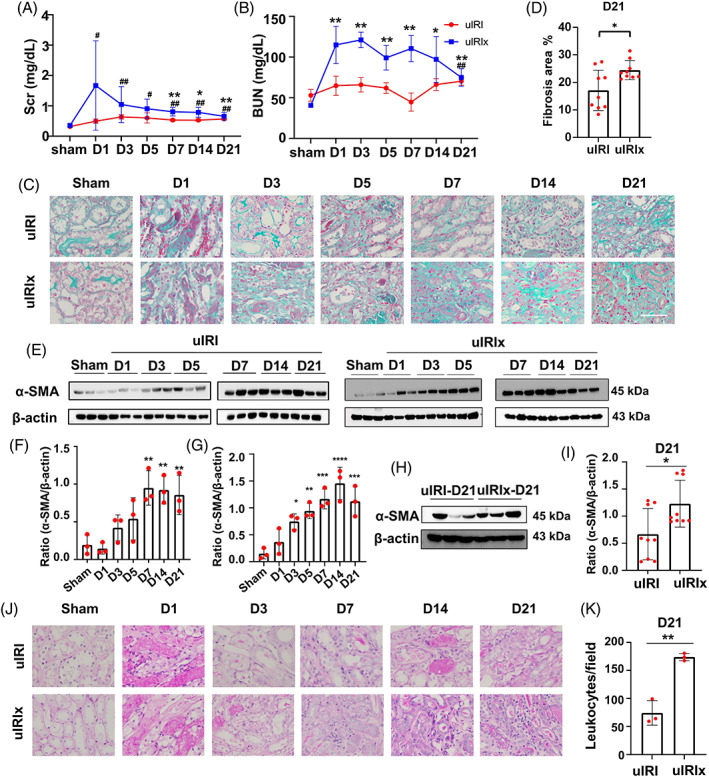
Objectives: Ischemia-reperfusion injury (IRI) is a major cause of chronic renal fibrosis. Currently, numerous therapies have shown a minimal effect on the blockade of fibrosis progression. Here, the therapeutic potential of peptide-based TGF-β1 inhibitor P144 in IRI-induced renal fibrosis and the underlying mechanism were analyzed.

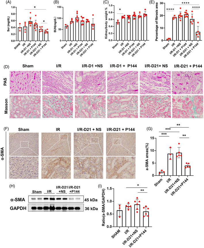
Materials and methods: The unilateral ischemia-reperfusion injury with the contralateral nephrectomy model was established, and the P144 was administered intravenously 1d/14d after the onset of IRI. The histopathology and immunofluorescence staining were used to detect renal fibrosis and macrophage infiltration. The in vivo fluorescence imaging was used to measure the bio-distribution of P144. The transwell assays were used to observe the migration of macrophages. RT-qPCR and western blot were used to analyze TGF-β1 signaling.

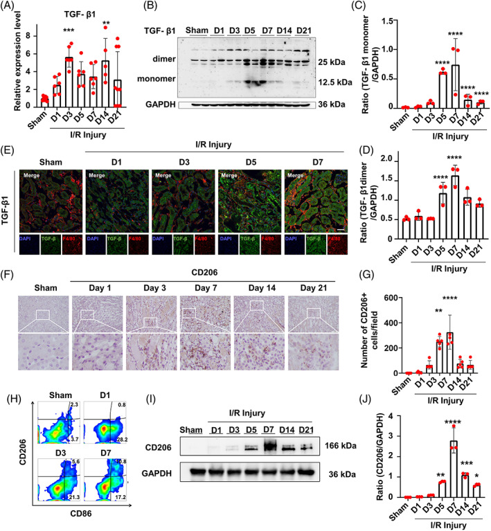
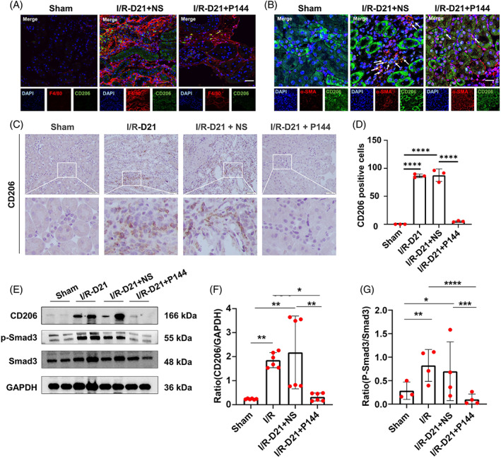
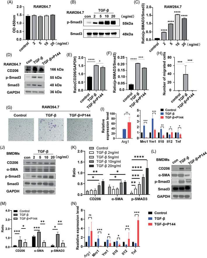
Results: P144 ameliorated the accumulation of extracellular matrix in the kidney and improved the renal function in the unilateral ischemia-reperfusion injury plus contralateral nephrectomy model. Mechanistically, P144 downregulated the TGF-β1-Smad3 signaling at both the transcriptional and translational levels and further reduced the TGF-β1-dependent infiltration of macrophages to the injured kidney. Additionally, P144 blocked the polarization of macrophages to an M2-like phenotype induced by TGF-β1 in vitro, but showed no effect on their proliferation.

Conclusions: Our study showed that the TGF-β1 peptide-based inhibitor P144 decreased renal fibrosis through the blockade of the TGF-β1-Smad3 signaling pathway and the modulation of macrophage polarization, suggesting its potential therapeutic use in IRI-induced renal fibrosis.


Conflict of interest statement

The author declares that there are no conflict of interest.

Figures

FIGURE 1
FIGURE 1
The renal function and pathological characteristics of uIRIx and uIRI models. (A, B) The levels of Scr and Bun in mice in the uIRIx and uIRI models were measured by ELISA. (C, D) Masson staining of uIRIx and uIRI samples at different time points after reperfusion; the percentage of the fibrotic area was quantified. (E–G) Expression of α‐SMA in the kidney after reperfusion detected by western blotting in uIRI and uIRIx mice. (H, I) Expression of α‐SMA in the kidney at D21 after reperfusion detected by western blotting in uIRIx and uIRI mice. (J, K) PAS staining of uIRIx and uIRI samples at different time points after reperfusion; the number of infiltrated leukocytes/field was quantified. Data are presented as the mean ± SD, n = 3–9. *p < 0.05, **p < 0.01. Scale bar = 50 μm. α‐SMA, α‐smooth muscle actin; Bun, blood urea nitrogen; PAS, Periodic acid–Schiff; Scr, serum creatinine; uIRI, unilateral ischemia–reperfusion injury model group; uIRIx, unilateral ischemia–reperfusion injury and contralateral nephrectomy
FIGURE 2
FIGURE 2
Expression pattern of TGF‐β1 and infiltration of macrophages in the kidney of uIRIx mice. (A) mRNA expression of TGF‐β1 in uIRIx mice measured by RT‐qPCR. (B–D) Expression of TGF‐β1 dimers and TGF‐β1 monomers in uIRIx mice at D1, D3, D5, D7, D14, and D21 after reperfusion detected by western blotting. (E) Expression of F4/80 and TGF‐β1 in uIRIx mice at D1, D3, D5, and D7 after reperfusion detected by immunofluorescence. (F,G) Expression of CD206 in the kidney of uIRIx mice detected by immunohistochemical staining. (H) Expression of CD86 and CD206 in the kidney of uIRIx mice detected by flow cytometry. (I, J) Expression of CD206 in the kidney of uIRIx mice detected by western blotting in uIRIx mice. Data are presented as the mean ± SD, n = 3–9. *p < 0.05, **p < 0.01, ***p < 0.001, ****p < 0.0001. Scale bar = 50 μm. TGF‐β1, transforming growth factor‐β; uIRIx, unilateral ischemia–reperfusion injury and contralateral nephrectomy
FIGURE 3
FIGURE 3
The antifibrotic effect of P144 in IRI‐induced renal fibrosis in vivo. (A, B) The levels of Scr and Bun in uIRIx mice treated with P144 were measured by ELISA. (C) The ratio of kidney/body weight in uIRIx mice treated with P144. (D) PAS staining and Masson staining of kidneys of uIRIx mice treated with P144. (E) The percentage of the fibrotic area was quantified. (F, G) Immunohistochemical staining of α‐SMA in the kidneys of uIRIx mice treated with P144 at D21 after reperfusion. (H, I) Expression of α‐SMA in the kidneys of uIRIx mice treated with P144 at D21 after reperfusion detected by western blotting. Data are presented as the mean ± SD, n = 3–8.*p < 0.05, **p < 0.01, ***p < 0.001, ****p < 0.0001. Scale bar = 50 μm. Bun, blood urea nitrogen; IRI, ischemia–reperfusion injury; Scr, serum creatinine; TGF‐β1, transforming growth factor‐β; uIRIx, unilateral ischemia–reperfusion injury and contralateral nephrectomy
FIGURE 4
FIGURE 4
In vivo fluorescence imaging of P144 distribution. (A, B) Mice were used to generate a uIRIx model and were then injected with 1 mg/kg of the labeled P144 at D21 postsurgery. Fluorescence imaging was performed after intravenous injection of labeled P144 at indicated times. (C, D) Biodistribution of Alexa Fluor 647‐labeled P144 in different organs at indicated times after intravenous injection. Mice were sacrificed at the indicated time after P144 injection and different tissues were isolated for biodistribution evaluation. n = 3–4, ****p < 0.0001. uIRIx, unilateral ischemia–reperfusion injury and contralateral nephrectomy
FIGURE 5
FIGURE 5
M2 microphage polarization and TGF‐β1 signaling pathway after P144 treatment. (A) CD206 positive macrophages in the kidney of uIRIx mice treated with P144 detected by immunofluorescence staining. (B) Expression of α‐SMA in CD206 positive macrophages in the kidney of uIRIx mice treated with P144 detected by immunofluorescence staining. The white arrows indicate the α‐SMA+ CD206+ macrophages. (C, D) Immunohistochemical staining of CD206 in the kidney of uIRIx mice treated with P144. (E–G) Expression of CD206, p‐Smad3, and Smad3 in the kidney of uIRIx mice treated with P144 detected by western blotting. Data are presented as the mean ± SD, n = 3–6. *p < 0.05, **p < 0.01, ***p < 0.001, ****p < 0.0001. Scale bar = 50 μm. TGF‐β1, transforming growth factor‐β; uIRIx, unilateral ischemia–reperfusion injury and contralateral nephrectomy
FIGURE 6
FIGURE 6
Migration and polarization of macrophages after P144 treatment in vitro. (A) The proliferation of RAW264.7 cells treated with TGF‐β1 was measured using a CCK‐8 assay. (B, C) Expression of p‐Smad3 in RAW264.7 cells treated with different doses of TGF‐β1 measured by western blotting. (D–F) Expression of CD206 and p‐Smad3 in RAW264.7 cells treated with TGF‐β1 and P144 measured by western blotting. (G, H) Migration of RAW264.7 cells treated with TGF‐β1 and P144 measured using the transwell migration assay; the number of migrated cells was quantified. (I) mRNA expression of M1 and M2 macrophage effectors in RAW264.7 cells measured by RT‐qPCR. (J, K) Expression of CD206, α‐SMA, and p‐Smad3 in BMDMs treated with TGF‐β1 measured by western blotting. (L, M) Expression of CD206, α‐SMA, and p‐Smad3 in BMDMs treated with TGF‐β1 and P144 measured by western blotting. (N) mRNA expression of M1 and M2 macrophage effectors in BMDMs measured by RT‐qPCR. Data are presented as the mean ± SD, n = 3, *p < 0.05, ***p < 0.001, ****p < 0.0001. BMDMs, bone marrow‐derived macrophages; TGF‐β1,transforming growth factor‐β

References

    1. Ronco C, Bellomo R, Kellum JA. Acute kidney injury. Lancet. 2019;394:1949‐1964. doi:10.1016/S0140-6736(19)32563-2 - DOI - PubMed
    1. Webster AC, Nagler EV, Morton RL, Masson P. Chronic kidney disease. Lancet. 2017;389:1238‐1252. doi:10.1016/S0140-6736(16)32064-5 - DOI - PubMed
    1. Gu YY, Liu XS, Huang XR, Yu XQ, Lan HY. Diverse role of TGF‐beta in kidney disease. Front Cell Dev Biol. 2020;8:123. doi:10.3389/fcell.2020.00123 - DOI - PMC - PubMed
    1. Broekema M, Harmsen MC, van Luyn MJ, et al. Bone marrow‐derived myofibroblasts contribute to the renal interstitial myofibroblast population and produce procollagen I after ischemia/reperfusion in rats. J Am Soc Nephrol. 2007;18:165‐175. doi:10.1681/ASN.2005070730 - DOI - PubMed
    1. Wollin L, Wex E, Pautsch A, et al. Mode of action of nintedanib in the treatment of idiopathic pulmonary fibrosis. Eur Respir J. 2015;45:1434‐1445. doi:10.1183/09031936.00174914 - DOI - PMC - PubMed

MeSH terms

Substances