. 2022 Sep 1;323(3):L372-L389.
doi: 10.1152/ajplung.00136.2022. Epub 2022 Jun 28.

Exploring antiviral and anti-inflammatory effects of thiol drugs in COVID-19

Affiliations

Exploring antiviral and anti-inflammatory effects of thiol drugs in COVID-19

Kritika Khanna et al. Am J Physiol Lung Cell Mol Physiol. .

Abstract

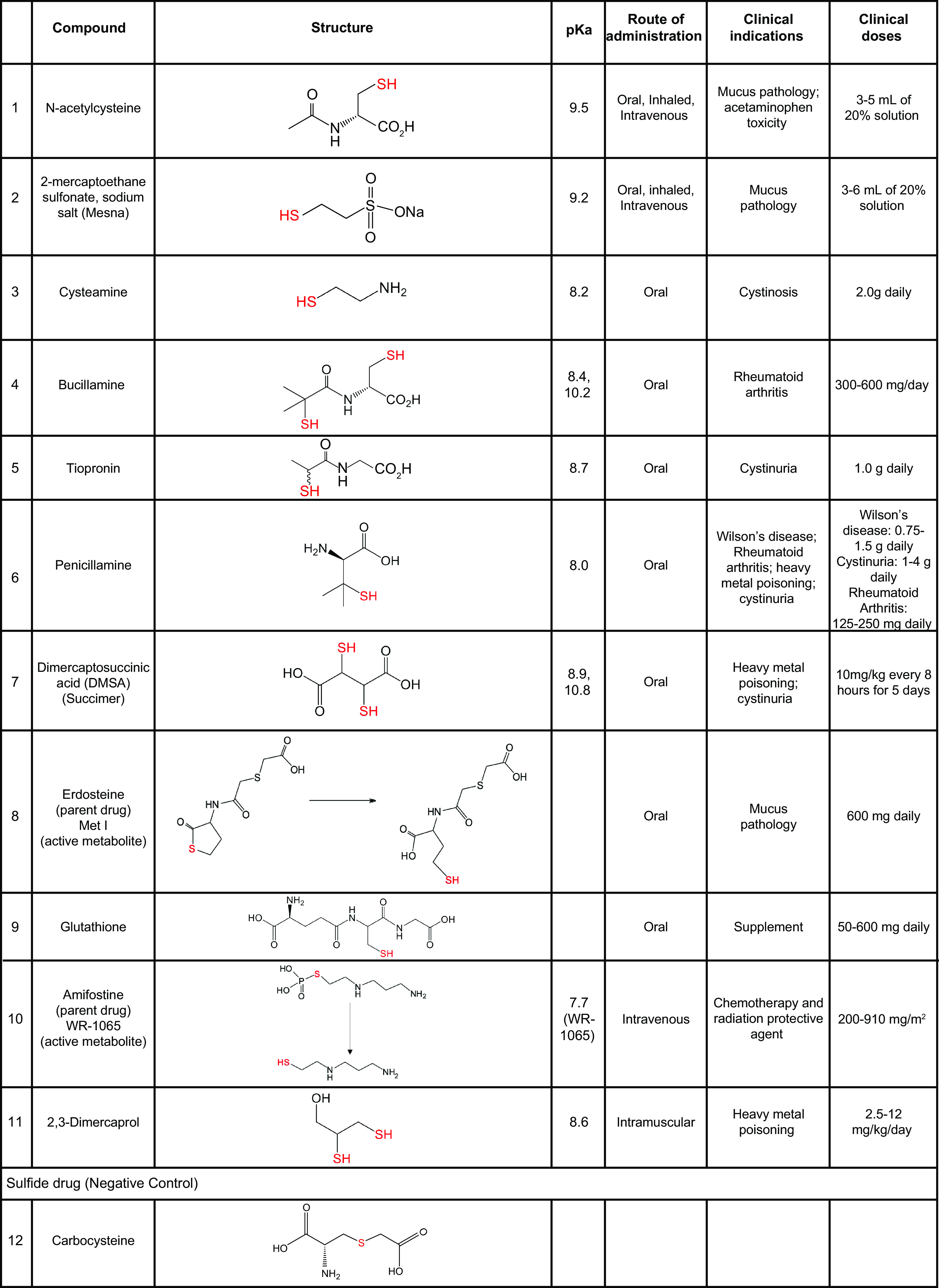
The redox status of the cysteine-rich SARS-CoV-2 spike glycoprotein (SARS-2-S) is important for the binding of SARS-2-S to angiotensin-converting enzyme 2 (ACE2), suggesting that drugs with a functional thiol group ("thiol drugs") may cleave cystines to disrupt SARS-CoV-2 cell entry. In addition, neutrophil-induced oxidative stress is a mechanism of COVID-19 lung injury, and the antioxidant and anti-inflammatory properties of thiol drugs, especially cysteamine, may limit this injury. To first explore the antiviral effects of thiol drugs in COVID-19, we used an ACE-2 binding assay and cell entry assays utilizing reporter pseudoviruses and authentic SARS-CoV-2 viruses. We found that multiple thiol drugs inhibit SARS-2-S binding to ACE2 and virus infection. The most potent drugs were effective in the low millimolar range, and IC50 values followed the order of their cystine cleavage rates and lower thiol pKa values. To determine if thiol drugs have antiviral effects in vivo and to explore any anti-inflammatory effects of thiol drugs in COVID-19, we tested the effects of cysteamine delivered intraperitoneally to hamsters infected with SARS-CoV-2. Cysteamine did not decrease lung viral infection, but it significantly decreased lung neutrophilic inflammation and alveolar hemorrhage. We speculate that the concentration of cysteamine achieved in the lungs with intraperitoneal delivery was insufficient for antiviral effects but sufficient for anti-inflammatory effects. We conclude that thiol drugs decrease SARS-CoV-2 lung inflammation and injury, and we provide rationale for future studies to test if direct (aerosol) delivery of thiol drugs to the airways might also result in antiviral effects.

Keywords: ACE2; COVID-19; SARS-CoV-2; cysteamine; thiol drugs.


Conflict of interest statement

J. V. Fahy, S. Oscarson, I. Gitlin, and W. W. Raymond are inventors on patent applications related to the use of thiol drugs as treatments for COVID-19. None of the other authors has any conflicts of interest, financial or otherwise, to disclose.

Figures

None
Graphical abstract
Figure 1.
Figure 1.
Cystine mapping and conservation of cystines in RBD of SARS-1-S and SARS-2-S. A: cystine map for SARS-2-S domain S1 (UniProt: P0DTC2). Ten cystine linkages are denoted by dashed lines with amino acid residue number above. The dark gray region is the RBD, and the lighter gray box highlights the ACE2 binding motif, a cluster of amino acids that make contact with ACE2. B: amino acid alignment of SARS-2-S RBD domain (aa 319-541, PDB: 6M0J) and SARS-1-S RBD domain (aa 306-517, PDB: 3SCI). Residues that are shared are highlighted by black boxes, and residues that represent a similar amino acid class replacement are bound by gray boxes. The solid lines link cystine-forming cysteines. The solid red line and red numbers highlight the conserved critical cystine bridge in the RBDs for both viruses. *Amino acids that are within 4 Å of ACE2 in their respective solved structures. C: a surface rendering of SARS-2-RBD (PDB: 6M0J) oriented with the ACE2 binding region (blue) facing forward. D: amino acid sequence of SARS-2-S RBD highlighting the RBD mutations identified in the circulating SARS-CoV-2 variants. Amino acids are noted with single letter code and sequence number. The conserved RBD cystine (C480-C488) is highlighted in red lines. The amino acid mutations are highlighted in red with the variant designations in blue. BA.1 and BA.2 represent the subvariants of B.1.1.529 Omicron variant. ACE2, angiotensin-converting enzyme 2; RBD, receptor binding domain.
Figure 2.
Figure 2.
Currently approved thiol drug compounds or drugs that generate an active thiol-containing drug metabolite. The chemical name along with the chemical structures, thiol pKa values (from published literature and PubChem), routes of administration, clinical indications, and clinical doses are provided. Two thiol pKa values are provided for bucillamine and succimer because these drugs have two thiol groups. The clinical doses provided for NAC and Mesna are those used for inhalation route. Two compounds, amifostine and erdosteine, are prodrugs that can convert to active metabolites. Carbocysteine is not a thiol drug but it is a sulfur containing drug that we use in our experiments as a negative control because it does not have a free thiol warhead. Not shown here are three thiol containing drugs (captopril, zofenopril, and racecadotril) in which primary mechanisms of action is not through reactions with the thiol group. Mesna, 2-mercaptoethane sulfonate sodium salt; NAC, n-acetylcysteine.
Figure 3.
Figure 3.
Binding of SARS-CoV-2 RBD to ACE2 is inhibited by thiol drugs. A: schematic representation of the SARS-CoV-2 RBD to ACE2 binding assay. B: percentage of binding of RBDWuhan-1 to ACE2 in the presence of the drugs (n = 4–6 independent experiments). C: histograms denoting areas under the curve (AUC) analyses for binding studies in B, in which the AUC is the percent binding of ACE2 to RBD in the presence of each drug. The reference AUC is determined from the percent binding of ACE to RBD in the absence of drug. The dashed line represents 50% of reference AUC. D: binding of RBDWuhan-1 to ACE2 at 1 and 2 h after drug exposure and washout (n = 4–5 independent experiments). Statistical significance for C was analyzed by one-way ANOVA followed by Dunnett’s post hoc analysis. Data are means ± SE. Significance indicates differences from reference AUC. **P ≤ 0.01, ****P ≤ 0.0001. ACE2, angiotensin-converting enzyme 2; HRP, horseradish peroxidase; RBD, receptor binding domain.
Figure 4.
Figure 4.
Thiol drugs inhibit ancestral SARS-CoV-2 in vitro. Pseudovirus (PV) entry efficiency after pseudovirus exposure to carbocysteine (sulfide drug, negative control; A), amifostine (parent drug of WR-1065, negative control; B), thiol drugs NAC (C), tiopronin (D), Mesna (E), bucillamine (F), WR-1065 (G), and cysteamine (H) before transduction into HEK293T-ACE2-TMPRSS2 cells (n = 3–4 independent experiments). The effects of drugs on cell viability were quantified using Cell Titer Glo 2.0 with lower drug dose exposures, reflecting 66-fold dilution of drugs when pseudovirus/drug mixture was incubated with cells (n = 3 independent experiments). I: IC50 values of the different thiol drugs in the PV assay. Cytopathic effects (CPE) quantified by visual inspection when authentic SARS-CoV-2 is exposed to carbocysteine (negative control; J) and cysteamine (thiol drug; K) before infection in Vero E6-TMPRSS2 cells (n = 3 independent experiments). The effects of drugs on VeroE6-TMPRSS2 cell viability were quantified with exposure of cells to lower drug doses, reflecting the 24-fold dilution of drugs when virus/drug mixture was incubated with cells (n = 3 independent experiments). The X-axes are scaled to log10—the lower X-axis refers to the concentration of drugs on the PV or authentic virus, and the upper X-axis refers to concentration of drugs on the cells post dilution. Percentage changes are with respect to no drug control which is set as 100%. IC50 of the drugs was determined using the nonlinear regression fitting with a variable slope. Data are means ± SD. Mesna, 2-mercaptoethane sulfonate sodium salt; NAC, n-acetylcysteine; TMPRSS2, transmembrane protease, serine 2.
Figure 5.
Figure 5.
Thiol drugs inhibit Delta SARS-CoV-2 in vitro. Pseudovirus (PV) entry efficiency when the B.1.617.1, B.1.617.2, and B.1.1.529 BA.1 pseudoviruses were exposed to carbocysteine (sulfide drug, negative control; A), thiol drugs Mesna (B), bucillamine (C), TDG (D), cysteamine (E), and WR-1065 (F) before transduction into HEK293T-ACE2-TMPRSS2 cells (n = 3–5 independent experiments). The X-axes are scaled to log10. Percentage changes are with respect to no drug control which is set as 100%. IC50 of the drugs was determined using the nonlinear regression fitting with a variable slope. Data are means ± SD. G: IC50 values of the thiol drugs in the PV transduction assay and their respective thiol pKa values. Bucillamine has 2 pKa values corresponding to the two thiol groups in its structure. Data are means ± SE. Cytopathic effects (CPE) quantified by visual inspection when authentic Delta SARS-CoV-2 is exposed to carbocysteine (sulfide drug, negative control; H), thiol drugs Mesna (I), bucillamine (J), TDG (K), cysteamine (L), and WR-1065 (M) before infection in Vero E6-TMPRSS2 cells (n = 3 independent experiments). The effects of drugs on VeroE6-TMPRSS2 cell viability were quantified with exposure of cells to lower drug doses, reflecting the 24-fold dilution of drugs when virus/drug mixture was incubated with cells (n = 3 independent experiments). The X-axes are scaled to log10—the lower X-axis refers to the concentration of drugs on the authentic virus, and the upper X-axis refers to concentration of drugs on the cells post dilution. Percentage changes are with respect to no drug control which is set as 100%. IC50 of the drugs was determined using the nonlinear regression fitting with a variable slope. Data are means ± SD. Mesna, 2-mercaptoethane sulfonate sodium salt; TDG, methyl 6-thio-6-deoxy-α-d-galactopyranoside; TMPRSS2, transmembrane protease, serine 2.
Figure 6.
Figure 6.
Thiol pKa influences the cystine cleaving rates of the thiol drugs and their efficacy in B.1.617.1 PV transduction assay. PV entry efficiency when B.1.617.1 pseudoviruses were treated with three cysteine derivates n-acetyl cysteine (A), l-cysteine (B), and l-cysteine methyl ester (CME; C) having variable thiol pKa (n = 3–6 independent experiments). The effects of cysteine derivatives on cell viability were quantified using Cell Titer Glo 2.0 with lower drug dose exposures, reflecting 66-fold dilution of drugs when pseudovirus/drug mixture was incubated with cells (n = 3 independent experiments). Data are means ± SD. IC50 of the drugs was determined using the nonlinear regression fitting with a variable slope. D: schematic representation of the BODIPY cystine assay. E: rate of BODIPY FL l-cystine cleavage when exposed to thiol drugs or controls (carbocysteine and amifostine; n = 3 independent experiments). Dotted lines indicate SE. F: maximum slope (Max V) for the reactivity of thiol drugs with the BODIPY FL cystine reagent (based on data in E). Numbers on graph (E) correspond with numbers designated to thiol compounds in F. Data in F are means ± SE. Pseudovirus, PV.
Figure 7.
Figure 7.
Effects of cysteamine on a Syrian hamster model of SARS-CoV-2 infection. A: study design for assessing the effect of cysteamine in Syrian hamster model of COVID-19. Cysteamine (100 mg/kg) was administered twice daily via intraperitoneal injection for 5 days, with the first dose given 2 h before the virus inoculation on Day 0. B: viral RNA levels in the lungs of animals treated with cysteamine relative to the vehicle control group. C: lung weights, normalized to the terminal body weights, of the animals. Total protein (D) and total cell counts (E) in the BAL fluid of hamsters treated with cysteamine with respect to the vehicle controls. Differential leukocyte counts in the BAL fluid of animals, showing neutrophil (F), macrophage (G), lymphocyte (H), and eosinophil (I) counts in treated and vehicle control groups. Histopathology scores for mixed cell (macrophages and neutrophils) inflammation in the peribronchovascular and the centriacinar regions of the lung (J) and alveolar hemorrhage (K) in the lungs of animals treated with cysteamine relative to vehicle control group. L: representative images of the lung sections of the animals highlighting the extent of alveolar hemorrhage in the animals from the two groups. Scale bars for ×4 images equal 200 µm; scale bars for ×10 images equal 500 µm. Control, intraperitoneal vehicle control group. CYS, cysteamine treated group. Each group had n = 10 animals. Two BAL samples from the cysteamine group could not be analyzed due to a technical error. Data are means ± SE. Statistical significance was analyzed by two tailed, unpaired t test between cysteamine treated (CYS) and respective control group (control). BAL, bronchoalveolar lavage.

References

    1. Wiersinga WJ, Rhodes A, Cheng AC, Peacock SJ, Prescott HC. Pathophysiology, transmission, diagnosis, and treatment of coronavirus disease 2019 (COVID-19): a review. JAMA 324: 782–793, 2020. doi:10.1001/jama.2020.12839. - DOI - PubMed
    1. Zhu N, Zhang D, Wang W, Li X, Yang B, Song J, Zhao X, Huang B, Shi W, Lu R, Niu P, Zhan F, Ma X, Wang D, Xu W, Wu G, Gao GF, Tan W; China Novel Coronavirus Investigating and Research Team. A novel coronavirus from patients with pneumonia in China, 2019. N Engl J Med 382: 727–733, 2020. doi:10.1056/NEJMoa2001017. - DOI - PMC - PubMed
    1. Hoffmann M, Kleine-Weber H, Schroeder S, Krüger N, Herrler T, Erichsen S, Schiergens TS, Herrler G, Wu NH, Nitsche A, Müller MA, Drosten C, Pöhlmann S. SARS-CoV-2 cell entry depends on ACE2 and TMPRSS2 and is blocked by a clinically proven protease inhibitor. Cell 181: 271–280.e8, 2020. doi:10.1016/j.cell.2020.02.052. - DOI - PMC - PubMed
    1. Weissenhorn W, Dessen A, Calder LJ, Harrison SC, Skehel JJ, Wiley DC. Structural basis for membrane fusion by enveloped viruses. Mol Membr Biol 16: 3–9, 1999. doi:10.1080/096876899294706. - DOI - PubMed
    1. Gallagher TM. Murine coronavirus membrane fusion is blocked by modification of thiols buried within the spike protein. J Virol 70: 4683–4690, 1996. doi:10.1128/jvi.70.7.4683-4690.1996. - DOI - PMC - PubMed

Publication types

MeSH terms