. 2022 Jul;31(7):e4358.
doi: 10.1002/pro.4358.

Ca2+ attenuates nucleation activity of leiomodin

Affiliations

Ca2+ attenuates nucleation activity of leiomodin

Garry E Smith et al. Protein Sci. 2022 Jul.

Abstract

A transient increase in Ca2+ concentration in sarcomeres is essential for their proper function. Ca2+ drives striated muscle contraction via binding to the troponin complex of the thin filament to activate its interaction with the myosin thick filament. In addition to the troponin complex, the myosin essential light chain and myosin-binding protein C were also found to be Ca2+ sensitive. However, the effects of Ca2+ on the function of the tropomodulin family proteins involved in regulating thin filament formation have not yet been studied. Leiomodin, a member of the tropomodulin family, is an actin nucleator and thin filament elongator. Using pyrene-actin polymerization assay and transmission electron microscopy, we show that the actin nucleation activity of leiomodin is attenuated by Ca2+ . Using circular dichroism and nuclear magnetic resonance spectroscopy, we demonstrate that the mostly disordered, negatively charged region of leiomodin located between its first two actin-binding sites binds Ca2+ . We propose that Ca2+ binding to leiomodin results in the attenuation of its nucleation activity. Our data provide further evidence regarding the role of Ca2+ as an ultimate regulator of the ensemble of sarcomeric proteins essential for muscle function. SUMMARY STATEMENT: Ca2+ fluctuations in striated muscle sarcomeres modulate contractile activity via binding to several distinct families of sarcomeric proteins. The effects of Ca2+ on the activity of leiomodin-an actin nucleator and thin filament length regulator-have remained unknown. In this study, we demonstrate that Ca2+ binds directly to leiomodin and attenuates its actin nucleating activity. Our data emphasizes the ultimate role of Ca2+ in the regulation of the sarcomeric protein interactions.

Keywords: actin; circular dichroism; leiomodin; nuclear magnetic resonance; pyrene-actin polymerization; transmission electron microscopy.


Figures

FIGURE 1
FIGURE 1
Domain structure of Lmod2. Lmod2 is comprised of several functionally distinguished regions: TpmBS – Tropomyosin‐binding site, ABS – Actin‐binding site, EDRR – Glu/Asp rich region
FIGURE 2
FIGURE 2
Effect of Ca2+ on Lmod2 nucleation observed by pyrene‐actin polymerization assay. (a) Representative time courses of each condition with their sigmoidal fits shown by solid lines. (b) Lmod2/control maximum polymerization rate ratios. (c) Lmod2/control lag time ratios. Bars and error bars are the average and standard deviation, respectively. Values of each repeat are represented by circles, and the P‐values were calculated by a paired t‐test, n = 4
FIGURE 3
FIGURE 3
Spontaneous and Lmod2‐nucleated polymerization of G‐actin observed by electron microscopy of negatively stained samples at low and high Ca2+ levels. G‐actin was polymerized in the absence (a‐f) or presence (g‐l) of 50 nM Lmod. Each trial was done at either low (a‐c and g‐i) or high (d‐f and j‐l) levels of Ca2+, at 30 sec, 2 min and 10 min time points
FIGURE 4
FIGURE 4
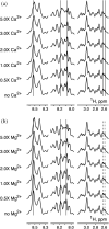
NMR titrations of the Lmod2 EDRR with Ca2+ and Mg2+. Displayed are NMR spectral changes in three chemical shift regions upon addition of (a) Ca2+ and (b) Mg2+. Molar excesses (0, 0.5, 1.0, 2.0, 3.0, and 5.0) of [cation] over [peptide] are shown to the left of the relevant spectra. Left and middle panels in both titrations (chemical shift regions 8.25–8.65 ppm and 7.95–8.35 ppm, respectively) correspond to resonance peaks of amide (backbone) protons. Right panels (chemical shift region 2.5–3.2 ppm) correspond to resonance peaks of side‐chain protons. Solid vertical lines show initial (prior to cation addition) positions of the most affected peaks. Dashed vertical lines at ~2.6 ppm in (b) correspond to ~2.6 ppm solid lines in (a). The dashed lines demonstrate that side‐chain‐resonance peaks affected by Ca2+ are shifted markedly less when the peptide is titrated with Mg2+
FIGURE 5
FIGURE 5
Effect of pre‐incubation of Lmod2 with Ca2+ on nucleation of Mg‐actin observed by pyrene‐actin polymerization assay. (a) Representative time courses of each condition with sigmoidal fits represented by solid lines. (b) Lmod2/control maximum polymerization rate ratios. (c) Lmod2/control lag time ratios. Values of each repeat are represented by circles, and the P‐values were calculated by a paired t‐test, n = 4

References

    1. DuVall MM, Gifford JL, Amrein M, Herzog W. Altered mechanical properties of titin immunoglobulin domain 27 in the presence of calcium. Eur Biophys J. 2013;42:301–307. - PubMed
    1. Kuhn ER, Naik AR, Lewis BE, et al. Nanothermometry reveals calcium‐induced remodeling of myosin. Nano Lett. 2018;18:7021–7029. - PMC - PubMed
    1. Previs MJ, Mun JY, Michalek AJ, et al. Phosphorylation and calcium antagonistically tune myosin‐binding protein C's structure and function. Proc Natl Acad Sci U S A. 2016;113:3239–3244. - PMC - PubMed
    1. Marston S, Zamora JE. Troponin structure and function: A view of recent progress. J Muscle Res Cell Motil. 2020;41:71–89. - PMC - PubMed
    1. Heissler SM, Sellers JR. Myosin light chains: Teaching old dogs new tricks. Bioarchitecture. 2014;4:169–188. - PMC - PubMed

Publication types