. 2022 Jul;11(7):e202200110.
doi: 10.1002/open.202200110. Epub 2022 Jun 28.

An Activatable 19 F MRI Molecular Probe for Sensing and Imaging of Norepinephrine

Affiliations

An Activatable 19 F MRI Molecular Probe for Sensing and Imaging of Norepinephrine

Lingxuan Li et al. ChemistryOpen. 2022 Jul.

Abstract

Norepinephrine (NE), acting as both a neurotransmitter and hormone, plays a significant role in regulating the action of the brain and body. Many studies have demonstrated a strong correlation between mental disorders and aberrant NE levels. Therefore, it is of urgent demand to develop in vivo analytical methods of NE for diagnostic assessment and mechanistic investigations of mental diseases. Herein, we report a 19 F MRI probe (NRFP) for sensing and imaging NE, which is constructed by conjugating a gadolinium chelate to a fluorine-containing moiety through a NE-responsive aromatic thiocarbonate linkage. The capacity and specificity of NRFP for detecting NE is validated with in vitro detecting/imaging experiments. Furthermore, the feasibility of NRFP for visualizing NE in animals is illustrated by ex vivo and in vivo imaging experiments, demonstrating the promising potential of NRFP for selective detection and specific imaging of NE in deep tissues of living subjects.

Keywords: 19F NMR/MRI; deep-tissue sensing; imaging agents; in vivo imaging; norepinephrine.


Conflict of interest statement

The authors declare no conflict of interest.

Figures

Figure 1
Figure 1
(a) In vivo metabolic pathways of catecholamines, including dopamine (DA), norepinephrine (NE), and epinephrine (E). MAO: monoamine oxidase; PMNT: phenylethanolamine N‐methyltransferase. (b) A schematic illustration showing the functional mechanism of our norepinephrine‐responsive 19F MRI probe (NRFP). 19F NMR/MRI signals of NRFP are “OFF” due to the strong paramagnetic relaxation enhancement (PRE) effect exerted by the chelated Gd3+ ion. The effect is significantly weakened when norepinephrine (NE) selectively cleaves the aromatic thiocarbonate linkage, which releases 3,5‐bis(trifluoromethyl)benzene‐thiol (BTFBT), leading to the switching “ON” of 19F NMR/MRI signals.
Figure 2
Figure 2
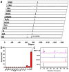
(a) Representative 19F NMR spectra of NRFP (−62.7 ppm) in 500 μL 10 % D2O/H2O (final concentration: 0.4 mm), which were incubated with various analytes for 3 h at 25 °C. PBS: 50 mm, pH 7.4; FBS: 10 % in PBS; dopamine (DA), norepinephrine (NE) and epinephrine (E): 10 mm in PBS; Glu: 5.0 mm glucose in PBS; GSH: 2.0 mm glutathione in PBS; all other analytes: 0.40 mm in PBS for 3 h at 25 °C. CF3COONa (at −75.4 ppm) was used as an internal reference. (b) Signal‐to‐noise ratio (SNR) analysis on the results of the experiment as indicated in (a) (n=5). The SNR for the peak of NRFP in PBS was set as 1.0 and the other SNRs were normalized accordingly. (c) HPLC chromatograms showing NRFP (0.2 mm) alone and the products of NRFP incubated with 50 equiv. NE for 3 h. The expected products, Gd‐DOTA‐Tyramine (GDTy) and 3,5‐bis(trifluoromethyl)benzene‐thiol (BTFBT) were also analyzed. See Figure S4 for HR‐ESI‐MS analysis of the fractions corresponding to the peaks indicated by an asterisk and a triangle.
Figure 3
Figure 3
(a) Representative 19F MR images of NRFP (at indicated concentrations) before and after specific activation towards NE (0.3 m). (b) Representative 19F MR images of NRFP (60 mm 19F) before and after incubated with: dopamine (DA), norepinephrine (NE) and epinephrine (E): 0.3 m in PBS; glucose (Glu), H2O2 and glutathione (GSH): 10 mm in PBS for 3 h at 25 °C. (c) SNR analysis on the results of the experiment as indicated in (a) (n=5). The SNR for the 19F MR image of 0.83 mm NRFP (i. e., 10 mm 19F) in PBS was set as 1.0 and the other SNRs were normalized accordingly. (d) SNR analysis on the results of the experiment as indicated in (b) (n=5). The SNR for the 19F MR image of NRFP with glucose in PBS was set as 1.0 and the other SNRs were normalized accordingly.
Figure 4
Figure 4
(a) A schematic illustration showing the protocol for ex vivo 19F MRI. The center frequency corresponding to the 19F chemical shift at −62.7 ppm was chosen for 19F MRI. (b) Representative 1H and 19F MR images of a piece of pork at pre‐injection (0 min) or indicated time points after subcutaneous injection of prepared NRFP solutions (200 μL), which were 10 mm NRFP in 1 × PBS incubated with 50 equiv. NE (Top) and 10 mm NRFP in 1 × PBS alone (Bottom). The sites of injection are indicated by white circles. (c) SNR analysis on the results of the experiment as indicated in (b) (n=3). The SNR for the 19F MR image of NRFP without NE treatment at 0 min was set as 1.0 and the other SNRs were normalized accordingly.
Figure 5
Figure 5
(a) A schematic illustration showing the protocol for in vivo 19F MRI. The center frequency corresponding to the 19F chemical shift at −62.7 ppm was chosen for 19F MRI. (b) Representative 1H and 19F MR images of living mice (BALB/c) at pre‐injection (0 min) or indicated time points after subcutaneous injection of prepared NRFP solution (200 μL), which contains 10 mm NRFP in 1 × PBS alone (left hinder limbs) and 10 mm NRFP in 1 × PBS incubated with 50 equiv. NE (right hinder limbs) for 3 h. (c) SNR analysis on the results of the experiment as indicated in (b) (n=3). The average SNR for the left thigh regions of the 19F MR images at 0 min was set as 1.0 and the other SNRs were normalized accordingly.

Similar articles

Cited by

References

    1. Robertson S. D., Plummer N. W., de Marchena J., Jensen P., Nat. Neurosci. 2013, 16, 1016–1023. - PMC - PubMed
    1. Liu Y. U., Ying Y. L., Li Y. J., Eyo U. B., Chen T. J., Zheng J. Y., Umpierre A. D., Zhu J., Bosco D. B., Dong H. L., Wu L. J., Nat. Neurosci. 2019, 22, 1771–1781. - PMC - PubMed
    1. Richards D. J., Li Y., Kerr C. M., Yao J., Beeson G. C., Coyle R. C., Chen X., Jia J., Damon B., Wilson R., Hazard E. S., Hardiman G., Menick D. R., Beeson C. C., Yao H., Ye T., Mei Y., Nat. Biomed. Eng. 2020, 4, 446–462. - PMC - PubMed
    1. Schwarz L. A., Miyamichi K., Gao X. J. J., Beier K. T., Weissbourd B., DeLoach K. E., Ren J., Ibanes S., Malenka R. C., Kremer E. J., Luo L. Q., Nature 2015, 524, 88. - PMC - PubMed
    1. Zahalka A. H., Arnal-Estape A., Maryanovich M., Nakahara F., Cruz C. D., Finley L. W. S., Frenette P. S., Science 2017, 358, 321–326. - PMC - PubMed

Publication types

LinkOut - more resources