

A concise history of echocardiography: timeline, pioneers, and landmark publications
- PMID: 35762885
- PMCID: PMC9365309
- DOI: 10.1093/ehjci/jeac111
A concise history of echocardiography: timeline, pioneers, and landmark publications
Abstract
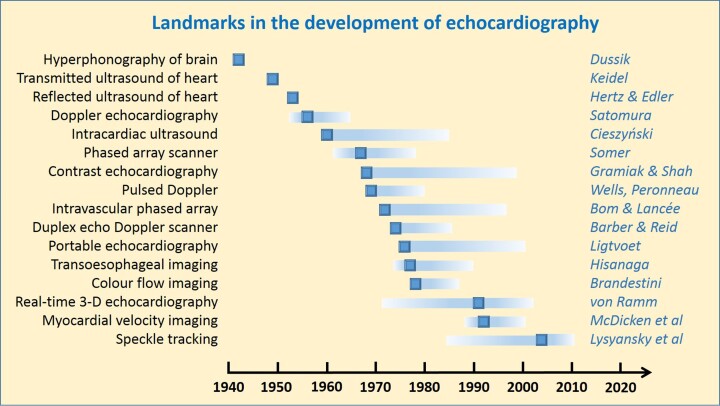
Echocardiography is less than 70 years old, and many major advances have occurred within living memory, but already some pioneering contributions may be overlooked. In order to consider what circumstances have been common to the most successful innovations, we have studied and here provide a timeline and summary of the most important developments in transthoracic and transoesophageal ultrasound imaging and Doppler techniques, as well as in intravascular ultrasound and imaging in paediatric cardiology. The entries are linked to a comprehensive list of first publications and to a collection of first-hand historical accounts published by early investigators. Review of the original manuscripts highlights that it is difficult to establish unequivocal precedence for many new imaging methods, since engineers were often working independently but simultaneously on similar problems. Many individuals who are prominently linked with particular developments were not the first in their field. Developments in echocardiography have been highly dependent on technological advances, and most likely to be successful when engineers and clinicians were able to collaborate with open exchange between centres and disciplines. As with many other new medical technologies, initial responses were sceptical and introduction into clinical practice required persistence and substantial energy from the first adopters. Current developments involve advances in software as much as in equipment, and progress will depend on continuing collaborations between engineers and clinical scientists, for example to identify unmet needs and to investigate the clinical impact of particular imaging approaches.
Keywords: echocardiography; history; key publications; pioneers.
© The Author(s) 2022. Published by Oxford University Press on behalf of the European Society of Cardiology.
Conflict of interest statement
Conflict of interest: None declared.
Figures

References
-
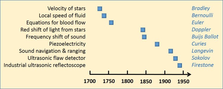
- Bradley J. An account of a new discovered motion of the fix’d stars. Philos Trans R Soc Lond 1727;35:637–660.
-
- Bernoulli D. Hydrodynamica, sive de viribus et motibus fluidorum commentarii. Basel: Johannis Reinholdi Dulseckeri; 1738.
-
- Euler L. Principes généraux de l'état d'équilibre d'un fluide. Mémoires de l'académie des sciences de Berlin (Royal Prussian Academy of Sciences) 1757;11:217–273.
-
- Euler L. Principia pro motu sanguinis per arterias determinando, 1775. In: Fuss PH and Fuss N (eds.), Opera posthuma mathematica et physica anno 1844 detecta: Euler Archive Eneström Number 855; 1862. p814–823.
-
- Doppler C. Über das farbige Licht der Dopplersterne und einiger anderen Gestirne des Himmels. Gesellschaft der Wissenschaften 1843;2:465–482.
MeSH terms
LinkOut - more resources
Full Text Sources
Research Materials
