

Interobserver agreement between eight observers using IOTA simple rules and O-RADS lexicon descriptors for adnexal masses
- PMID: 35763052
- PMCID: PMC9388428
- DOI: 10.1007/s00261-022-03580-8
Interobserver agreement between eight observers using IOTA simple rules and O-RADS lexicon descriptors for adnexal masses
Abstract
Purpose: To evaluate interobserver agreement in assigning imaging features and classifying adnexal masses using the IOTA simple rules versus O-RADS lexicon and identify causes of discrepancy.
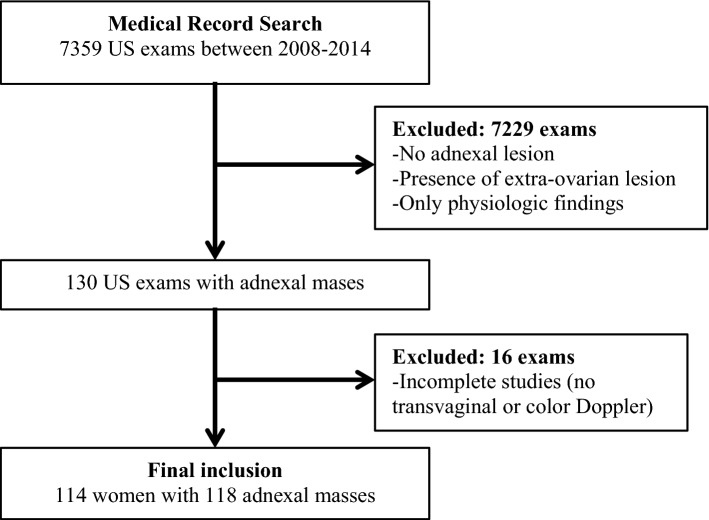
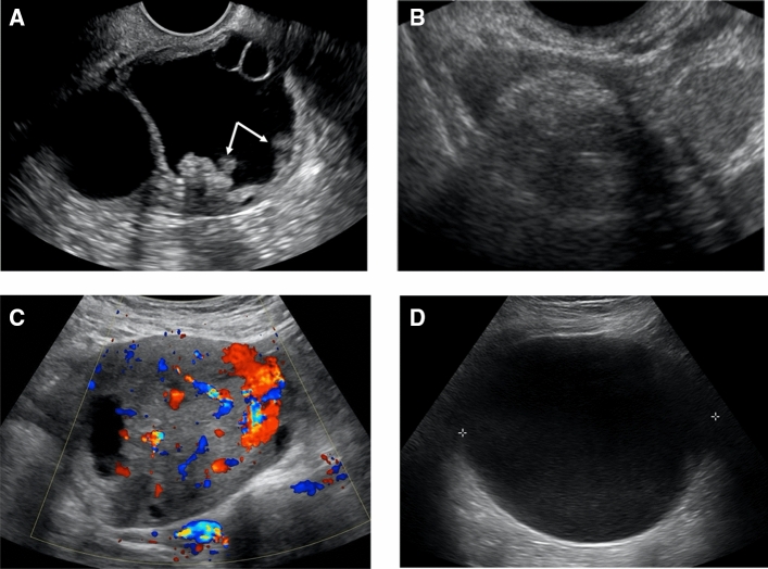
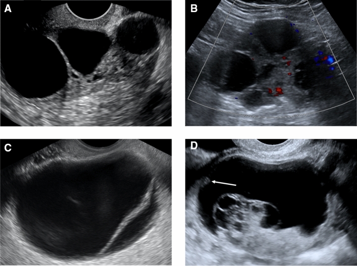
Methods: Pelvic ultrasound (US) examinations in 114 women with 118 adnexal masses were evaluated by eight radiologists blinded to the final diagnosis (4 attendings and 4 fellows) using IOTA simple rules and O-RADS lexicon. Each feature category was analyzed for interobserver agreement using intraclass correlation coefficient (ICC) for ordinal variables and free marginal kappa for nominal variables. The two-tailed significance level (a) was set at 0.05.
Results: For IOTA simple rules, interobserver agreement was almost perfect for three malignant lesion categories (M2-4) and substantial for the remaining two (M1, M5) with k-values of 0.80-0.82 and 0.68-0.69, respectively. Interobserver agreement was almost perfect for two benign feature categories (B2, B3), substantial for two (B4, B5) and moderate for one (B1) with k-values of 0.81-0.90, 0.69-0.70 and 0.60, respectively. For O-RADS, interobserver agreement was almost perfect for two out of ten feature categories (ascites and peritoneal nodules) with k-values of 0.89 and 0.97. Interobserver agreement ranged from fair to substantial for the remaining eight feature categories with k-values of 0.39-0.61. Fellows and attendings had ICC values of 0.725 and 0.517, respectively.
Conclusion: O-RADS had variable interobserver agreement with overall good agreement. IOTA simple rules had more uniform interobserver agreement with overall excellent agreement. Greater reader experience did not improve interobserver agreement with O-RADS.
Keywords: Adnexal; IOTA; O-RADS; Ovarian; Pelvic; Ultrasound.
© 2022. The Author(s).
Conflict of interest statement
All authors declare that they have no conflict of interest.
Figures
Comment in
-
Letter to the Editor.Abdom Radiol (NY). 2024 Oct;49(10):3758. doi: 10.1007/s00261-024-04220-z. Epub 2024 Jun 24. Abdom Radiol (NY). 2024. PMID: 38913138 No abstract available.
References
-
- Timmerman D, Ameye L, Fischerova D, Epstein E, Melis GB, Guerriero S, Van Holsbeke C, Savelli L, Fruscio R, Lissoni AA, Testa AC, Veldman J, Vergote I, Van Huffel S, Bourne T, Valentin L. Simple ultrasound rules to distinguish between benign and malignant adnexal masses before surgery: prospective validation by IOTA group. BMJ. 2010;14(341):c6839. doi: 10.1136/bmj.c6839. - DOI - PMC - PubMed
-
- Timmerman D, Van Calster B, Testa A, Savelli L, Fischerova D, Froyman W, et al. Predicting the risk of malignancy in adnexal masses based on the simple rules from the international ovarian tumor analysis group. Am J Obstet Gynecol. 2016;214(4):424–437. doi: 10.1016/j.ajog.2016.01.007. - DOI - PubMed
