. 2022 Jun 28;18(6):e1010272.
doi: 10.1371/journal.pgen.1010272. eCollection 2022 Jun.

Loss of the cleaved-protamine 2 domain leads to incomplete histone-to-protamine exchange and infertility in mice

Affiliations

Loss of the cleaved-protamine 2 domain leads to incomplete histone-to-protamine exchange and infertility in mice

Lena Arévalo et al. PLoS Genet. .

Abstract

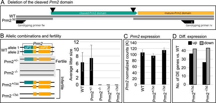
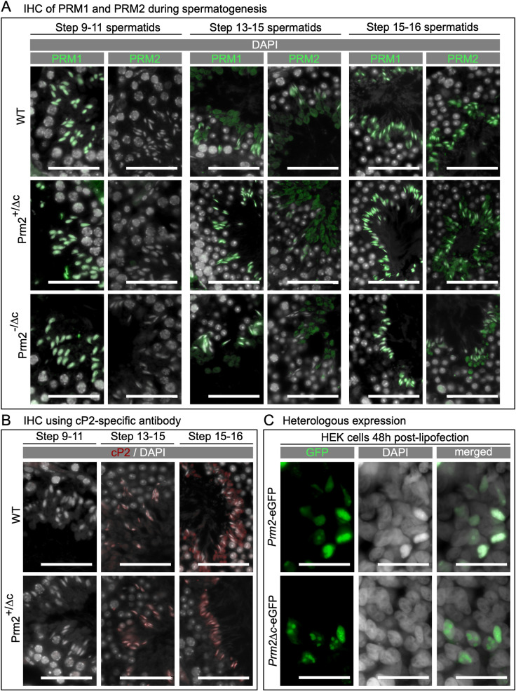
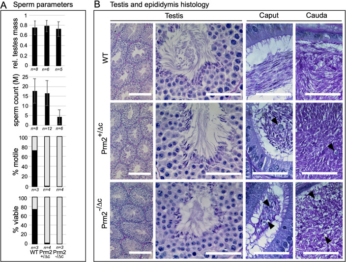
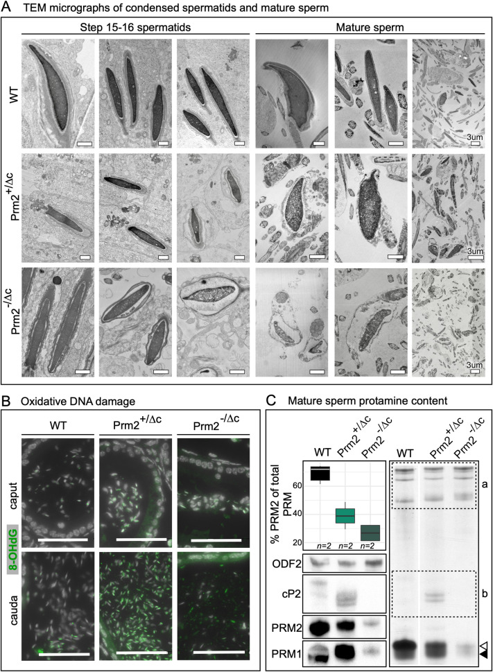
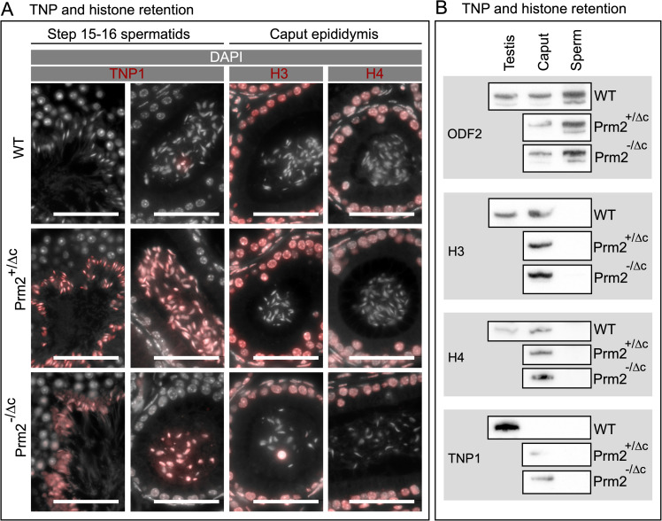
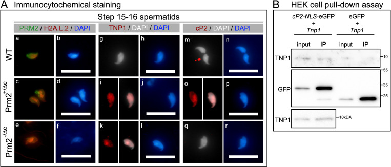
Protamines are unique sperm-specific proteins that package and protect paternal chromatin until fertilization. A subset of mammalian species expresses two protamines (PRM1 and PRM2), while in others PRM1 is sufficient for sperm chromatin packaging. Alterations of the species-specific ratio between PRM1 and PRM2 are associated with infertility. Unlike PRM1, PRM2 is generated as a precursor protein consisting of a highly conserved N-terminal domain, termed cleaved PRM2 (cP2), which is consecutively trimmed off during chromatin condensation. The carboxyterminal part, called mature PRM2 (mP2), interacts with DNA and together with PRM1, mediates chromatin-hypercondensation. The removal of the cP2 domain is believed to be imperative for proper chromatin condensation, yet, the role of cP2 is not yet understood. We generated mice lacking the cP2 domain while the mP2 is still expressed. We show that the cP2 domain is indispensable for complete sperm chromatin protamination and male mouse fertility. cP2 deficient sperm show incomplete protamine incorporation and a severely altered protamine ratio, retention of transition proteins and aberrant retention of the testis specific histone variant H2A.L.2. During epididymal transit, cP2 deficient sperm seem to undergo ROS mediated degradation leading to complete DNA fragmentation. The cP2 domain therefore seems to be a key aspect in the complex crosstalk between histones, transition proteins and protamines during sperm chromatin condensation. Overall, we present the first step towards understanding the role of the cP2 domain in paternal chromatin packaging and open up avenues for further research.


Conflict of interest statement

The authors have declared that no competing interests exist.

Figures

Fig 1
Fig 1. Gene editing, fertility and expression.
(A) Schematic representation of the generation of cP2 deletion. Double strand breaks induced by Cas9 indicated by black triangles. (B) Schematic overview of analyzed genotypes and fertility. Prm2+/- and Prm2-/- [6] were included as a comparison. Bar plot of average litter size for WT, Prm2+/-, Prm2-/-, Prm2+/Δc and Prm2-/Δc, n = 5 for each genotype. (C) Bar plot showing average DESeq2 normalized read counts of Prm2 for WT, Prm2+/Δc and Prm2-/Δc. (D) Bar plot showing comparison between number of differentially higher and lower expressed genes for Prm2+/Δc and Prm2-/Δc compared to wildtype.
Fig 2
Fig 2. Localization and translation timing of PRM1, PRM2, unprocessed PRM2 and DNA condensing ability of Prm2Δc.
(A) Immunohistochemical fluorescent staining of PRM2 (WT, Prm2+/Δc) or mP2 (Prm2+/Δc, Prm2-/Δc) (green) and PRM1 (all genotypes) (green) in testis sections, counterstaining with DAPI (pseudo-colored grey). Scale bar = 50μm. (B) Immunohistochemical fluorescent staining of unprocessed PRM2 (red) using a cP2-specific antibody in testis sections, counterstained with DAPI (pseudo-colored grey). Scale bar = 50μm. C) Fluorescent images of human embryonic kidney 293 (HEK) cells 48 hours post-transfection with plasmids encoding eGFP tagged PRM2 (Prm2-eGFP) or Prm2Δc (Prm2Δc-eGFP) (green), counterstained with Hoechst (pseudo-colored grey). Scale bar = 50μm.
Fig 3
Fig 3. Sperm and testis parameters and histology.
(A) Bar plots showing data for relative testes mass, mature sperm count, percentage of viable mature sperm (eosin-nigrosin assay) and percentage of motile mature sperm in Prm2+/Δc and Prm2-/Δc mice compared to wildtype. (B) PAS staining of testis and epididymal sections of Prm2+/Δc, Prm2-/Δc and WT males. Scale bar = 50μm (200μm for left column).
Fig 4
Fig 4. Chromatin integrity, oxidative damage and protamine content.
(A) Electron micrographs of condensed spermatids and mature sperm of Prm2+/Δc, Prm2-/Δc and WT. Scale bars = 1μm unless otherwise indicated. (B) Immunohistochemical fluorescent staining of 8-Oxo-2’-deoxyguanosine (8-OHdG) (green) in caput epididymis (upper row) and cauda epididymis (lower row) of Prm2+/Δc, Prm2-/Δc and WT. Counterstained with DAPI (pseudo-colored grey). Scale bar = 50μm. (C) Boxplot showing the percentage of PRM2 (including PRM2 precursors) of total protamine by band density analysis of Coomassie stained acid urea gel electrophoresis (AU-PAGE) (S8C Fig). Asterisk indicates significant difference. To the right: AU-PAGE of WT, Prm2+/Δc and Prm2-/Δc mature sperm basic nuclear protein extractions, equal amounts of pooled sample loaded per genotype. a = non-protamine basic proteins, b = PRM2 precursors, open arrowhead indicates mature PRM2 band, solid arrowhead indicates PRM1 band. Below: Immunoblot of PRM1, PRM2, cP2 (indicating the presence of PRM2 precursor) and ODF2.
Fig 5
Fig 5. Histone H3, histone H4 and transition protein 1 IHC staining and western blot detection.
(A) Two left columns: Immunohistochemical fluorescent staining of TNP1 (red) in WT, Prm2+/Δc and Prm2-/Δc step 15–16 spermatids and caput epididymis sections, counterstained with DAPI (pseudo-colored grey). Scale bar = 50μm. Two right columns: Immunohistochemical fluorescent staining of Histone H3 (H3) or Histone H4 (H4) (red) in WT, Prm2+/Δc and Prm2-/Δc caput epididymis sections counterstained with DAPI (pseudo-colored grey). Scale bar = 50μm. (B) Immunoblots against TNP1 and H3 and H4 and ODF2 as a control in testis (WT), caput epididymis (caput) and mature sperm (sperm) basic nuclear protein extractions (WT, Prm2+/Δc and Prm2-/Δc).
Fig 6
Fig 6. Differential abundance of mature sperm basic nuclear proteins.
(A) volcano plots showing differential abundance (DA) of basic nuclear proteins in Prm2+/Δc compared to WT (upper plot) and Prm2-/Δc compared to WT (lower plot). Significantly DA proteins are indicated in color (teal = lower abundant, yellow = higher abundant). Top DA proteins and proteins of interest are labeled with their corresponding gene symbol. (B) Venn diagram showing the overlap between DA proteins found in the three different comparisons (WT vs. Prm2+/Δc, WT vs. Prm2-/Δc and WT vs. Prm2-/-). Proteins present in overlaps of interest are listed with their corresponding gene symbol. H2A.L.2 is marked in red. (C) Boxplot of median normalized log2 abundance of H2A.L.2 in WT, Prm2-/-, Prm2+/Δc and Prm2-/Δc. (D) Immunoblots against H2A.L.2 and ODF2 as a control in testis (WT), caput epididymis (caput) and mature sperm (sperm) basic nuclear protein extractions (WT, Prm2+/Δc and Prm2-/Δc).
Fig 7
Fig 7. Immunocytochemical staining of H2A.L.2 and PRM2 as well as TNP1 and unprocessed PRM2.
(A) (a-f) Immunocytochemical fluorescent staining of H2A.L.2 (red) and PRM2 (green) in WT, Prm2+/Δc and Prm2-/Δc step 15–16 spermatids from tubule preparations. Counterstained with DAPI (blue). (g-l) Immunocytochemical fluorescent staining of TNP1 (red) in WT, Prm2+/Δc and Prm2-/Δc step 15–16 spermatids from tubule preparations. Counterstained with DAPI (pseudo-colored grey or blue). (m-r) Immunocytochemical fluorescent staining of cP2 indicating the PRM2 precursor (red) in WT, Prm2+/Δc and Prm2-/Δc step 15–16 spermatids from tubule preparations. Counterstained with DAPI (pseudo-colored grey or blue). Scale bar = 20μm. (B) Immunoblots against GFP and TNP1 of GFP pull-down assay of HEK293 cells co-transfected with cP2-NLS-eGFP (pCP2-NLS-eGFP-N3) and untagged TNP1 (pTnp1-STOP-mCherry-N1) or eGFP (pEGFP-N3) and untagged TNP1. Immunoblot was repeated for cP2-NLS-eGFP and untagged TNP1 co-transfected samples (lower blot).

References

    1. Chen H, Cheung M, Chow P, Cheung A, Liu W, O W. Protection of sperm DNA against oxidative stress in vivo by accessory sex gland secretions in male hamsters. Reproduction. 2002;124: 491–499. doi: 10.1530/rep.0.1240491 - DOI - PubMed
    1. Tourmente M, Gomendio M, Roldan ER. Sperm competition and the evolution of sperm design in mammals. Bmc Evol Biol. 2011;11: 12. doi: 10.1186/1471-2148-11-12 - DOI - PMC - PubMed
    1. Balhorn R. The protamine family of sperm nuclear proteins. Genome Biol. 2007;8: 227. doi: 10.1186/gb-2007-8-9-227 - DOI - PMC - PubMed
    1. Rathke C, Baarends WM, Awe S, Renkawitz-Pohl R. Chromatin dynamics during spermiogenesis. Biochimica et Biophysica Acta. 2014;1839: 155–168. doi: 10.1016/j.bbagrm.2013.08.004 - DOI - PubMed
    1. Aoki VW, Carrell DT. Human protamines and the developing spermatid: their structure, function, expression and relationship with male infertility. Asian J Androl. 2003;5: 315–24. - PubMed

Publication types