. 2022 Nov;530(16):2881-2900.
doi: 10.1002/cne.25382. Epub 2022 Jun 28.

Oxytocin receptors are widely distributed in the prairie vole (Microtus ochrogaster) brain: Relation to social behavior, genetic polymorphisms, and the dopamine system

Affiliations

Oxytocin receptors are widely distributed in the prairie vole (Microtus ochrogaster) brain: Relation to social behavior, genetic polymorphisms, and the dopamine system

Kiyoshi Inoue et al. J Comp Neurol. 2022 Nov.

Abstract

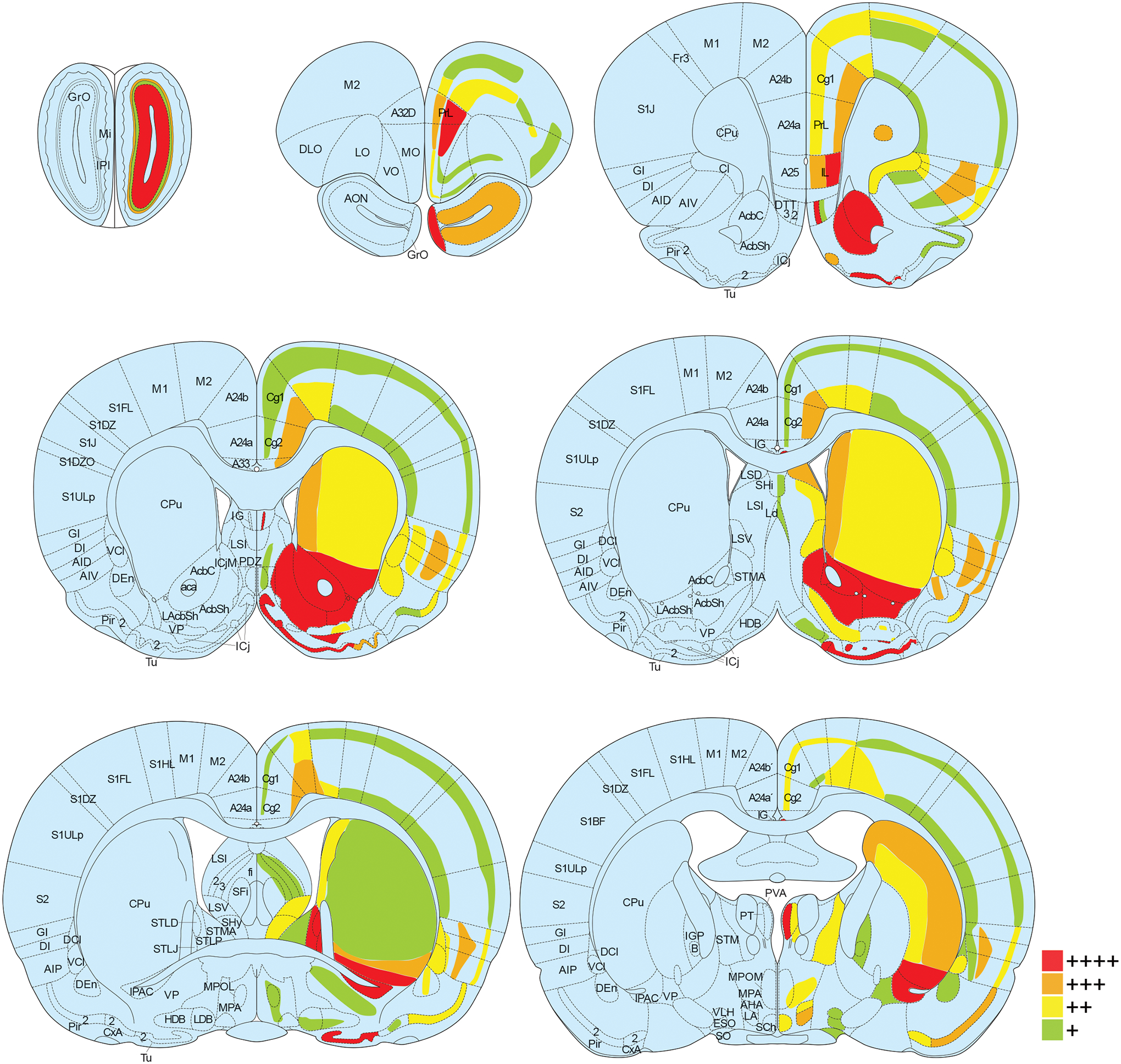
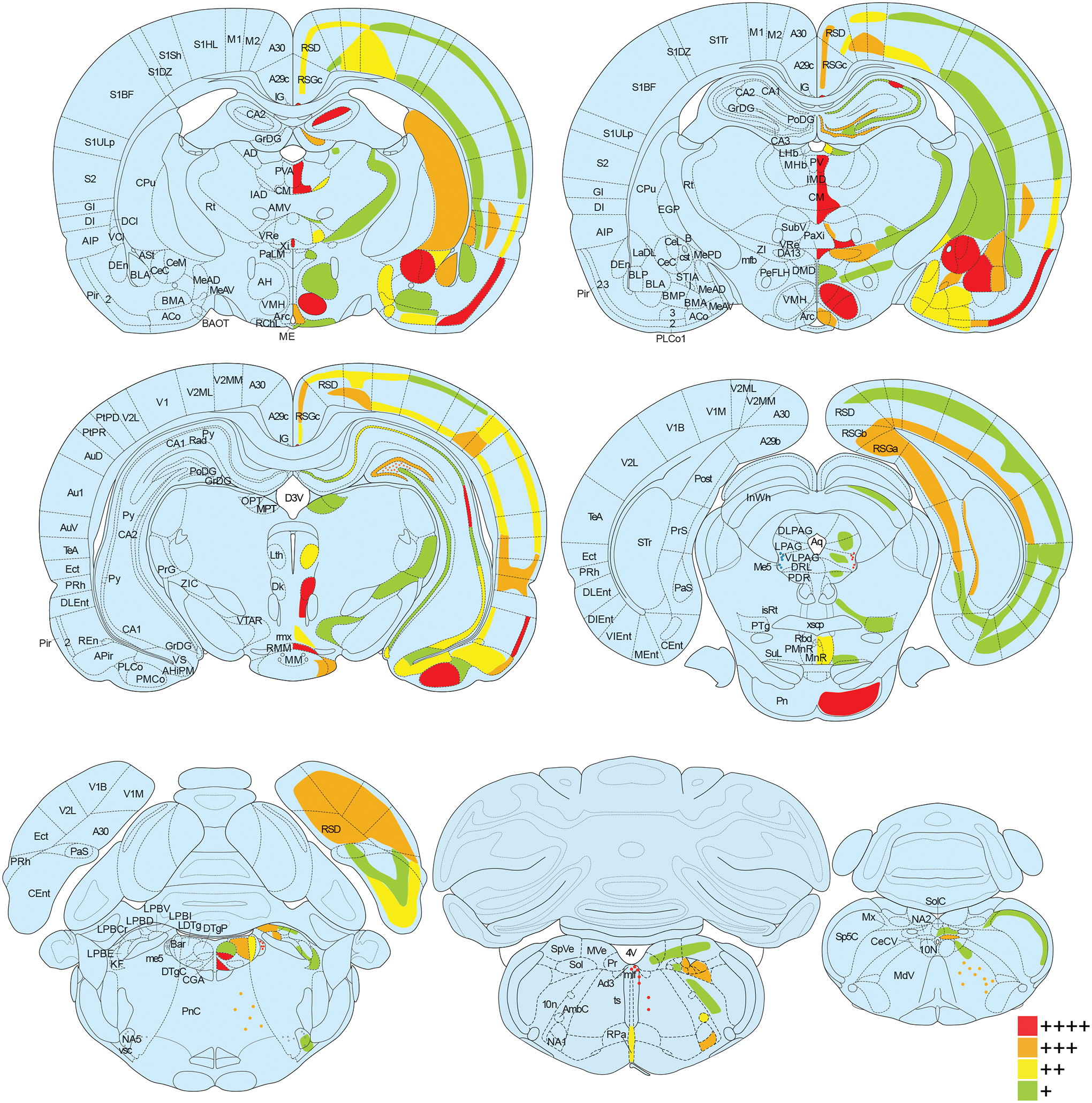
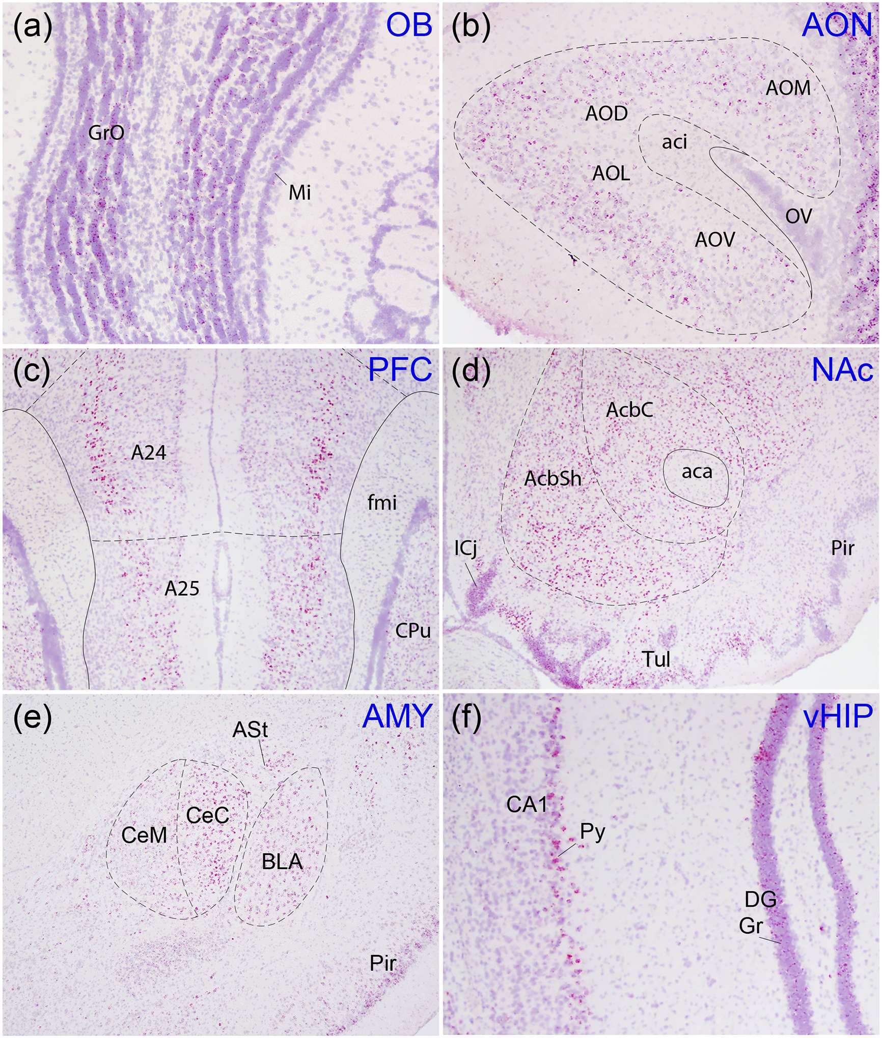
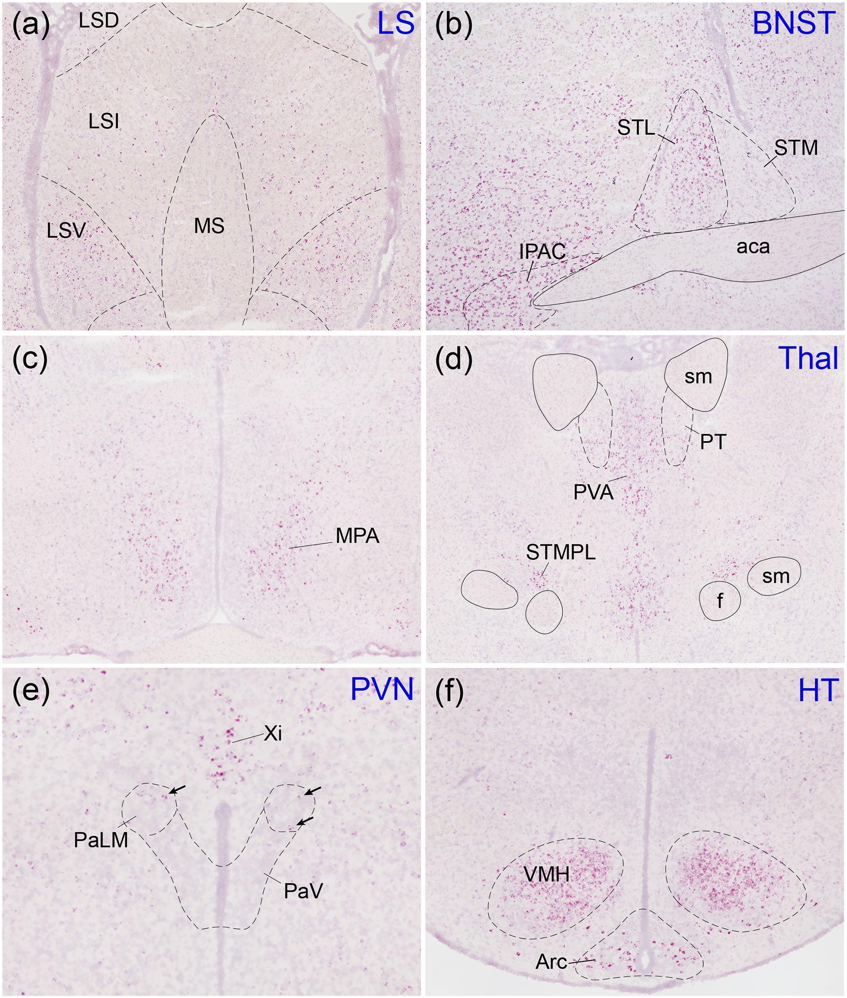
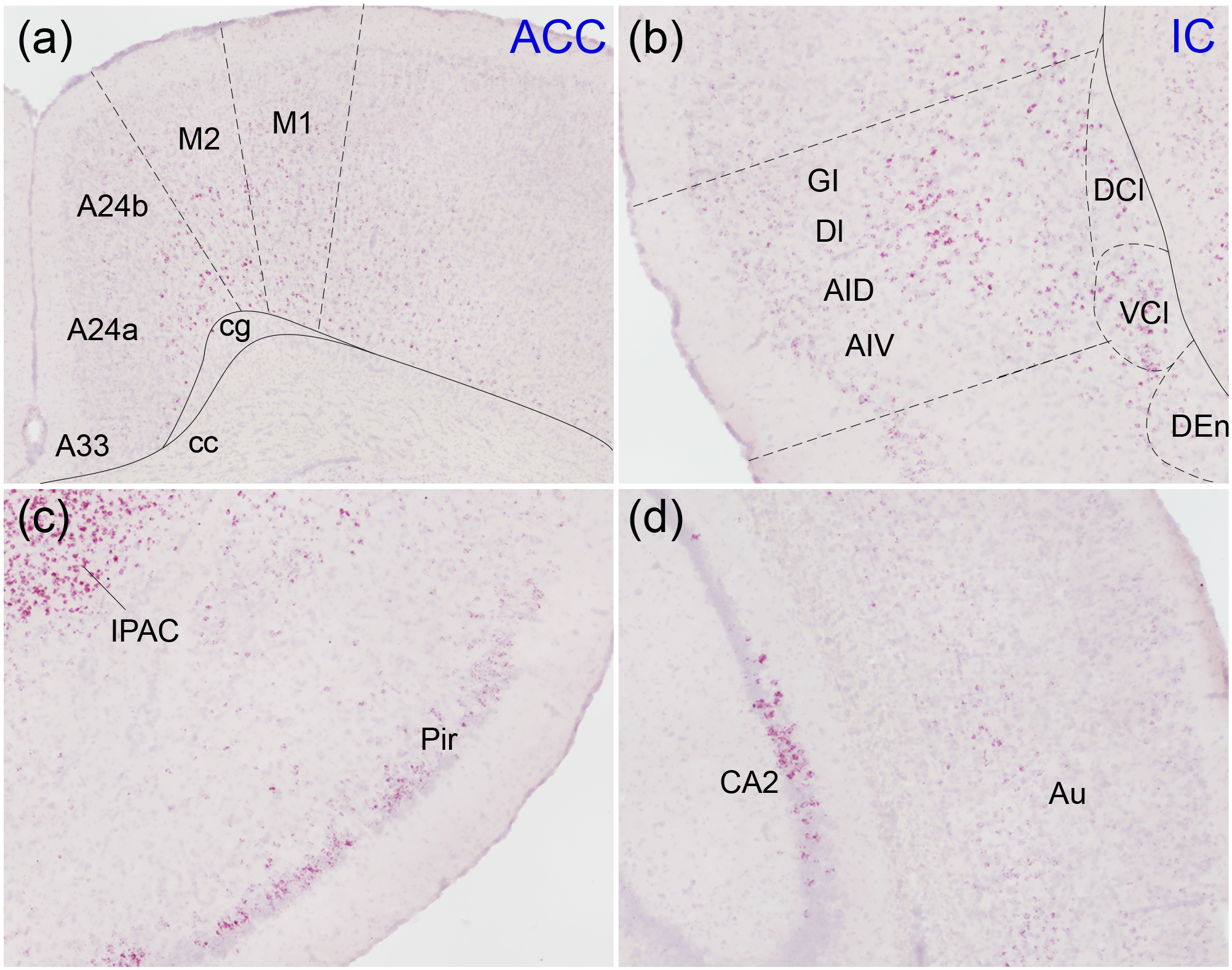
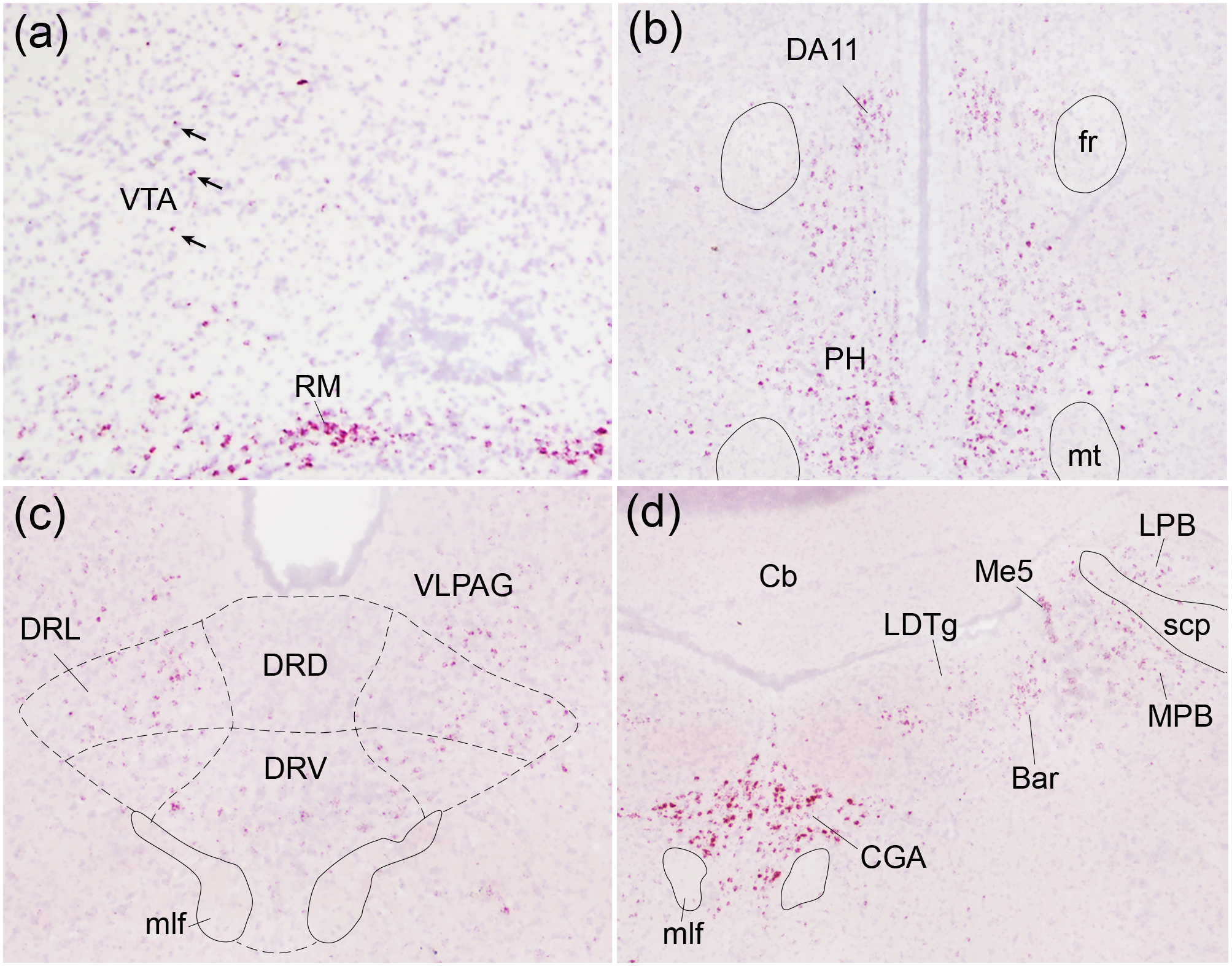
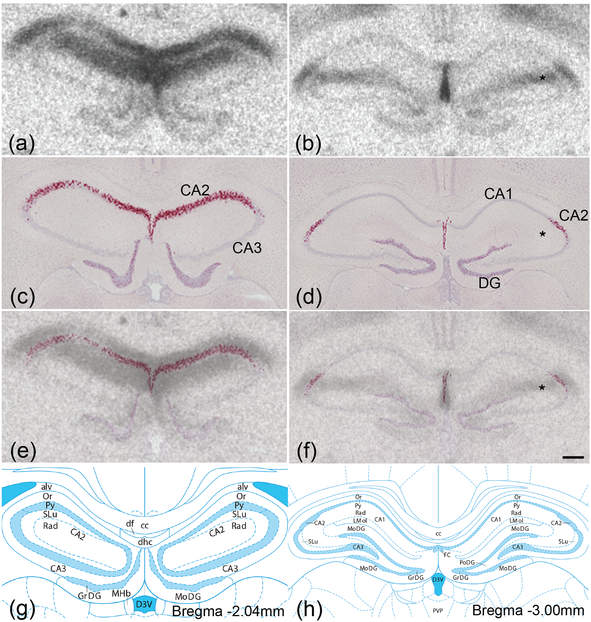
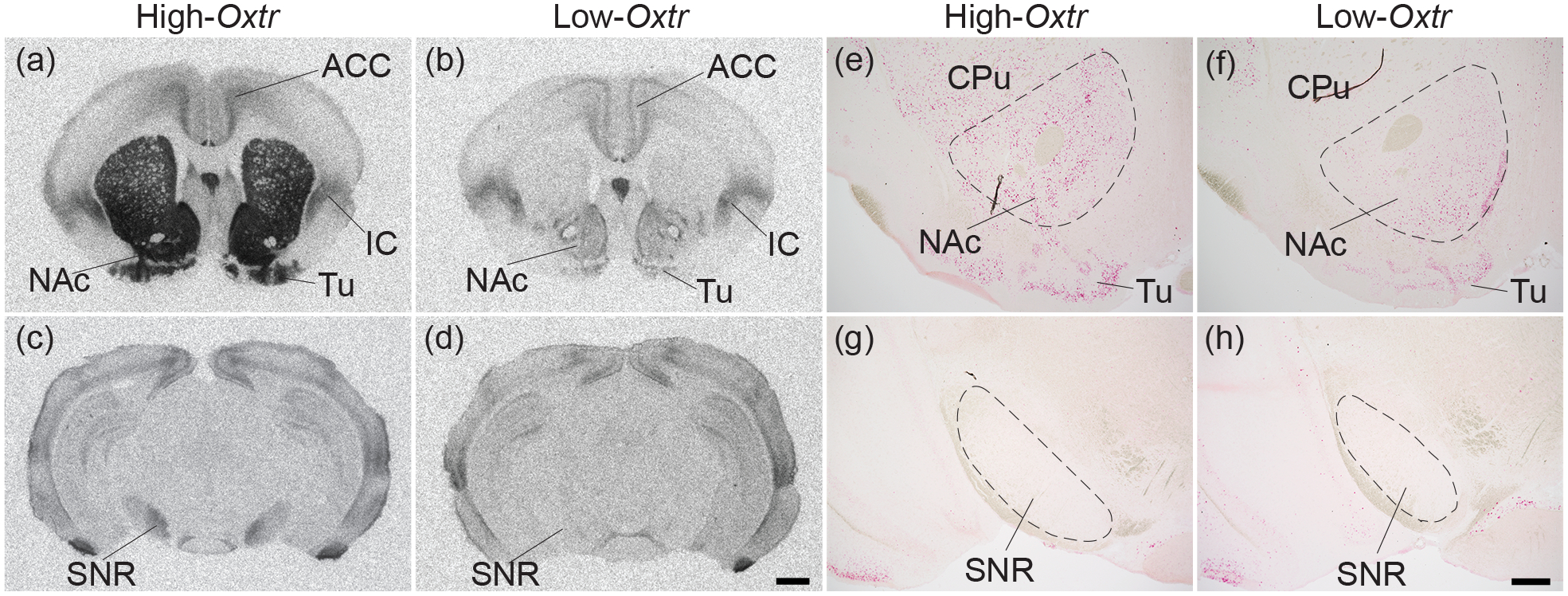
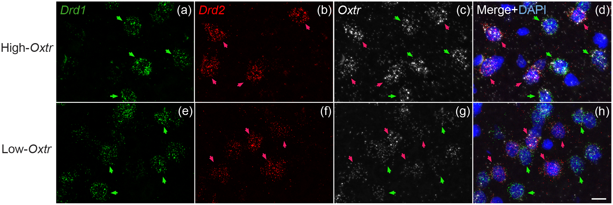
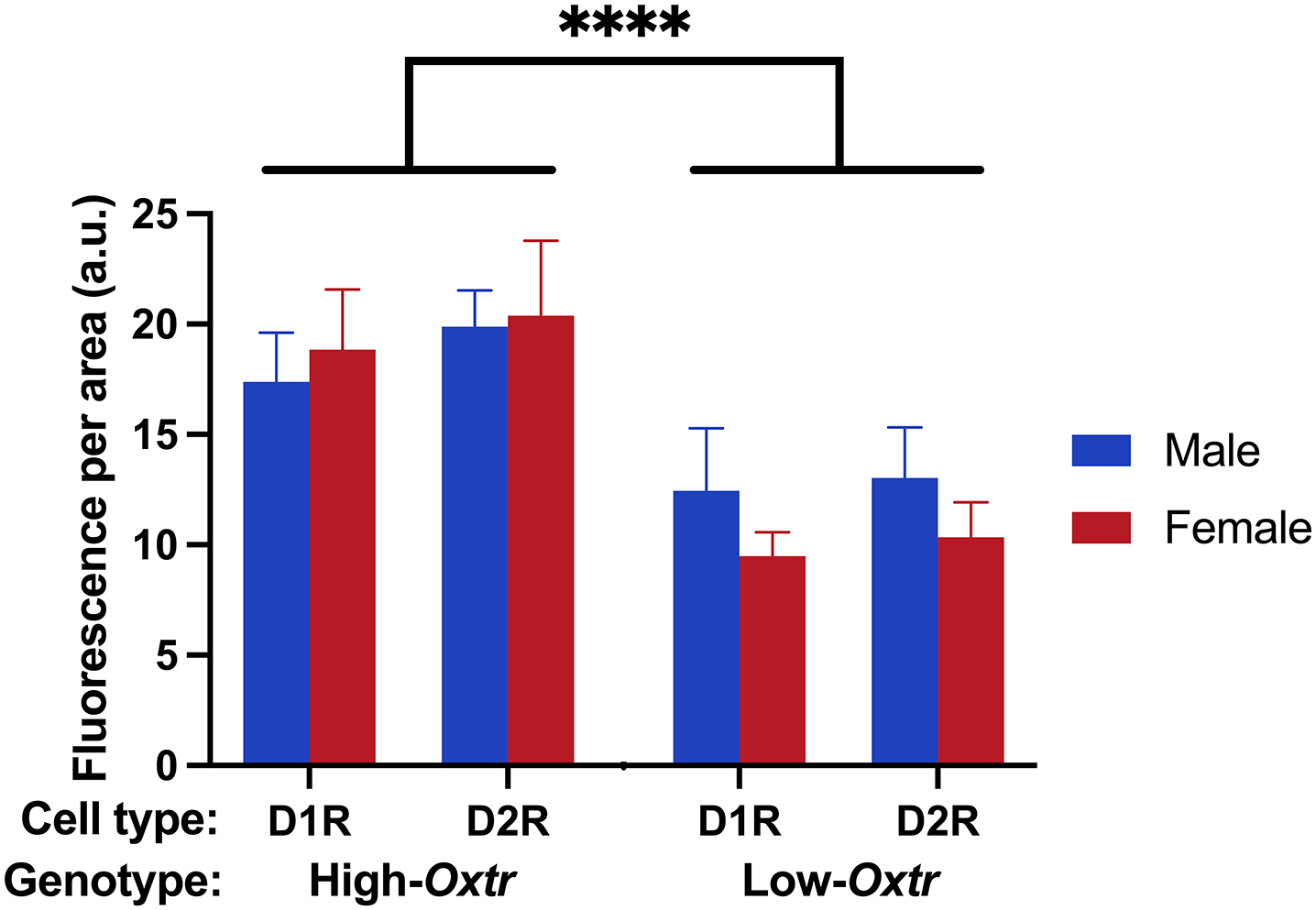
Oxytocin regulates social behavior via direct modulation of neurons, regulation of neural network activity, and interaction with other neurotransmitter systems. The behavioral effects of oxytocin signaling are determined by the species-specific distribution of brain oxytocin receptors. The socially monogamous prairie vole has been a useful model organism for elucidating the role of oxytocin in social behaviors, including pair bonding, response to social loss, and consoling. However, there has been no comprehensive mapping of oxytocin receptor-expressing cells throughout the prairie vole brain. Here, we employed a highly sensitive in situ hybridization, RNAscope, to construct an exhaustive, brain-wide map of oxytocin receptor mRNA-expressing cells. We found that oxytocin receptor mRNA expression was widespread and diffused throughout the brain, with specific areas displaying a particularly robust expression. Comparing receptor binding with mRNA revealed that regions of the hippocampus and substantia nigra contained oxytocin receptor protein but lacked mRNA, indicating that oxytocin receptors can be transported to distal neuronal processes, consistent with presynaptic oxytocin receptor functions. In the nucleus accumbens, a region involved in oxytocin-dependent social bonding, oxytocin receptor mRNA expression was detected in both the D1 and D2 dopamine receptor-expressing subtypes of cells. Furthermore, natural genetic polymorphisms robustly influenced oxytocin receptor expression in both D1 and D2 receptor cell types in the nucleus accumbens. Collectively, our findings further elucidate the extent to which oxytocin signaling is capable of influencing brain-wide neural activity, responses to social stimuli, and social behavior. KEY POINTS: Oxytocin receptor mRNA is diffusely expressed throughout the brain, with strong expression concentrated in certain areas involved in social behavior. Oxytocin receptor mRNA expression and protein localization are misaligned in some areas, indicating that the receptor protein may be transported to distal processes. In the nucleus accumbens, oxytocin receptors are expressed on cells expressing both D1 and D2 dopamine receptor subtypes, and the majority of variation in oxytocin receptor expression between animals is attributable to polymorphisms in the oxytocin receptor gene.

Keywords: Oxtr single nucleotide polymorphisms; autoradiography; dopamine receptor; in situ hybridization; nucleus accumbens; oxytocin receptors mRNA; phenotypic plasticity.


Conflict of interest statement

Conflict of interest: The authors declare no conflicts of interest.

Figures

Figure 1.
Figure 1.. Oxtr mRNA expression throughout prairie vole brain.
Oxtr mRNA was labeled with in situ hybridization and the percent of cells positive for Oxtr was calculated for each brain region. Brain regions are color coded according to Oxtr expression density (green for 1–25% of cells expressing Oxtr, yellow for 26–50%, orange for 51–75%, and red for 76–100%) in coronal sections from a rat brain atlas at 14 points along the rostral-caudal axis (Paxinos, 2014). For some regions (i.e. reticular formation, molecular layer of the hippocampus) with sparse cells containing exceptionally strong expression, red dots were used. Abbreviations on the left half of the images correspond to new nomenclature used in the seventh edition of “The Rat Brain Atlas in Stereotaxic Coordinates” (Paxinos, 2014), while the abbreviations on the right half of the images correspond to traditional nomenclature used in previous editions. See Supplementary Table 1 for definitions of abbreviations.
Figure 2.
Figure 2.. Oxtr expression in regions involved in social salience and memory.
Oxtr mRNA (red) was labeled with in situ hybridization against a Hematoxylin counterstain (purple). The olfactory bulb (a), anterior olfactory area (AO; AOV, ventral; AOL, lateral; AOD, dorsal; AOM, medial; b), prefrontal cortex (c), nucleus accumbens (AcbSh, shell; AcbC, core; d), amygdala (e), and ventral hippocampus (f) all showed strong labeling. GrO, granule cell layer of olfactory bulb; Mi, mitral cell layer of olfactory bulb; aci, anterior commissure, intrabulbar; OV, olfactory ventricle; A24, cingulate cortex previously known as prelimbic cortex; A25, cingulate cortex previously known as infralimbic cortex; fmi, forceps minor of the corpus callosum; CPu, caudate putamen; ICj, island of Calleja; Tul, olfactory tubercle; aca, anterior commissure, anterior part; CeC, central amygdaloid nucleus, capsular; CeM, central amygdaloid nucleus, medial; BLA, basolateral amygdaloid nucleus, anterior; ASt, amygdalostriatal transition area; Pir, piriform cortex; Py, stratum pyramidale; DG, dentate gyrus; Gr, granule cell layer of the dentate gyrus.
Figure 3.
Figure 3.. Oxtr expression in hypothalamic regions.
Oxtr mRNA (red) was labeled with in situ hybridization against a Hematoxylin counterstain (purple). The lateral septum (a) contained moderate expression concentrated mostly in the ventral (LSV) portion. Oxtr was robustly expressed in the lateral division of the bed nucleus of the stria terminalis (STL) and the interstitial nucleus of the posterior limb of the anterior commissure (IPAC; b) as well as the ventromedial nucleus of the hypothalamus (VMH; f). The medial preoptic area (MPA; c), thalamus (d), and arcuate nucleus of the hypothalamus (Arc; f) displayed moderate expression while the paraventricular nucleus of the hypothalamus (PaLM, lateral magnocellular; PaV, ventral part; e; arrows indicate Oxtr-positive cells) displayed sparse expression. LSI, lateral septal nucleus, intermediate; LSD, lateral septal nucleus, dorsal; MS, medial septal nucleus; STM, bed nucleus of the stria terminalis, medial division; aca, anterior commissure, anterior part; PT, paratenial thalamic nucleus; PVA, paraventricular thalamic nucleus, anterior; sm, stria medullaris; f, fornix; STMPL, bed nucleus of the stria terminalis, medial, posterolateral; Xi, xiphoid thalamic nucleus.
Figure 4.
Figure 4.. Oxtr expression in neuromodulatory areas.
Oxtr mRNA (red) was labeled with in situ hybridization against a Hematoxylin counterstain (purple). The anterior cingulate cortex (A24a, A24b, A33; a), insular cortex (b), piriform cortex (Pir; c), and secondary auditory cortex (Au; d) all contained Oxtr primarily restricted to cortical layers 2, 5, and 6. M1, primary motor cortex; M2, secondary motor cortex; cg, cingulum; cc, corpus callosum; GI, granular insular cortex; DI, dysgranular insular cortex; AID, agranular insular cortex, dorsal; AIV, agranular insular cortex, ventral; DCl, dorsal claustrum; VCl, ventral claustrum; DEn, dorsal endopiriform nucleus; IPAC, interstitial nucleus of the posterior limb of the anterior commissure.
Figure 5.
Figure 5.. Oxtr expression in other regions of interest.
Oxtr mRNA (red) was labeled with in situ hybridization against a Hematoxylin counterstain (purple). The ventral tegmental area (VTA; a) and dorsal raphe nucleus (DR; c) displayed spare Oxtr labeling while the retromammillary nucleus (RM; a), posterior hypothalamic nucleus (PH; b), and central gray, alpha part (CGA; d) displayed robust labeling. Arrows indicate Oxtr-positive cells within VTA; DA11, DA11 dopamine cells; fr, fasciculus retroflexus; mt, mammillothalamic tract; DRD, dorsal raphe nucleus, dorsal part; DRL, dorsal raphe nucleus, lateral part; DRV, dorsal raphe nucleus, ventral part; VLPAG, ventrolateral periaqueductal gray; mlf, medial longitudinal fasciculus; CG, central gray, LDTg, laterodorsal tegmental nucleus; LPB, lateral parabrachial nucleus; MPB, medial parabrachial nucleus; Me5, mesencephalic trigeminal nucleus; Bar, Barrington’s nucleus; Cb, cerebellum; scp, superior cerebellar peduncle.
Figure 6.
Figure 6.. Oxtr mRNA and OXTR protein mismatch in hippocampus.
OXTR protein (black; a,b) was labeled with autoradiography and, in adjacent sections, Oxtr mRNA (red; c,d) was labeled with in situ hybridization against a Hematoxylin counterstain (purple). Images from adjacent autoradiography and in situ hybridization sections were merged (e,f) to compare protein and mRNA localization. Oxtr mRNA was restricted to stratum pyramidale, the principal cell layer containing the somas of pyramidal cells, while robust OXTR protein binding was present superficial and basal to stratum pyramidale, most likely in the stratum radiatum, stratum lacunosum-moleculare, and stratum oriens. Illustrations from the Rat brain Atlas indicate that Oxtr mRNA was enriched in the CA2 in the Ammon’s horn (g,h). DG, dentate gyrus; * denotes OXTR protein in the absence of Oxtr mRNA. Scale bar =300μm shown in (f).
Figure 7.
Figure 7.. Genotype-dependent Oxtr mRNA and OXTR protein mismatch in substantia nigra pars reticulata.
OXTR protein (black; a-d) was labeled with autoradiography and, in adjacent sections, Oxtr mRNA (red; e-h) was labeled with in situ hybridization against a Hematoxylin counterstain (purple) in High-Oxtr voles (a,c,e,g) and Low-Oxtr voles (b,d,f,h). High-Oxtr and Low-Oxtr genotypes predicted protein binding in the nucleus accumbens (NAc) and substantia nigra pars reticulata (SNR), and mRNA expression in NAc. However, no Oxtr mRNA was detected in the SNR of either genotype. CPu, caudate putamen; ACC, anterior cingulate cortex; Tu, olfactory tubercle; IS, insular cortex. Scale bars for (a-d) =1mm shown in (d), for (e-h) =400μm shown in (h).
Figure 8.
Figure 8.. Oxtr mRNA colocalized with both Drd1 and Drd2 mRNA in NAc.
Drd1 (green; a,e), Drd2 (red; b,f), and Oxtr (white; c,g) mRNA were labeled with in situ hybridization in both High- and Low-Oxtr genotypes (a-d, e-h, respectively). Oxtr was expressed in both Drd1- and Drd2-expressing cells (d,h) in both genotypes. Scale bar =10μm shown in (h).
Figure 9.
Figure 9.. Genotype predicts Oxtr mRNA expression in both D1R and D2R cells in NAc.
The intensity of Oxtr labeling was quantified in D1R and D2R cells of both genotypes (High-Oxtr and Low-Oxtr) and sexes. A three-way mixed model ANOVA revealed a significant main effect of genotype accounting for 70.81% of variation in Oxtr expression. Post hoc tests revealed significant differences in Oxtr expression between High- and Low-Oxtr genotypes in each sex-by-cell-type comparison. ****, p < 0.0001.

Similar articles

Cited by

References

    1. Ahern TH, Olsen S, Tudino R, & Beery AK (2021). Natural variation in the oxytocin receptor gene and rearing interact to influence reproductive and nonreproductive social behavior and receptor binding. Psychoneuroendocrinology, 128, 105209. doi:10.1016/j.psyneuen.2021.105209 - DOI - PMC - PubMed
    1. Althammer F, & Grinevich V (2017). Diversity of oxytocin neurons: beyond magno- and parvocellular cell types? J Neuroendocrinol. doi:10.1111/jne.12549 - DOI - PubMed
    1. Andari E, Hurlemann R, & Young LJ (2018). A Precision Medicine Approach to Oxytocin Trials. Curr Top Behav Neurosci, 35, 559–590. doi:10.1007/7854_2017_29 - DOI - PMC - PubMed
    1. Andari E, Massa NM, Fargotstein MD, Taylor NB, Halverson DM, Owens AV, … Duncan EJ (2021). Effects of Oxytocin on Emotion Recognition in Schizophrenia: A Randomized Double-Blind Pilot Study. J Clin Psychopharmacol, 41(2), 103–113. doi:10.1097/jcp.0000000000001367 - DOI - PMC - PubMed
    1. Aragona BJ, Liu Y, Yu YJ, Curtis JT, Detwiler JM, Insel TR, & Wang Z (2006). Nucleus accumbens dopamine differentially mediates the formation and maintenance of monogamous pair bonds. Nat Neurosci, 9(1), 133–139. doi:10.1038/nn1613 - DOI - PubMed

Publication types