. 2022 Aug:54:102377.
doi: 10.1016/j.redox.2022.102377. Epub 2022 Jun 18.

Ablation of long noncoding RNA MALAT1 activates antioxidant pathway and alleviates sepsis in mice

Affiliations

Ablation of long noncoding RNA MALAT1 activates antioxidant pathway and alleviates sepsis in mice

Jingshu Chen et al. Redox Biol. 2022 Aug.

Abstract

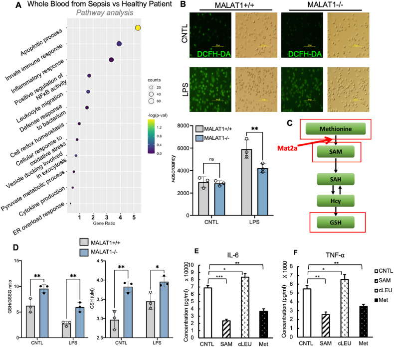
The metastasis-associated lung adenocarcinoma transcript1 (MALAT1) is a long noncoding RNA (lncRNA) and is known for its role in cancer development and prognosis. In this study, we report that MALAT1 plays an important role in regulating acute inflammatory responses in sepsis. In patient samples, MALAT1 expression was positively correlated with severity of sepsis. In cultured macrophages, LPS treatment significantly induced MALAT1 expression, while genetic ablation of MALAT1 greatly reduced proinflammatory cytokine levels. Furthermore, MALAT1-ablated mice had significantly increased survival rates in cecal ligation and puncture (CLP)-induced sepsis and LPS-induced endotoxemia. One novel and salient feature of MALAT1-ablated mice is greatly reduced ROS level in macrophages and other cell types and increased glutathione/oxidized glutathione (GSH/GSSG) ratio in macrophages, suggesting an increased antioxidant capacity. We showed a mechanism for MALAT1 ablation leading to enhanced antioxidant capacity is through activation of methionine cycle by epitranscriptomical regulation of methionine adenosyltransferase 2A (MAT2A). MAT2A 3'UTR can be methylated by METTL16 which was known to directly bind to MALAT1. MALAT1 ablation was found to reduce methylation in MAT2A hairpin1 and increase MAT2A protein levels. Our results suggest a MALAT1-METTL16-MAT2A interactive axis which may be targeted for treatments of sepsis.


Conflict of interest statement

The authors have declared that no conflict of interest exists.

Figures

Image 1
Graphical abstract
Fig. 1
Fig. 1
MALAT1 is induced by sepsis process in both cells and sepsis patients. A, MALAT1 expression level in mouse BMDM M0 (resting phase) and M1 (pro-inflammatory phase) determined by qRT-PCR. B, MALAT1 expression level in mouse PBMC treated with LPS (5 μg/mL, 12 h) determined by qRT-PCR. C, MALAT1 expression level in THP-1 derived macrophage treated with LPS (5 μg/mL, 12h) determined by qRT-PCR. D, Time-dependent MALAT1 expression level in mouse PM treated with LPS (5 μg/mL) determined by qRT-PCR. E, MALAT1 expression in transcriptomic analysis of whole blood sample from patient diagnosed with late-onset sepsis. F, MALAT1 expression visualized in t-distributed stochastic neighbor embedding (t-SNE) plot from in alveolar macrophages of COVID-19 with severe symptom than those with mild symptom.
Fig. 2
Fig. 2
Ablation of MALAT1 leads to inhibited proinflammatory response in peritoneal macrophages. A-C, LPS induced pro-inflammatory gene expression in wild type (MALAT1+/+) and MALAT1 ablated (MALAT1−/−) PM treated with LPS (5 μg/mL) determined by qRT-PCR. D-F, Pro-inflammatory cytokine levels in the supernatant of PM isolated from MALAT1+/+ and MALAT1−/− mice treated with LPS (5 μg/mL, 0–8 h). G, Cytokine levels from culture supernatant of PM treated with LPS (5 μg/mL, 8 h) from both MALAT1+/+ and MALAT1−/− mice were determined using mouse cytokine array. H, NF-κB and NLRP3 inflammasome signaling pathway in PM treated with LPS (5 μg/mL, 0, 0.5, 1, 2, 4, 6 h) from MALAT1+/+ and MALAT1−/− mice. I and J, NLRP3 and Caspase1 gene expression level in MALAT1+/+ and MALAT1−/− PM treated with LPS (5 μg/mL, 6 h) determined by qRT-PCR.
Fig. 3
Fig. 3
MALAT1 ablation alleviates septic shock and increases the survival rate in both LPS-induced and CLP-induced mouse sepsis model. A, Kaplan-Meier survival curves of MALAT1 ablated mice (n = 13) and wild type controls (n = 8) in response to i.p. injection of LPS, p = 0.0016 determined by Gehan-Breslow-Wilcoxon test. B, CLP induced mouse sepsis model. The ligation percentage of the cecum is 50%. C, Kaplan-Meier survival curves of MALAT1 ablated mice (n = 6) and wild type mice (n = 6) in 50% CLP-induced sepsis model, p = 0.0013 determined by Gehan-Breslow-Wilcoxon test. D, Kaplan-Meier survival curves of MALAT1 ablated mice (n = 16) and wild type mice (n = 11) in the CLP-induced sepsis model with antibiotics treatment, p = 0.0281 determined by Gehan-Breslow-Wilcoxon test.
Fig. 4
Fig. 4
MALAT1 ablation enhanced antioxidant capacity through regulation of methionine/SAM pathway and attenuated proinflammatory response. A, Enriched pathway analysis in aortic intima from sepsis patients versus healthy controls. B, ROS levels in MALAT1 null and wild type PM challenged with LPS (5 μg/mL, 12 h) were determined by both immunofluorescence microscopy and fluorescence quantification with plate reader (BioTek) using H2-DCFH-DA as a fluorescent dye. C, Schematic illustration of methionine/SAM/GSH pathway. D, GSH/GSSG ratio and total GSH level in MALAT1 null and wild type PM. E-F, IL-6 and TNF-α level in the supernatant of PM co-treated with LPS (5 μg/mL) and methionine (1 mM), SAM (4 mM), cLEU (30 mM) for 6 h.
Fig. 5
Fig. 5
MALAT1 regulate MAT2A expression level through m6A methylation on MAT2A mRNA. A, MAT2A as a key enzyme to catalyze methionine to SAM. B, MAT2A gene and protein expression in PM treated with LPS (5 μg/mL) in MALAT1+/+ and MALAT1−/− mice. p value was determined two-way ANOVA with Fisher's test. C, METTL16 catalyzes methylation of MAT2A, leading to mRNA decay. D, m6A methylation enrichment on MAT2A mRNA under treatment of LPS in wild type and MALAT1 ablated PM.
Fig. 6
Fig. 6
MALAT1 epitranscriptomically regulates MAT2A expression level through m6A methylation on MAT2A mRNA. A, Schematic illustration of T3 ligation-dependent PCR assay for m6A detection on the MAT2A gene. B, Gel electrophoresis for identifying m6A event using T3 ligation-dependent PCR of MAT2A hairpin1 in BMDM from wild type and MALAT1 null mice. C, m6A methylation enrichment on MAT2A hairpin1 under treatment of LPS in wild type and MALAT1 ablated BMDM under a time-dependent manner. D, Gel electrophoresis for identifying m6A event using T3 ligation-dependent PCR of MAT2A hairpin1 in THP-1 derived macrophages treated with LPS (5 μg/mL). E, m6A methylation enrichment on MAT2A hairpin1 under treatment of LPS in THP-1 derived macrophages under a time-dependent manne.
Fig. 7
Fig. 7
MALAT1 ablation alleviates cytokine storm and increase the survival rate during sepsis through regulating methionine/SAM pathway. A, Kaplan-Meier survival curves of wild type and MALAT1 null mice administered with methionine (20 mg/kg) via gavage in the CLP-induced sepsis model (WT n = 8, MALAT1−/− n =13, WT methionine n = 6, MALAT1−/− methionine n = 8. p < 0.0001, determined by Gehan-Breslow-Wilcoxon test). B, Illustration of MALAT1 ablation alleviates cytokine storm and increased survival rate in mouse sepsis model.

References

    1. Yang Q., et al. The role of tyrosine phosphorylation of protein kinase C delta in infection and inflammation. Int. J. Mol. Sci. 2019;20(6):1498. - PMC - PubMed
    1. Hippensteel J.A., et al. Circulating heparan sulfate fragments mediate septic cognitive dysfunction. J. Clin. Invest. 2019;129(4):1779–1784. - PMC - PubMed
    1. Nguyen H.B., et al. Severe sepsis and septic shock: review of the literature and emergency department management guidelines. Ann. Emerg. Med. 2006;48(1):54. e1. - PubMed
    1. Prescott H.C., Angus D.C. Enhancing recovery from sepsis: a review. JAMA. 2018;319(1):62–75. - PMC - PubMed
    1. Galley H.F. Oxidative stress and mitochondrial dysfunction in sepsis. Br. J. Anaesth. 2011;107(1):57–64. - PubMed