Review
. 2022 Aug;298(8):102198.
doi: 10.1016/j.jbc.2022.102198. Epub 2022 Jun 25.

Mechanisms orchestrating the enzymatic activity and cellular functions of deubiquitinases

Affiliations
Review

Mechanisms orchestrating the enzymatic activity and cellular functions of deubiquitinases

Benjamin Estavoyer et al. J Biol Chem. 2022 Aug.

Abstract

Deubiquitinases (DUBs) are required for the reverse reaction of ubiquitination and act as major regulators of ubiquitin signaling processes. Emerging evidence suggests that these enzymes are regulated at multiple levels in order to ensure proper and timely substrate targeting and to prevent the adverse consequences of promiscuous deubiquitination. The importance of DUB regulation is highlighted by disease-associated mutations that inhibit or activate DUBs, deregulating their ability to coordinate cellular processes. Here, we describe the diverse mechanisms governing protein stability, enzymatic activity, and function of DUBs. In particular, we outline how DUBs are regulated by their protein domains and interacting partners. Intramolecular interactions can promote protein stability of DUBs, influence their subcellular localization, and/or modulate their enzymatic activity. Remarkably, these intramolecular interactions can induce self-deubiquitination to counteract DUB ubiquitination by cognate E3 ubiquitin ligases. In addition to intramolecular interactions, DUBs can also oligomerize and interact with a wide variety of cellular proteins, thereby forming obligate or facultative complexes that regulate their enzymatic activity and function. The importance of signaling and post-translational modifications in the integrated control of DUB function will also be discussed. While several DUBs are described with respect to the multiple layers of their regulation, the tumor suppressor BAP1 will be outlined as a model enzyme whose localization, stability, enzymatic activity, and substrate recognition are highly orchestrated by interacting partners and post-translational modifications.

Keywords: BAP1; DUB activity; deubiquitinase; folding; multiprotein complex; post-translational modifications; quality control; ubiquitin.


Conflict of interest statement

Conflicts of interest The authors declare that they have no conflicts of interest with the content of this article.

Figures

Figure 1
Figure 1
The deubiquitinase (DUB) superfamily. DUBs are classified into seven families: USP, OTU, MJD, UCH, MINDY, JAMM, and ZUP1. The JAMM family are zinc metalloproteases DUBs. The other DUB families are cysteine proteases. JAMM, JAB1/MPN/MOV34 metalloenzymes; MINDY, motif-interacting with ubiquitin-containing novel DUB family; MJD, Machado–Josephin domain-containing proteases; OTU, OTU domain-containing proteins; UCH, ubiquitin carboxy-terminal hydrolases; USP, ubiquitin-specific proteases; ZUP1, zinc finger-containing ubiquitin peptidase 1. AMSH PDB (3RZU), UCHL5 PDB (4UEM).
Figure 2
Figure 2
DUB regulation by intramolecular interactions and self-assembly. A, schematic representation of USP7 domain organization and boundaries. USP7 catalytic triad is shown in red as well as the C-terminal tail (K1084 to N1102) required for its activation in black. B, USP7 self-activation. USP7 adopts two conformations respectively associated with an activated and an inactivated state. The switch from the inactive to the active conformation requires USP7 C-terminal tail binding into an activating cavity located in the catalytic domain (CD). This leads to a conformational rearrangement of the UBLs that takes place in the presence of the ubiquitin-conjugated substrate. This rearrangement might also involve a long flexible charged α-helix positioned at the interface between the CD and the UBLs. Once the C-terminal tail is engaged into the activation cleft, the catalytic domain is stabilized and fully active. The “plus” signs show the charged helix. C, schematic representation of Calypso and ASX domain organization and boundaries. Calypso catalytic triad is shown in red. D, Calypso dimerization promotes its recruitment to the nucleosomes. The Drosophila Calypso and ASX proteins form a 2:2 stochiometric complex. This assembly is needed for chromatin recruitment and catalytic activity toward H2AK118ub. ASX, additional-sex comb; CC1/2, coiled-coil 1/2; CD, catalytic domain; CTE, C-terminal extension, DEUBAD, DEUBiquitinase ADaptator; NLS, nuclear localization signal; NTE, N-terminal extension; PHD, plant homeo-domain; TRAF, tumor necrosis factor receptor–associated factor; Ub, ubiquitin; UBL, ubiquitin-like; UCH, ubiquitin carboxy-terminal hydrolase; ULD, UCHL5-like domain.
Figure 3
Figure 3
DUB complexes: composition, regulation, and complexes.,,, , , , , , , ,,,,,,,,,,, , ,, , , , , , , , , ,, , ,,,,,, , ,,, , , , , , , , ABRO1, abraxas brother 1; ADRM1, adhesion-regulating molecule 1; AR, androgen receptor; ASXL, additional sex-combs like; BRCA1, breast cancer 1; BAP1, BRCA1-associated protein 1; BRCC36, BRCA1/BRCA2-containing complex subunit 3; BRISC, BRCC36-containing isopeptidase complex; FANCI, Fanconi anemia complementation group I; FANCD2, Fanconi anemia complementation group D2; HAT, histone acetyltransferase; HCF-1, host cell factor 1; HOIP, HOIL-1L interaction protein; IFNAR, interferon alpha and beta receptor; JAMM, JAB1/MPN/MOV34 metalloprotease; LUBAC, linear ubiquitin assembly complex; MERIT40, mediator of RAP80 interactions and targeting subunit of 40 kDa; NFRKB, nuclear factor related to kappaB binding protein; NOD2, nucleotide binding oligomerization domain containing 2; OGT, O-linked N-acetylglucosamine transferase; PHLPP, PH domain leucine-rich repeat protein phosphatase; RAP80, receptor-associated protein 80; RBR, RING between RING; SHMT2, serine hydroxymethyltransferase 2; RIPK2, receptor interacting serine/threonine kinase 2; SAGA, Spt-Ada-Gcn5 acetyltransferase; Sgf11/73, SAGA-associated factor 11/73; Tra1, transcription-associated protein 1; TNKS, tankyrase; UAF1, USP1 associated factor 1; UCH, ubiquitin C-terminal hydrolase; USP, ubiquitin-specific protease; WDR20, WD repeat domain 20.
Figure 3
Figure 3
DUB complexes: composition, regulation, and complexes.,,, , , , , , , ,,,,,,,,,,, , ,, , , , , , , , , ,, , ,,,,,, , ,,, , , , , , , , ABRO1, abraxas brother 1; ADRM1, adhesion-regulating molecule 1; AR, androgen receptor; ASXL, additional sex-combs like; BRCA1, breast cancer 1; BAP1, BRCA1-associated protein 1; BRCC36, BRCA1/BRCA2-containing complex subunit 3; BRISC, BRCC36-containing isopeptidase complex; FANCI, Fanconi anemia complementation group I; FANCD2, Fanconi anemia complementation group D2; HAT, histone acetyltransferase; HCF-1, host cell factor 1; HOIP, HOIL-1L interaction protein; IFNAR, interferon alpha and beta receptor; JAMM, JAB1/MPN/MOV34 metalloprotease; LUBAC, linear ubiquitin assembly complex; MERIT40, mediator of RAP80 interactions and targeting subunit of 40 kDa; NFRKB, nuclear factor related to kappaB binding protein; NOD2, nucleotide binding oligomerization domain containing 2; OGT, O-linked N-acetylglucosamine transferase; PHLPP, PH domain leucine-rich repeat protein phosphatase; RAP80, receptor-associated protein 80; RBR, RING between RING; SHMT2, serine hydroxymethyltransferase 2; RIPK2, receptor interacting serine/threonine kinase 2; SAGA, Spt-Ada-Gcn5 acetyltransferase; Sgf11/73, SAGA-associated factor 11/73; Tra1, transcription-associated protein 1; TNKS, tankyrase; UAF1, USP1 associated factor 1; UCH, ubiquitin C-terminal hydrolase; USP, ubiquitin-specific protease; WDR20, WD repeat domain 20.
Figure 4
Figure 4
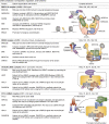
Regulation of BAP1 by intramolecular interactions and self-deubiquitination. A, BAP1 intramolecular interactions are required for its ASXLs-mediated regulation. Interactions between the UCH-CC1 and the CTD domains of BAP1 as well as between the CTD and the DEUBAD of ASXLs are essential for the formation of the CUBI (composite ubiquitin-binding interface). This composite interface enables binding of the VLI hydrophobic patch and the RQDR charged patch of ubiquitin to BAP1 UCH domain and CTD, respectively. This assembly results in the activation of BAP1 DUB activity. B, regulation of BAP1 by self-deubiquitination. UBE2O is a E2 Ub-conjugating-E3 Ub ligase hybrid enzyme that multimonoubiquitinates BAP1 in its NLS region. The CC2 region of BAP1 interacts with the UCH-CC1 domains. This event brings the NLS close enough to the UCH catalytic domain facilitating NLS deubiquitination. BAP1 is then able to translocate into the nucleus and act as a tumor suppressor. Cancer mutations localized in the CC2 domain promote BAP1 association with UBE2O and lead to the disruption of BAP1 self-deubiquitination. As a consequence, BAP1 is sequestered in the cytoplasm. BAP1, BRCA1-associated protein 1; CC1/2, coiled-coil 1/2; CTD, C-terminal domain; DEUBAD, DEUBiquitinase ADaptator; HBM, HCF-1 binding motif; NLS, nuclear localization signal; PHD, plant homeo-domain; Ub, ubiquitin; UCH, ubiquitin carboxy-terminal hydrolase.
Figure 5
Figure 5
Post-translational modifications of DUBs.,,,,,,,,, , , , , , , , , , , , , , AT3, ataxin3; BAP1, BRCA1-associated protein 1; CC, coiled-coil; CFTR, cystic fibrosis transmembrane conductance regulator; CK2, casein kinase 2, CTD, C-terminal domain; DTT, dithiothreitol; DUB, deubiquitinase; FOXK, forkhead box class K; Glu, glutamylation; GSK3, glycogen synthase kinase 3; HBM, HCF-1 binding motif; MALT1, mucosa-associated lymphoid tissue lymphoma translocation protein 1; MJD, Machado–Joseph deubiquitinases; NFκB, nuclear factor kappa B; NES, nuclear exporting signal; NLS, nuclear localization signal; P, phosphorylation; QQQ, poly-Q region; SUMO, SUMOylation; SIM, SUMO-interacting motif; Ub, ubiquitin; UBA, Ub-associated domain; UCH, Ub-C terminal hydrolase; UIM, Ub-interacting motif; USP, ubiquitin carboxyl-terminal hydrolase; ZF, zing finger.
Figure 6
Figure 6
DUB regulation by post-translational modifications. A, phosphorylation of BAP1 during the DNA damage response. Upon ionizing-irradiation, DNA double-stand breaks (DSBs) are recognized by the MRN complex resulting in ATM activation. ATM then phosphorylates BAP1 leading to its recruitment at the DSB sites. In turn, BAP1 recruits homologous recombination (HR) proteins such as BRCA1 and RAD51 to promote HR. B, acetylation of OTUD3 in innate immunity. Under normal conditions, OTUD3 is acetylated on its lysine 129 (K129-Ac). K129-Ac enhances OTUD3 DUB activity toward the poly-K63 ubiquitin chains of the immune protein MAVS. This mechanism prevents the aggregation of MAVS and activation of the innate immune response. However, upon viral infection, the deacetylase SIRT1 is recruited to OTUD3 to catalyze its deacetylation and inactivation. As a consequence, MAVS becomes polyubiquitinated and interacts with RIG-I, leading to its aggregation. This ultimately leads to the activation of downstream immune signaling pathways involved in the antiviral response. ATM, ataxia telangiectasia mutated; BAP1, BRCA1-associated protein 1; IKK, IκB kinase; MAVS, mitochondrial anti-viral signaling protein; MRN, MRE11/RAD50/NBS1; OTUD3, OTU-domain containing protein 3; RIG-I, retinoic acid-inducible gene I; SIRT1, sirtuin 1; TBK1, TANK-binding kinase 1; Ub, ubiquitin.

Similar articles

Cited by

References

    1. Komander D., Rape M. The ubiquitin code. Annu. Rev. Biochem. 2012;81:203–229. - PubMed
    1. Yau R., Rape M. The increasing complexity of the ubiquitin code. Nat. Cell Biol. 2016;18:579–586. - PubMed
    1. Swatek K.N., Komander D. Ubiquitin modifications. Cell Res. 2016;26:399–422. - PMC - PubMed
    1. Rape M. Ubiquitylation at the crossroads of development and disease. Nat. Rev. Mol. Cell Biol. 2018;19:59–70. - PubMed
    1. Popovic D., Vucic D., Dikic I. Ubiquitination in disease pathogenesis and treatment. Nat. Med. 2014;20:1242–1253. - PubMed

Publication types

MeSH terms

Grants and funding

LinkOut - more resources