Review
. 2022 Sep:112:51-67.
doi: 10.1016/j.reprotox.2022.06.011. Epub 2022 Jun 25.

Best practices to quantify the impact of reproductive toxicants on development, function, and diseases of the rodent mammary gland

Affiliations
Review

Best practices to quantify the impact of reproductive toxicants on development, function, and diseases of the rodent mammary gland

Klara Matouskova et al. Reprod Toxicol. 2022 Sep.

Abstract

Work from numerous fields of study suggests that exposures to hormonally active chemicals during sensitive windows of development can alter mammary gland development, function, and disease risk. Stronger links between many environmental pollutants and disruptions to breast health continue to be documented in human populations, and there remain concerns that the methods utilized to identify, characterize, and prioritize these chemicals for risk assessment and risk management purposes are insufficient. There are also concerns that effects on the mammary gland have been largely ignored by regulatory agencies. Here, we provide technical guidance that is intended to enhance collection and evaluation of the mammary gland in mice and rats. We review several features of studies that should be controlled to properly evaluate the mammary gland, and then describe methods to appropriately collect the mammary gland from rodents. Furthermore, we discuss methods for preparing whole mounted mammary glands and numerous approaches that are available for the analysis of these samples. Finally, we conclude with several examples where analysis of the mammary gland revealed effects of environmental toxicants at low doses. Our work argues that the rodent mammary gland should be considered in chemical safety, hazard and risk assessments. It also suggests that improved measures of mammary gland outcomes, such as those we present in this review, should be included in the standardized methods evaluated by regulatory agencies such as the test guidelines used for identifying reproductive and developmental toxicants.

Keywords: Developmental abnormality; Differentiation; Ductal hyperplasia; Lactation; Puberty; Terminal end bud.


Figures

Figure 1:
Figure 1:. Stages of rodent mammary gland development and the role of hormones.
The mammary gland begins developing prior to birth and undergoes several specific phases of change as indicated by the timeline on the bottom of this schematic. The major morphological changes to the epithelial compartment are illustrated in the middle of the figure at the fetal, neonatal, pubertal, adult, pregnant, lactational, and involution/aging stages. These periods are not only times of change in the gland, they are also windows of vulnerability when the gland is especially sensitive to environmental insults. At the top of the figure are several of the hormones involved in mammary gland development at these stages. GH = growth hormone; IGF-1 = Insulin-like growth factor-I.
Figure 2:
Figure 2:. Sexual dimorphism of the mouse mammary gland.
Although it is more subtle prior to puberty, there is a clear sexually dimorphic development of the mouse mammary gland. First, the male mouse lacks areola and nipples. Yet, a small rudimentary epithelial structure is found in the mammary fat pad, located approximately in the area where a nipple would have formed in the absence of androgen signaling. In this figure, panels indicated with F represent females and M represent males. The third column of images is a higher magnification view of the male mammary epithelium. In all panels, scalebar = 1 mm.
Figure 3:
Figure 3:. Creating whole mounted mammary glands.
This figure represents the major steps to removing and mounting the mammary gland in mice. Similar approaches are followed in rats, although a more complex fascia connects the skin to the abdominal muscle wall. A) An incision is made in the skin starting at the genitals and moving upward to the neck, making sure not to cut into the body cavity. The location of the nipples are marked with green hexagons and the mammary glands are numbered (1–5) based on the standard convention. Glands 1–3 are thoracic glands whereas glands 4–5 are inguinal. B) Once the incision has been made, the skin is separated from the underlying abdominal muscle wall using forceps and scissors. C) The skin is pinned down to a necropsy board, gently stretching the full skin fold away from the body. D) Three pins are strategically placed to allow the 4th and 5th inguinal mammary glands to be visualized. The approximate location of the 4th mammary gland’s outer boundaries (yellow), lymph node (organ) and ductal epithelium (purple) are indicated. E) The fat pad of the 5th mammary gland is grasped with forceps and a first cut is made parallel to the skin. F) The 4th mammary gland is gently lifted away from the skin while additional cuts to the connective tissue are made at the edges of the fat pad. G) Once the most dorsal aspect of the mammary gland is reached, pushing the abdominal muscles out of the way to reveal the gland which wraps around the dorsal side of the hind limb, a final cut is made with scissors to remove the gland from the skin. H) The mammary gland is placed on a positively-charged slide. I) Forceps are used to gently tug at the connective tissue at the edge of the gland, stretching it only to the shape and size it maintained in the body. At this stage, make sure not to put the forceps inside the fat pad to avoid damage to the ducts. J) The fully stretched mammary gland resembles the gland’s shape and size in situ. The approximate location of the gland’s outer boundaries (yellow), lymph node (organ) and ductal epithelium (purple) are indicated.
Figure 4:
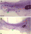
Figure 4:. Morphometric parameters analyzed in the prepubertal (A) and pubertal (B) female mouse mammary gland.
Examples of morphometric parameters that can be collected in female mice prior to adulthood. The blue polygon represents the subtended ductal area. The orange arrow indicates a measure of ductal extension compared to the lymph node. Branching points are typically only counted prior to puberty; a few example branching points are indicated by red stars. The area of TEBs, as indicated by the yellow outlines, can also be measured and these structures can be counted; only a few example TEBs are indicated here. LN = lymph node, green hexagon indicates the location of the nipple. In both panels, scalebar = 1 mm.
Figure 5:
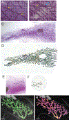
Figure 5:. Methods to evaluate mammary gland morphology.
A) In unbiased stereology methods, which are typically used to evaluate the morphology of whole mounted mammary glands collected from adult animals, an image is collected from each animal at a consistent position in the gland (e.g., just anterior to the lymph node). A grid is placed on each representative image and the presence/absence of specific structures is noted at each crosshair. Compare this panel to B) a sample with significantly more epithelium and alveolar buds. In both panels, yellow arrows indicate crosshairs at ducts, and blue arrowheads indicate crosshairs at alveolar buds. C) This is an example of the use of the Sholl method to evaluate complexity of a pubertal mouse mammary gland. A skeletonized outline of the mammary epithelium is created in ImageJ for the Sholl analysis. The outline is placed over an image of the whole mounted gland to ensure that there are no errors in the process of creating the skeletonized image. D) A series of concentric rings are placed over the skeletonized image with the nipple positioned at the center of these rings. An ImageJ plug-in can be used to count the number of intersections in each ring. These values are used to calculate the Sholl coefficient (a k-value), which is a measurement of the rate of decay of epithelial branching. E) To evaluate complexity of the male mouse mammary gland, a skeletonized outline of the mammary epithelium is created in ImageJ and superimposed over an image of the whole mounted mammary gland. F) Concentric rings are placed over the skeletonized image, similar to what is done with the female gland. However, in this case, because of the absence of a nipple, judgement must be used to determine which part of the skeletonized image should be centered in the concentric rings. G) An image of a pre-pubertal rat mammary gland collected using confocal microscopy and assembled using 3D stacking. The green overlay indicates the mammary epithelium after segmentation. H) Confocal analysis of the mammary epithelium reveals differences in epithelial thickness. Here, warmer colors correspond to thicker parts of the epithelium. In panels G and H, scalebar = 1 mm. Panels G and H from [123], reproduced with permission from the authors.
Figure 6:
Figure 6:. Example pathologies that have been observed in whole mounted mammary glands.
Abnormalities can be detected with ease in whole mounted mammary glands. In panels A and B, blue arrows indicated ducts with a beaded appearance. These are intraductal hyperplasias. Areas outlined with white boxes are other hyperplastic structures, most likely hyperplastic alveolar buds. Abnormal structures can be excised from whole mounted glands, embedded in paraffin, and evaluated histologically. Panel C illustrates a lesion in a whole mounted mammary gland collected from BALB/c mice following transplantation of mammary epithelium from p53−/− tumors. The area indicated by the dotted box was excised and evaluated for histopathology, as indicated in panel D. A pathologist determined that this lesion was a mammary intraepithelial neoplasia.

Similar articles

Cited by

References

    1. Rudel RA, et al., Environmental exposures and mammary gland development: state of the science, public health implications, and research recommendations. Environ Health Perspect, 2011. 119(8): p. 1053–61. - PMC - PubMed
    1. Russo J and Russo IH, DNA labeling index and structure of the rat mammary gland as determinants of its susceptibility to carcinogenesis. J Nat Cancer Inst, 1978. 61: p. 1451–1459. - PubMed
    1. Russo J and Russo IH, Experimentally induced mammary tumors in rats. Breast Cancer Res Treat, 1996. 39(1): p. 7–20. - PubMed
    1. Fenton SE, Endocrine-disrupting compounds and mammary gland development: early exposure and later life consequences. Endocrinology, 2006. 147(6 Suppl): p. S18–24. - PubMed
    1. Vandenberg LN, Najmi A, and Mogus JP, Agrochemicals with estrogenic endocrine disrupting properties: Lessons Learned? Mol Cell Endocrinol, 2020: p. 110860. - PMC - PubMed

Publication types

Substances

LinkOut - more resources