. 2022 Jun;10(6):e004646.
doi: 10.1136/jitc-2022-004646.

Therapeutic efficacy of antibody-drug conjugates targeting GD2-positive tumors

Affiliations

Therapeutic efficacy of antibody-drug conjugates targeting GD2-positive tumors

Daniel V Kalinovsky et al. J Immunother Cancer. 2022 Jun.

Abstract

Background: Both ganglioside GD2-targeted immunotherapy and antibody-drug conjugates (ADCs) have demonstrated clinical success as solid tumor therapies in recent years, yet no research has been carried out to develop anti-GD2 ADCs against solid tumors. This is the first study to analyze cytotoxic activity of clinically relevant anti-GD2 ADCs in a wide panel of cell lines with varying GD2 expression and their effects in mouse models of GD2-positive solid cancer.

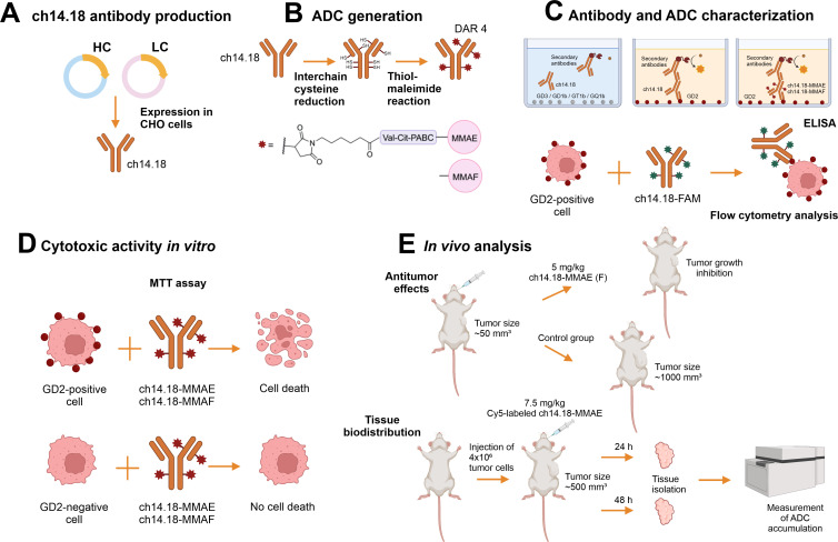
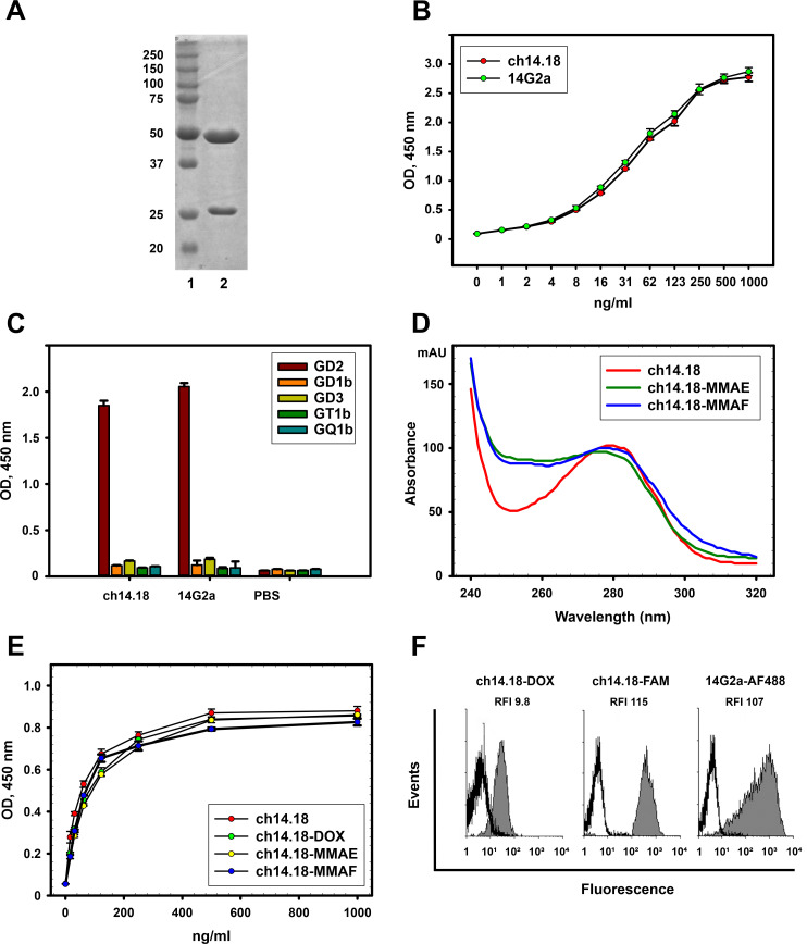
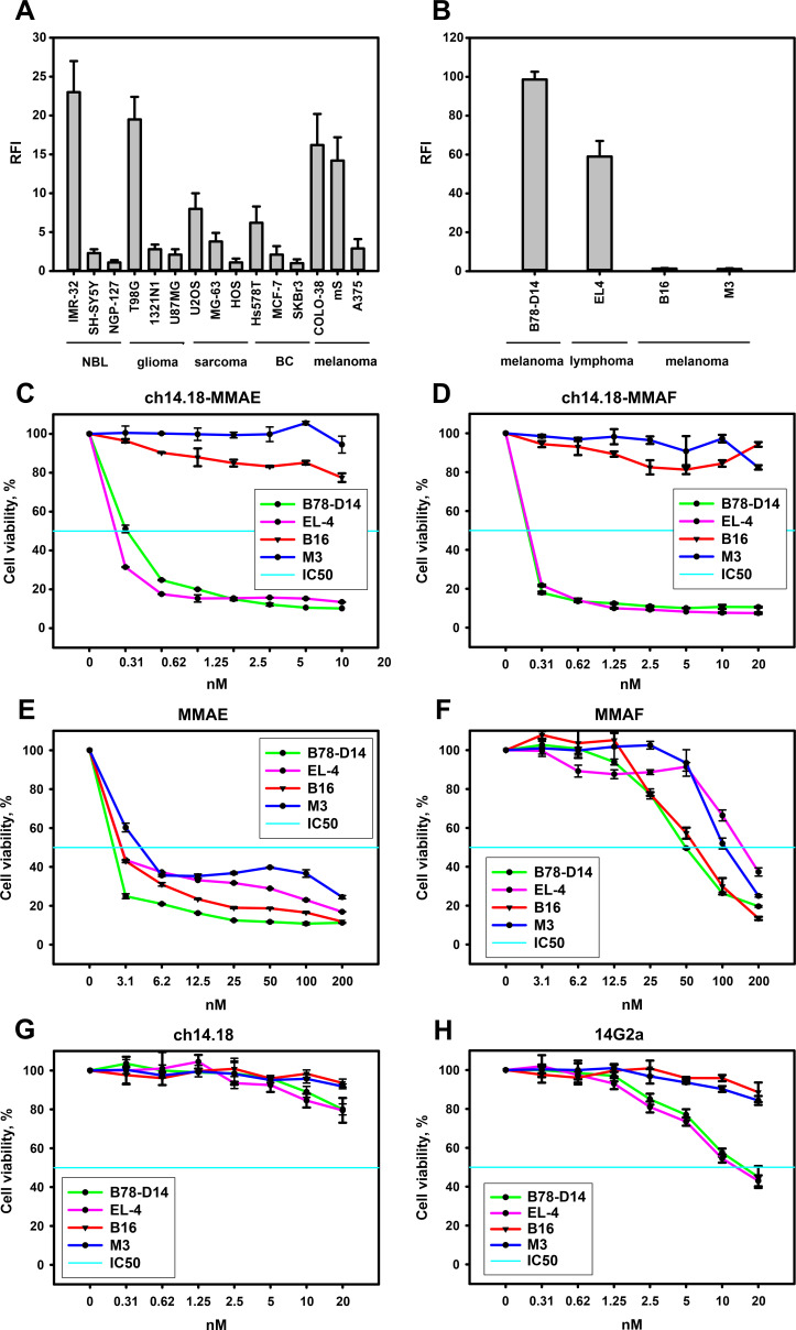
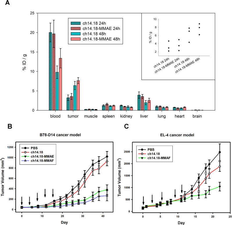
Methods: Anti-GD2 ADCs were generated based on the GD2-specific antibody ch14.18 approved for the treatment of neuroblastoma and commonly used drugs monomethyl auristatin E (MMAE) or F (MMAF), conjugated via a cleavable linker by thiol-maleimide chemistry. The antibody was produced in a mammalian expression system, and its specific binding to GD2 was analyzed. Antigen-binding properties and biodistribution of the ADCs in mice were studied in comparison with the parent antibody. Cytotoxic effects of the ADCs were evaluated in a wide panel of GD2-positive and GD2-negative tumor cell lines of neuroblastoma, glioma, sarcoma, melanoma, and breast cancer. Their antitumor effects were studied in the B78-D14 melanoma and EL-4 lymphoma syngeneic mouse models.

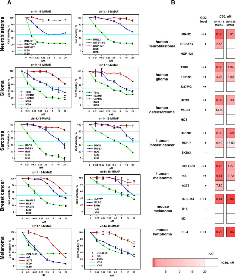
Results: The ch14.18-MMAE and ch14.18-MMAF ADCs retained antigen-binding properties of the parent antibody. Direct dependence of the cytotoxic effect on the level of GD2 expression was observed in cell lines of different origin for both ADCs, with IC50 below 1 nM for the cells with high GD2 expression and no cytotoxic effect for GD2-negative cells. Within the analyzed cell lines, ch14.18-MMAF was more effective in the cells overexpressing GD2, while ch14.18-MMAE had more prominent activity in the cells expressing low GD2 levels. The ADCs had a similar biodistribution profile in the B78-D14 melanoma model compared with the parent antibody, reaching 7.7% ID/g in the tumor at 48 hours postinjection. The average tumor size in groups treated with ch14.18-MMAE or ch14.18-MMAF was 2.6 times and 3.8 times smaller, respectively, compared with the control group. Antitumor effects of the anti-GD2 ADCs were also confirmed in the EL-4 lymphoma model.

Conclusion: These findings validate the potential of ADCs targeting ganglioside GD2 in treating multiple GD2-expressing solid tumors.

Keywords: antigens, tumor-associated, carbohydrate; brain neoplasms; breast neoplasms; immunotherapy; neuroblastoma.


Conflict of interest statement

Competing interests: None declared.

Figures

Figure 1
Figure 1
The main experimental stages of the work. The ch14.18 antibody was produced by stable expression in CHO cells (A), and its conjugation with the small molecules MMAE or MMAF was performed by thiol-maleimide chemistry (B). Specific binding of the parent antibody to ganglioside GD2 and preservation of the antigen-binding properties for the ADCs were analyzed by ELISA and flow cytometry (C). The cytotoxic effects of ch14.18-MMAE and ch14.18-MMAF were evaluated in vitro in a panel of tumor cell lines of neuroblastoma, glioma, sarcoma, melanoma, and breast cancer (D), while their antitumor effects and biodistribution in vivo were studied in the B78-D14 melanoma and EL-4 lymphoma mouse models (E). The illustrations were created using BioRender (BioRender.com). ADC, antibody-drug conjugate; DAR, drug-to-antibody ratio; MMAE or MMAF, monomethyl auristatin E or F.
Figure 2
Figure 2
Characterization of the ch14.18 antibody and anti-GD2 ADCs. (A) Reducing polyacrylamide gel electrophoresis of ch14.18 antibody; 1, molecular weight protein markers (Thermo Fisher Scientific); 2, ch14.18. (B) Antigen-binding properties of ch14.18 and 14G2a antibodies analyzed by ELISA; GD2 was adsorbed on the plate. (C) Cross-reactivity of ch14.18 and 14G2a antibodies to structurally similar gangliosides in direct ELISA. GD2, GD3, GD1b, GT1b, or GQ1b were adsorbed on the plate. Ch14.18 or 14G2a antibodies were added in 10 µg/mL concentration; (D) UV-VIS spectra of the ADCs and the parent ch14.18 antibody normalized at 280 nm. (E) Antigen-binding properties of the ADCs analyzed by ELISA; GD2 was adsorbed on the plate. (F) Flow cytometry analysis of EL-4 cells stained with ch14.18-DOX, ch14.18-FAM, and control 14G2a-AF488. Gray histograms represent staining with the corresponding conjugate and empty histograms represent autofluorescence of unstained cells. Relative fluorescence intensity (RFI) ratios of specific fluorescence of cells stained with fluorescently labeled antibodies to autofluorescence of control unstained cells are shown. While RFI values for cells stained with ch14.18-FAM and 14G2a-AF488 are practically equal (RFI 115 vs 107, respectively), a lower RFI for cells stained with ch14.18-DOX (RFI 9.8) is caused by poorer spectral characteristics of doxorubicin. ADC, antibody-drug conjugate; MMAE or MMAF, monomethyl auristatin E or F; UV-VIS, ultraviolet-visible.
Figure 3
Figure 3
GD2 expression level and cytotoxic activity of anti-GD2 ADCs in murine cell lines. (A, B) Flow cytometry analysis of ganglioside GD2 expression performed with ch14.18-FAM in a panel of human (A) and murine (B) tumor cell lines. For human neuroblastoma, osteosarcoma, and breast cancer (BC), cell lines with high (IMR-32, U2OS, and Hs578T—one per tumor type, respectively), low (SH-SY5Y, MG-63, and MCF-7) GD2 expression, or complete absence of GD2 expression (NGP-127, HOS, and SKBr3) were analyzed. All human glioma and melanoma cell lines expressed GD2 to some extent, but the level of expression varied significantly. Murine EL-4 lymphoma and B78-D14 melanoma exhibited GD2 overexpression, while the two other murine melanomas M3 and B16 were characterized by the absence of GD2 expression. (C–H) Viability of murine cell lines with varying expression of ganglioside GD2 analyzed by MTT assay following 72 hours incubation with ch14.18-MMAE (C), ch14.18-MMAF (D), MMAE (E), MMAF (F), ch14.18 antibody (G), or 14G2a antibody (H). ADC, antibody-drug conjugate; MMAE or MMAF, monomethyl auristatin E or F; NBL, neuroblastoma; RFI, relative fluorescence intensity.
Figure 4
Figure 4
Cytotoxic activity of anti-GD2 ADCs in human cell lines. (A) Viability of human cell lines with varying expression of ganglioside GD2 analyzed by MTT assay following 72 hours incubation with ch14.18-MMAE (left) or ch14.18-MMAF (right). (B) Correlation heatmap between ganglioside GD2 expression and IC50 values of ch14.18-MMAE and ch14.18-MMAF in the tumor cell line panel. ADC, antibody-drug conjugate; MMAE or MMAF, monomethyl auristatin E or F.
Figure 5
Figure 5
Tissue biodistribution and antitumor efficacy of anti-GD2 ADCs in vivo. (A) Tissue biodistribution following intravenous administration of fluorescently labeled ch14.18-MMAE (DAR 2.8) or its parent antibody ch14.18 in B78-D14 melanoma-bearing C57BL/6 mice at 24 hours and 48 hours post-injection. Data are presented as percentage of injected dose per gram of tissue type (% ID/g), and values represent the mean±SEM derived from groups of three animals. Additionally, for accumulation in the tumor, individual data points for both molecules are presented in a dot plot. In order to measure biodistribution in tissues with minimal blood contamination, residual blood was removed by transcardial perfusion with heparinized PBS following euthanasia. (B), Antitumor activity of ch14.18-MMAE, ch14.18-MMAF, and the parent antibody ch14.18 (each at 5 mg/kg body weight) in B78-D14 melanoma-bearing C57BL/6 mice. When the tumors in the control group reached approximately 1000 mm3, the tumor size in experimental groups constituted 266±97 mm3 for mice treated with ch14.18-MMAF, 381±93 mm3 for mice treated with ch14.18-MMAE, and 930±87 mm3 for those treated with the parent antibody; values represent the mean±SEM derived from groups of four animals. (C,) Antitumor activity of ch14.18-MMAF and the parent antibody ch14.18 (5 mg/kg) in EL-4 lymphoma-bearing C57BL/6 mice. Values represent the mean±SEM derived from groups of four animals. Arrows indicate the days of ADC administration. Error bars, SEM. ADC, antibody-drug conjugate; DAR, drug-to-antibody ratio; MMAE or MMAF, monomethyl auristatin E or F; PBS, phosphate-buffered saline.

References

    1. Dean AQ, Luo S, Twomey JD, et al. Targeting cancer with antibody-drug conjugates: promises and challenges. MAbs 2021;13:1951427. 10.1080/19420862.2021.1951427 - DOI - PMC - PubMed
    1. Kaplon H, Reichert JM. Antibodies to watch in 2021. MAbs 2021;13:1860476. 10.1080/19420862.2020.1860476 - DOI - PMC - PubMed
    1. Baah S, Laws M, Rahman KM. Antibody-drug conjugates-a tutorial review. Molecules 2021;26:2943. 10.3390/molecules26102943 - DOI - PMC - PubMed
    1. M. Bordeau B, P. Balthasar J. Strategies to enhance monoclonal antibody uptake and distribution in solid tumors. Cancer Biol Med 2021;18:649–64. 10.20892/j.issn.2095-3941.2020.0704 - DOI - PMC - PubMed
    1. Zhang X, Leng J, Zhou Y, et al. Efficacy and safety of anti-HER2 agents in combination with chemotherapy for metastatic HER2-positive breast cancer patient: a network meta-analysis. Front Oncol 2021;11:731210. 10.3389/fonc.2021.731210 - DOI - PMC - PubMed

Publication types