. 2022 Jun 28;7(1):70.
doi: 10.1038/s41541-022-00496-8.

The C3d-fused foot-and-mouth disease vaccine platform overcomes maternally-derived antibody interference by inducing a potent adaptive immunity

Affiliations

The C3d-fused foot-and-mouth disease vaccine platform overcomes maternally-derived antibody interference by inducing a potent adaptive immunity

Min Ja Lee et al. NPJ Vaccines. .

Abstract

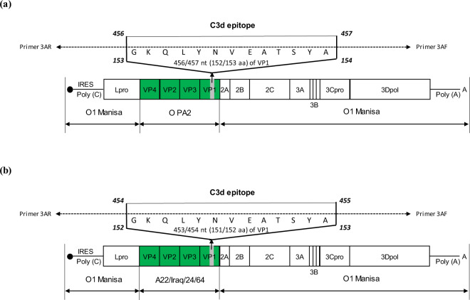
Vaccination prevents and controls foot-and-mouth disease (FMD). However, the current FMD vaccine remains disadvantageous since it cannot overcome maternally-derived antibody (MDA) interference in weeks-old animals, which suppress active immunity via vaccination. To address this, we developed the immune-enhancing O PA2-C3d and A22-C3d FMD vaccine strains that can stimulate receptors on the surface of B cells by inserting C3d (a B cell epitope) into the VP1 region of O PA2 (FMDV type O) and A22 (FMDV type A). We purified inactivated viral antigens from these vaccine strains and evaluated their immunogenicity and host defense against FMDV infection in mice. We also verified its efficacy in inducing an adaptive immune response and overcome MDA interference in MDA-positive (MDA(+), FMD-seropositive) and -negative (MDA(-), FMD-seronegative) pigs. These results suggest a key strategy for establishing novel FMD vaccine platform to overcome MDA interference and induce a robust adaptive immune response.


Conflict of interest statement

The authors declare no competing interests.

Figures

Fig. 1
Fig. 1. Construction of the immune-enhancing FMDV vaccine strains, O PA2-C3d and A22-C3d.
(a, b) O PA2-C3d (a); A22-C3d (b). The B cell epitope, C3d (with 13 amino acid residues in the VP1 region) is used to prepare the virus. The O PA2 (O PA2-R) or A22 (A22-R) P1 strains—where the P1 region of O1 Manisa is substituted with O PA2 P1 or A22 P1—are used as the backbone to prepare the immune-enhancing FMD vaccine strains that can overcome interference by maternally-derived antibodies.
Fig. 2
Fig. 2. Vaccine efficacy and protective effects of O PA2-C3d and A22-C3d in mice.
C57BL/6 mice (n = 5/group) were administered the test vaccine at 1/10, 1/40, 1/160, 1/640 doses of O PA2 or O PA2-C3d or A22 or A22-C3d antigen for cattle or pig use, ISA 206 (oil-based emulsion, 50%, w/w), 10% Al(OH)3, and 15 µg Quil-A. A negative control (NC) group was injected with the same volume of PBS. The test vaccines were injected intramuscularly into mice that were later challenged with FMDV type O (100 LD50 O/VET/2013) or FMDV type A (100 LD50 A/Malay/97) at 7 dpv. The survival rates and body weights were monitored for 7 dpc. (ai) Experimental strategy (a); survival rates post-challenge with O/VET/2013 (b, d) or A/Malay/97 (f, h); changes in body weight post-challenge with O/VET/2013 (c, e) or A/Malay/97 (g, i). The data represent the mean ± SEM of triplicate measurements (n = 5/group).
Fig. 3
Fig. 3. Vaccine efficacy and protective effects of a bivalent test vaccine containing the O PA2-C3d and A22-C3d antigens.
C57BL/6 mice (n = 4/group) were administered the test vaccine at 1/10, 1/40, 1/160, 1/640 doses of O PA2 + A22 antigen or O PA2-C3d + A22-C3d antigen for cattle or pig use, ISA 206 (oil-based emulsion, 50%, w/w), 10% Al(OH)3, and 15 µg Quil-A. A negative control (NC) group was injected with the same volume of PBS. The test vaccines were injected intramuscularly into mice that were later challenged with FMDV type O (100 LD50 O/VET/2013) or FMDV type A (100 LD50 A/Malay/97) at 7 dpv. The survival rates and body weights were monitored for 7 dpc. (ai) Experimental strategy (a); survival rates post-challenge with O/VET/2013 (b, d) or A/Malay/97 (f, h); changes in body weight post-challenge with O/VET/2013 (c, e) or A/Malay/97 (g, i). The data represent the mean ± SEM of triplicate measurements (n = 4/group).
Fig. 4
Fig. 4. Immune responses mediated by the immune-enhancing FMDV (O PA2-C3d and A22-C3d), as measured by SP O and SP A ELISA for overcoming interference by maternally-derived antibodies (MDA) in pigs.
Pigs (8–9 weeks old) that were FMD antibody-seropositive (MDA(+), n = 16) or FMD antibody-seronegative (MDA(−), n = 16) animals were divided into three groups, respectively: a negative control group (NC, n = 4/group), a positive control group (PC, n = 6/group), and an experimental group (Exp., n = 6/group). The Exp. group were administered the test vaccines containing 15 μg (1 dose for cattle and pig use) O PA2-C3d + A22-C3d antigen with ISA 206 (oil-based emulsion, 50%, w/w), 10% Al(OH)3, and 150 μg Quil-A. The positive control group received 15 μg (1 dose for cattle and pig use) O PA2 + A22 antigen with ISA 206 (oil-based emulsion, 50%, w/w), 10% Al(OH)3, and 150 μg Quil-A. A negative control (NC) group was injected with the same volume of PBS. The vaccination was performed twice at 28-day intervals, with 1 mL vaccine (1 dose) injected via a deep intramuscular route on the animals’ necks. Blood samples were collected at 0, 7, 14, 28, 42, 56, 70, and 84 days post vaccination in pigs for serological assays. (ai) Study strategy (a); SP O antibody titers (PrioCheckTM kit) in MDA(+) pigs (b); SP O antibody titers (VDPro® kit) in MDA(+) pigs (c); SP A antibody titers (PrioCheckTM kit) in MDA(+) pigs (d); SP A antibody titers (VDPro® kit) in MDA(+) pigs (e); SP O antibody titers (PrioCheckTM kit) in MDA(−) pigs (f); SP O antibody titers (VDPro® kit) in MDA(−) pigs (g); SP A antibody titers (PrioCheckTM kit) in MDA(−) pigs (h); SP A antibody titers (VDPro® kit) in MDA(−) pigs (i). The data represent the mean ± SEM of triplicate measurements (n = 4 or 6/group). Statistical analyses were performed using a two-way ANOVA followed by Tukey’s test. *p < 0.05; **p < 0.01; ***p < 0.001; and ****p < 0.001.
Fig. 5
Fig. 5. Immune responses mediated by the immune-enhancing FMDV (O PA2-C3d and A22-C3d), as measured by VN titers for overcoming interference of maternally-derived antibodies (MDA) in pigs.
Pigs (8–9 weeks old) that were FMD antibody-seropositive (MDA(+), n = 16) or FMD antibody-seronegative (MDA(−), n = 16) animals were divided into three groups, respectively: a negative control group (NC, n = 4/group), a positive control group (PC, n = 6/group), and an experimental group (Exp., n = 6/group). The Exp. group were administered the test vaccines containing 15 μg (1 dose for cattle and pig use) O PA2-C3d + A22-C3d antigen with ISA 206 (oil-based emulsion, 50%, w/w), 10% Al(OH)3, and 150 μg Quil-A. The positive control group received 15 μg (1 dose for cattle and pig use) O PA2 + A22 antigen with ISA 206 (oil-based emulsion, 50%, w/w), 10% Al(OH)3, and 150 μg Quil-A. A negative control (NC) group was injected with the same volume of PBS. The vaccination was performed twice at 28-day intervals, with 1 mL vaccine (1 dose) injected via a deep intramuscular route on the animals’ necks. Blood samples were collected at 0, 7, 14, 28, 42, 56, 70, and 84 days post vaccination in pigs for serological assays. (ae) O1 Campos, A2001 Argentina, and A24 Cruzeiro VN titers in MDA(+) or MDA(−) pigs (a); O PA2 VN titers in MDA(+) pigs (b); O PA2 VN titers in MDA(−) pigs (c); A22 VN titers in MDA(+) pigs (d); A22 VN titers in MDA(−) pigs (e). The data represent the mean ± SEM of triplicate measurements (n = 4 or 6/group). Statistical analyses were performed using two-way ANOVA followed by Tukey’s test. *p < 0.05; **p < 0.01; ***p < 0.001; and ****p < 0.001.
Fig. 6
Fig. 6. Immune responses mediated by the immune-enhancing FMDV (O PA2-C3d and A22-C3d), as measured by immunoglobulin subtypes such as IgG, IgM, and IgA in pigs.
Pigs (8–9 weeks old) that were FMD antibody-seropositive (MDA(+), n = 16) or FMD antibody-seronegative (MDA(−), n = 16) animals were divided into three groups, respectively: a negative control group (NC, n = 4/group), a positive control group (PC, n = 6/group), and an experimental group (Exp., n = 6/group). The Exp. group were administered the test vaccines containing 15 μg (1 dose for cattle and pig use) O PA2-C3d + A22-C3d antigen with ISA 206 (oil-based emulsion, 50%, w/w), 10% Al(OH)3, and 150 μg Quil-A. The positive control group received 15 μg (1 dose for cattle and pig use) O PA2 + A22 antigen with ISA 206 (oil-based emulsion, 50%, w/w), 10% Al(OH)3, and 150 μg Quil-A. A negative control (NC) group was injected with the same volume of PBS. The vaccination was performed twice at 28-day intervals, with 1 mL vaccine (1 dose) injected via a deep intramuscular route on the animals’ necks. Blood samples were collected at 0, 7, 14, 28, 42, 56, 70, and 84 days post vaccination in pigs for serological assays. (a–c) IgG concentration (a); IgM concentration (b); IgA concentration (c). The data represent the mean ± SEM of triplicate measurements (n = 4 or 6/group). Statistical analyses were performed using two-way ANOVA followed by Tukey’s test. *p < 0.05; **p < 0.01; ***p < 0.001; and ****p < 0.001.
Fig. 7
Fig. 7. O PA2-C3d and A22-C3d induced the gene expression of cytokine and co-stimulatory molecules in porcine peripheral blood mononuclear cells.
Porcine peripheral blood mononuclear cells (PBMCs) isolated from the whole blood of vaccinated pigs (n = 5/group) as described in Fig. 4a were used for qRT-PCR assays. Gene expression levels were normalized to HPRT levels and are presented as a relative ratio compared to control levels. (a–v) Gene expression levels of IFNα (a); IFNβ (b); IFNγ (c); IL-1β (d); IL-17A (e); IL-23p19 (f); IL-23R (g); IL-2 (h); IL-10 (i); TGFβ (j); IL-4 (k); IL-6 (l); CD40 (m); CD80 (n); CD86 (o); MHC class I (p); MHC class II (q); CTLA4 (r); CD21 (s); CD28 (t); ICOS (u); AHNAK (v). Statistical analyses were performed using one-way ANOVA followed by Tukey’s test. *p < 0.05; **p < 0.01; ***p < 0.001; and ****p < 0.001.

References

    1. Grubman MJ, Baxt BJC. m. r. Foot-and-mouth disease. Clin. Microbiol. Rev. 2004;17:465–493. - PMC - PubMed
    1. Alexandersen S, Quan M, Murphy C, Knight J, Zhang Z. Studies of quantitative parameters of virus excretion and transmission in pigs and cattle experimentally infected with foot-and-mouth disease virus. J. Comp. Pathol. 2003;129:268–282. - PubMed
    1. Kitching RP, Salt JS. The interference by maternally-derived antibody with active immunization of farm animals against foot-and-mouth disease. Br. Vet. J. 1995;151:379–389. - PMC - PubMed
    1. Elnekave E, et al. The long term effect of age and maternally derived antibodies against foot and mouth disease on the serological response following vaccination in young dairy calves. Vaccine. 2016;34:4927–4934. - PubMed
    1. Kim J, et al. The interference effect of maternally-derived antibodies on the serological performance of pigs immunized with a foot-and-mouth disease oil emulsion vaccine. Vaccine. 2020;38:1723–1729. - PubMed