. 2022 Aug;158(2):127-136.
doi: 10.1007/s00418-022-02119-8. Epub 2022 Jun 29.

Landmark-based retrieval of inflamed skin vessels enabled by 3D correlative intravital light and volume electron microscopy

Affiliations

Landmark-based retrieval of inflamed skin vessels enabled by 3D correlative intravital light and volume electron microscopy

Karina Mildner et al. Histochem Cell Biol. 2022 Aug.

Abstract

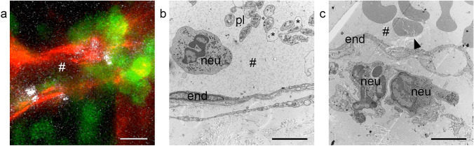
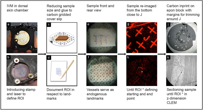
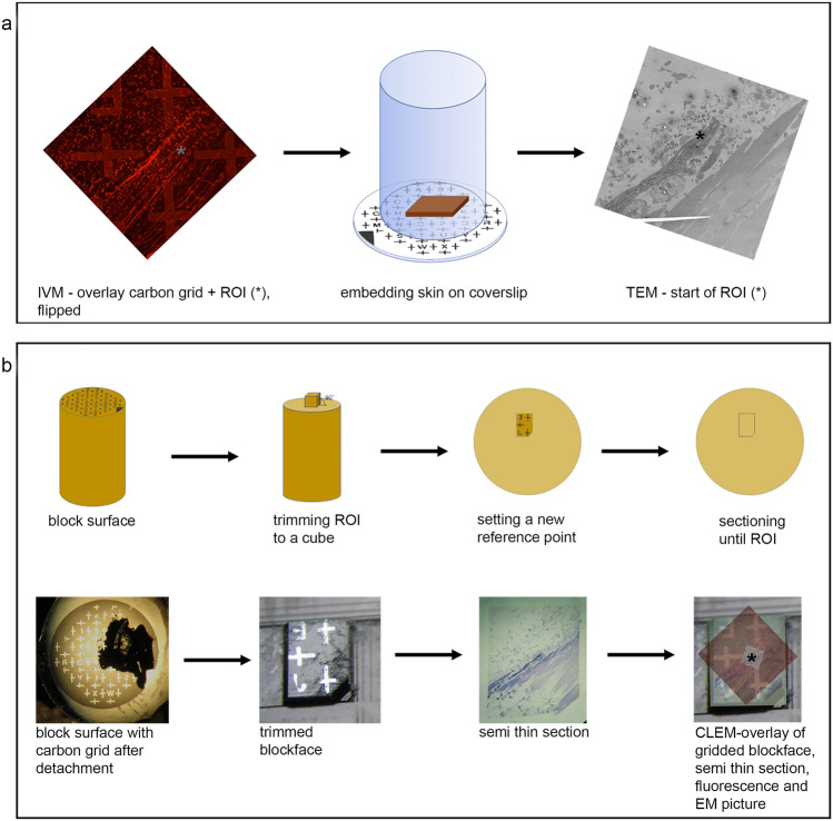
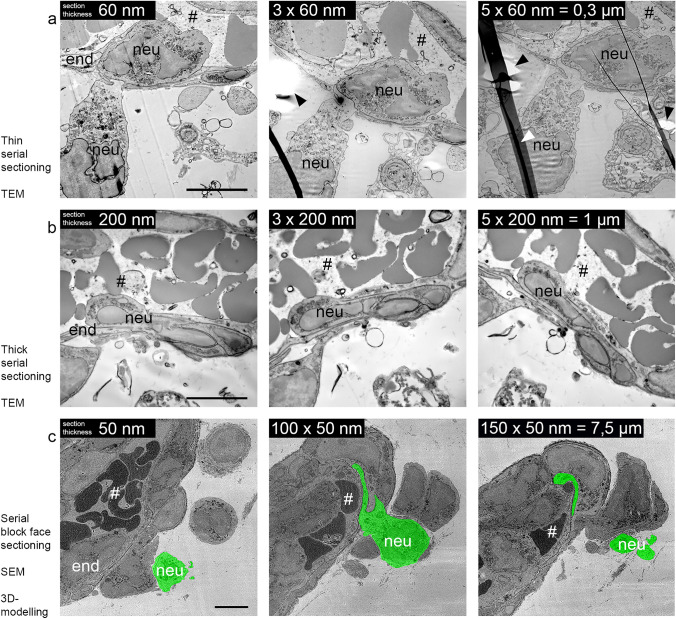
The nanometer spatial resolution of electron microscopy imaging remains an advantage over light microscopy, but the restricted field of view that can be inspected and the inability to visualize dynamic cellular events are definitely drawbacks of standard transmission electron microscopy (TEM). Several methods have been developed to overcome these limitations, mainly by correlating the light microscopical image to the electron microscope with correlative light and electron microscopy (CLEM) techniques. Since there is more than one method to obtain the region of interest (ROI), the workflow must be adjusted according to the research question and biological material addressed. Here, we describe in detail the development of a three-dimensional CLEM workflow for mouse skin tissue exposed to an inflammation stimulus and imaged by intravital microscopy (IVM) before fixation. Our aim is to relocate a distinct vessel in the electron microscope, addressing a complex biological question: how do cells interact with each other and the surrounding environment at the ultrastructural level? Retracing the area over several preparation steps did not involve any specific automated instruments but was entirely led by anatomical and artificially introduced landmarks, including blood vessel architecture and carbon-coated grids. Successful retrieval of the ROI by electron microscopy depended on particularly high precision during sample manipulation and extensive documentation. Further modification of the TEM sample preparation protocol for mouse skin tissue even rendered the specimen suitable for serial block-face scanning electron microscopy (SBF-SEM).

Keywords: Correlative light and electron microscopy; Dorsal skinfold chamber; Intravital microscopy; Live cell imaging; Serial block-face scanning electron microscopy; Transmission electron microscopy.


Conflict of interest statement

The authors declare no competing interest.

Figures

Fig. 1
Fig. 1
Vessel segment undergoing neutrophil extravasation during cutaneous inflammation. a Representative time-lapse still image of an inflamed vessel from intravital fluorescence microscopy showing the endothelial cell layer (red), transmigrating neutrophils (green), and platelets (white). Original movie in Supplementary Movie 1. b, c Electron micrographs of inflamed mouse skin sample, conventionally fixated and processed with CLEM techniques, exhibit poor preservation of the ultrastructure (asterisk marking bulging membranes of platelets) and distortion of the sections because of weak epon infiltration (arrowhead). # vessel lumen, end endothelial cell, neu neutrophil, pl platelet, *bulging membranes. Scale bar in a = 10 µm, scale bar in b, c = 5 µm
Fig. 2
Fig. 2
Introduction of landmarks for CLEM orientation. Illustrated workflow describing all steps introducing landmarks, which are essential to reposition ROI at the electron microscope exactly at the light microscopical location recorded by IVM in a dorsal skin chamber (a). The printed stamp on the skin supports to locate the laser point, which illuminates the last position of the IVM settings (ROI) b. After fixation, punching out, and resizing the skin to a minimum, the sample is glued to a carbon grid and marked on one corner c. The endogenous, visible vessels in the tissue serve as the next landmarks while repositioning the sample on the confocal stage f, where the fluorescently labeled vessel network in the fixed tissue serve as landmarks to relocate the ROI at the confocal microscope g, h. With the help of the carbon-gridded cover slip and its imprinted letters, the position of the ROI can be documented with respect to these very letters d, e. Together with the distance in the z-direction between the ROI and the coverslip, this information about the xy location defines the starting point for the microtome sectioning j further illustrated in Fig. 3. The measured z height in the sample thus guides this approach to successfully find the correlative view between light and electron microscope (CLEM) k
Fig. 3
Fig. 3
Target trimming and sectioning. a Outline of the CLEM challenge: retrieve a specific area from skin tissue, recorded by IVM (left panel), repositioned several times through the entire sample preparation (middle panel) and reimaging the area exactly at the ROI (*) in the electron microscope (right panel). The trimming and sectioning of the embedded sample block needs to be done in a controlled manner as further illustrated in b. After detachment from the coverslip, the block surface is covered with traces of the carbon grid (left). The resin material is trimmed away from the ROI, identified by the letter of the carbon imprint, resulting in a small square block (second image). One corner is cut off, defining a new reference point. Next, the ROI is approached by careful sectioning with µm/nm step size, until the z-height of the vessel of interest is reached. The approach is documented with 200-nm-thick sections, stained with toluidine blue, which are continuously compared with the pattern of the vessel network as imaged in the confocal microscope on the prefixed sample. The overlay of all corresponding layers (fluorescence image, carbon grid, and semithin and thin section) prove that the vessel has been retrieved in a correlative mode and allows further investigation at higher resolution in the electron microscope
Fig. 4
Fig. 4
Analyzing the ROI by thin sectioning, thick sectioning, or serial block-face sectioning. Electron micrographs of successfully recovered CLEM vessels of inflamed mouse skin. Throughout the vessel, several cellular interactions can be observed and studied in more detail with respect to the surrounding tissue components such as the endothelial cell (end) and transmigrating neutrophil (neu) leaving the lumen of the vessel (#). To provide more “volume” information, several techniques can be applied. Serial consecutive sections (60 nm thin or 200 nm thick) can be used to follow structures in 3D on a normal transmission electron microscope a, b. Physical distortions occur regularly, marked by arrowheads. An alternative approach is automated sectioning by SBF-SEM. The sample block is continuously cut via an ultramicrotome, accommodated within the instrument. The scanning detector scans the surface after every section, resulting in a large 3D volume of the selected ROI that can be recorded and analyzed as single sections c. Here, the contour of a transmigrating neutrophil is highlighted in green, and additionally annotated in Supplementary Movie 3. Scale bar = 5 µm

Similar articles

Cited by

  • The extracellular matrix dictates regional competence for tumour initiation.
    Bansaccal N, Vieugue P, Sarate R, Song Y, Minguijon E, Miroshnikova YA, Zeuschner D, Collin A, Allard J, Engelman D, Delaunois AL, Liagre M, de Groote L, Timmerman E, Van Haver D, Impens F, Salmon I, Wickström SA, Sifrim A, Blanpain C. Bansaccal N, et al. Nature. 2023 Nov;623(7988):828-835. doi: 10.1038/s41586-023-06740-y. Epub 2023 Nov 15. Nature. 2023. PMID: 37968399 Free PMC article.
  • In focus in HCB.
    Taatjes DJ, Roth J. Taatjes DJ, et al. Histochem Cell Biol. 2022 Aug;158(2):123-125. doi: 10.1007/s00418-022-02142-9. Histochem Cell Biol. 2022. PMID: 35859014 No abstract available.

References

    1. Belevich I, Joensuu M, Kumar D, Vihinen H, Jokitalo E. Microscopy image browser: a platform for segmentation and analysis of multidimensional datasets. PLoS Biol. 2016;14(1):e1002340. doi: 10.1371/journal.pbio.1002340. - DOI - PMC - PubMed
    1. Braun LJ, Stegmeyer RI, Schafer K, Volkery S, Currie SM, Kempe B, Nottebaum AF, Vestweber D. Platelets docking to VWF prevent leaks during leukocyte extravasation by stimulating Tie-2. Blood. 2020;136(5):627–639. doi: 10.1182/blood.2019003442. - DOI - PMC - PubMed
    1. Claesson-Welsh L, Dejana E, McDonald DM. Permeability of the endothelial barrier: identifying and reconciling controversies. Trends Mol Med. 2021;27(4):314–331. doi: 10.1016/j.molmed.2020.11.006. - DOI - PMC - PubMed
    1. Currie SM, Stegmeyer RI, Mildner K, Breitsprecher L, Zeuschner D, Psathaki OE, Schafer K, Wilkens M, Volkery S, Nieswandt B, Vestweber D. Confocal real-time analysis of cutaneous platelet recruitment during immune-complex-mediated inflammation. J Invest Dermatol. 2022 doi: 10.1016/j.jid.2022.03.011. - DOI - PubMed
    1. Deerinck TJ, Bushong EA, Thor A, Ellisman MH (2010) NCMIR methods for 3D EM: a new protocol for preparation of biological specimens for serial blockface scanning electron microscopy. https://ncmir.ucsd.edu/sbemprotocol

LinkOut - more resources