. 2022 Jun 28;10(1):94.
doi: 10.1186/s40478-022-01394-9.

Reassessment of Neuronal Tau Distribution in Adult Human Brain and Implications for Tau Pathobiology

Affiliations

Reassessment of Neuronal Tau Distribution in Adult Human Brain and Implications for Tau Pathobiology

Giavanna Paterno et al. Acta Neuropathol Commun. .

Abstract

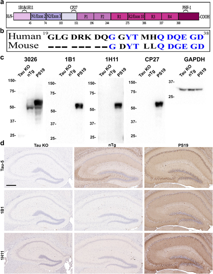
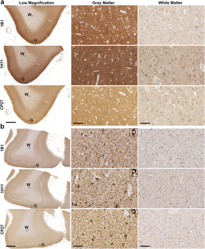
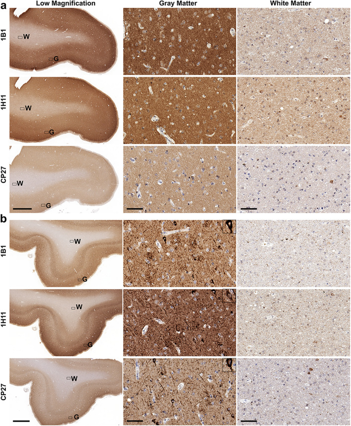
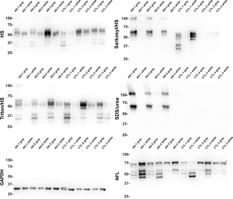
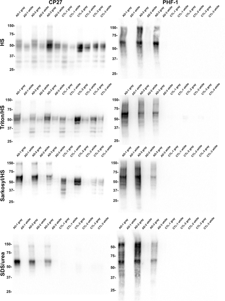
Tau is a predominantly neuronal, soluble and natively unfolded protein that can bind and stabilize microtubules in the central nervous system. Tau has been extensively studied over several decades, especially in the context of neurodegenerative diseases where it can aberrantly aggregate to form a spectrum of pathological inclusions. The presence of tau inclusions in the form of neurofibrillary tangles, neuropil threads and dystrophic neurites within senile plaques are essential and defining features of Alzheimer's disease. The current dogma favors the notion that tau is predominantly an axonal protein, and that in Alzheimer's disease there is a redistribution of tau towards the neuronal soma that is associated with the formation of pathological inclusions such as neurofibrillary tangles and neuropil threads. Using novel as well as previously established highly specific tau antibodies, we demonstrate that contrary to this overwhelmingly accepted fact, as asserted in numerous articles and reviews, in adult human brain, tau is more abundant in cortical gray matter that is enriched in neuronal soma and dendrites compared to white matter that is predominantly rich in neuronal axons. Additionally, in Alzheimer's disease tau pathology is significantly more abundant in the brain cortical gray matter of affected brain regions compared to the adjacent white matter regions. These findings have important implications for the biological function of tau as well as the mechanisms involved in the progressive spread of tau associated with the insidious nature of Alzheimer's disease.

Keywords: Alzheimer’s disease; Brain; Distribution; Human; Neuronal; Tau; Tauopathy.


Conflict of interest statement

We declare no conflict of interest in this manuscript.

Figures

Fig. 1
Fig. 1
Characterization of the specificity of the new tau antibodies 1B1 and 1H11. a Schematic of full-length human tau (2N4R) with the location of the antibody epitopes used in the study. Created with BioRender.com. b Amino acid sequence of human and mouse tau corresponding to residues 19 to 38 in human tau. Non-homologous amino acids between species are indicated in black lettering. Dashes indicate residues that are not present in mouse tau. c 10 µg of total brain lysate from tau KO, nTg, and PS19 mice were resolved by SDS-PAGE as indicated above each lane and used for immunoblotting as described in Materials and Methods. Blots were probed with antibodies 3026 (mouse and human tau), 1B1 (human tau), 1H11 (human tau), CP27 (human tau) and anti-GAPDH as a loading control. The mobility of protein markers with their molecular masses are indicated on the left side of each immunoblot. d IHC was performed as described in Materials and Methods. Brains of tau KO, nTg, and PS19 mice were stained with the total tau antibody Tau-5, and the two novel monoclonal total human tau antibodies, 1B1 and 1H11. Sections were counterstained with hematoxylin. Scale bar indicates 300 μm
Fig. 2
Fig. 2
Immunohistochemistry of the frontal cortex of a control individual and an AD individual with phospho-independent tau antibodies. IHC was performed as described in Materials and Methods. A representative control case (a) and a representative AD case (b) were stained with total human tau antibodies 1B1, 1H11, and CP27. Low magnification and high magnification images of gray matter and white matter were taken as indicated. “G” indicates gray matter region. “W” indicates white matter region. Scale bar for low magnification images is 2 mm. Scale bar for high magnification images is 60 µm; and insets 30 µm. Sections were counterstained with hematoxylin
Fig. 3
Fig. 3
Immunohistochemistry of the temporal cortex of a control individual and an AD individual with phospho-independent tau antibodies. IHC was performed as described in Materials and Methods. A representative control case (a) and a representative AD case (b) were stained with total human tau antibodies 1B1, 1H11, and CP27. Low magnification and high magnification images of gray matter and white matter were taken as indicated. “G” indicates gray matter region. “W” indicates white matter region. Scale bar for low magnification images is 2 mm. Scale bar for high magnification images is 60 µm; and insets 30 µm. Sections were counterstained with hematoxylin
Fig. 4
Fig. 4
Comparative biochemical fraction analyses of gray and white matter from the temporal cortex of AD and control cases with anti-tau antibody 1B1. Biochemical fractionation of AD and CTL cerebral temporal cortex was performed as described in Materials and Methods. Equal amount of protein (5 μg) from the high salt (HS) soluble, Triton/HS soluble, Sarkosyl/HS soluble, and Sarkosyl-insoluble SDS/urea soluble fractions were separated by SDS-PAGE and analyzed by immunoblotting with tau antibody, 1B1. The HS fractions were also probed for GAPDH and the SDS/urea fractions were probed for NFL. The mobility of protein markers with their molecular masses are indicated on the left side of each immunoblot
Fig. 5
Fig. 5
Comparative biochemical fraction analyses of gray and white matter from the temporal cortex of AD and control cases with tau antibodies CP27 and PHF-1. Biochemical fractionation of AD and CTL cerebral temporal cortex was performed as described in Materials and Methods. Equal amount of protein (5 μg) from the high salt (HS) soluble, Triton/HS soluble, Sarkosyl/HS soluble, and Sarkosyl-insoluble SDS/urea soluble fractions were separated by SDS-PAGE and analyzed by immunoblotting with antibodies CP27 or PHF-1. The mobility of protein markers with their molecular masses are indicated on the left side of each immunoblot

References

    1. De Ancos JG, Avila J. Differential distribution in white and grey matter of tau phosphoisoforms containing four tubulin-binding motifs. Biochem J. 1993;296:351–354. doi: 10.1042/bj2960351. - DOI - PMC - PubMed
    1. Aragão Gomes L, Uytterhoeven V, Lopez-Sanmartin D, Tomé SO, Tousseyn T, Vandenberghe R, Vandenbulcke M, von Arnim CAF, Verstreken P, Thal DR. Maturation of neuronal AD-tau pathology involves site-specific phosphorylation of cytoplasmic and synaptic tau preceding conformational change and fibril formation. Acta Neuropathol. 2021;141:173–192. doi: 10.1007/S00401-020-02251-6. - DOI - PubMed
    1. Arendt T, Stieler JT, Holzer M. Tau and tauopathies. Brain Res Bull. 2016;126:238–292. doi: 10.1016/J.BRAINRESBULL.2016.08.018. - DOI - PubMed
    1. Ayers JI, Giasson BI, Borchelt DR. Prion-like Spreading in Tauopathies. Biol Psychiatry. 2018;83:337–346. doi: 10.1016/J.BIOPSYCH.2017.04.003. - DOI - PMC - PubMed
    1. Baas PW, Rao AN, Matamoros AJ, Leo L. Stability properties of neuronal microtubules. Cytoskeleton (Hoboken) 2016;73:442–460. doi: 10.1002/CM.21286. - DOI - PMC - PubMed

Publication types