. 2022 Nov 1;42(6):638-649.
doi: 10.3343/alm.2022.42.6.638.

Natural Killer Cell Expansion and Cytotoxicity Differ Depending on the Culture Medium Used

Affiliations

Natural Killer Cell Expansion and Cytotoxicity Differ Depending on the Culture Medium Used

Seung Kwon Koh et al. Ann Lab Med. .

Abstract

Background: Adoptive cell therapy using umbilical cord blood (UCB)-derived allogeneic natural killer (NK) cells has shown encouraging results. However, because of the insufficient availability of NK cells and limited UCB volume, more effective culture methods are required. NK cell expansion and functionality are largely affected by the culture medium. While human serum is a major affecting component in culture media, the way it regulates NK cell functionality remains elusive. We elucidated the effects of different culture media and human serum supplementation on UCB NK cell expansion and functionality.

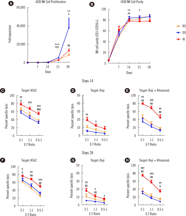
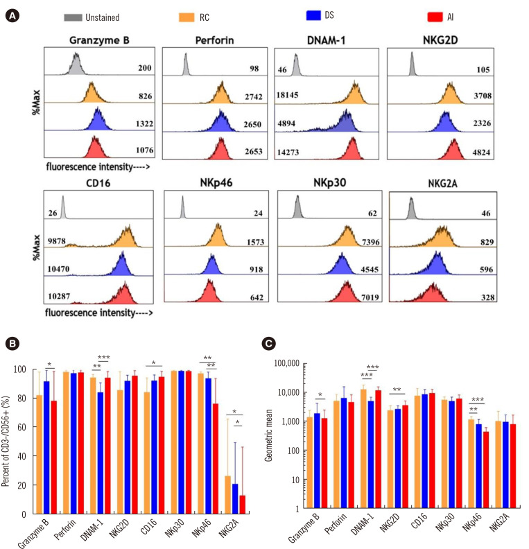
Methods: UCB NK cells were cultured under stimulation with K562-OX40L-mbIL-18/21 feeder cells and IL-2 and IL-15 in serum-containing and serum-free culture media. The effects of the culture media and human serum supplementation on NK cell expansion and cytotoxicity were evaluated by analyzing the expansion rate, activating and inhibitory receptor levels, and the cytotoxicity of the UCB NK cells.

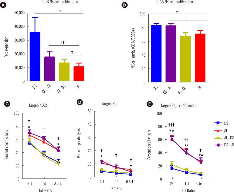
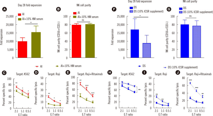
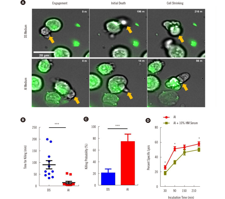
Results: The optimal medium for NK cell expansion was Dulbecco's modified Eagle's medium/Ham's F12 with supplements and that for cytotoxicity was AIM V supplemented with Immune Cell Serum Replacement. Shifting media is an advantageous strategy for obtaining several highly functional UCB NK cells. Live cell imaging and killing time measurement revealed that human serum enhanced NK cell proliferation but delayed target recognition, resulting in reduced cytotoxicity.

Conclusions: Culture medium supplementation with human serum strongly affects UCB NK cell expansion and functionality. Thus, culture media should be carefully selected to ensure both NK cell quantity and quality for adoptive cell therapy.

Keywords: Culture medium; Expansion; Feeder cells; Human serum; Natural killer cells; Umbilical cord blood.


Conflict of interest statement

CONFLICTS OF INTEREST

The authors declare that they have no known competing financial interests or personal relationships that could have appeared to influence the study reported in this paper.

Figures

Fig. 1
Fig. 1
The culture medium substantially affects NK cells expansion and functions. (A) Fold expansion of UCB NK cells cultured in RC (orange), DS (blue), or AI (AIM V + ICSR) (red) culture media (N=15). (B) Purity (CD56+/CD3– NK cells) of UCB NK cells cultured in RC, DS, or AI (N=15). (C–E) Flow cytometry-based assay of the cytotoxicity of UCB NK cells cultured in RC, DS, or AI media toward K562 cells (N=8) (C), Raji cells (N=6) (D), and Raji cells+rituximab (N=7) (E) on day 14. Cytotoxicity assay of UCB NK cells toward K562 cells (N=8) (F), Raji cells (N=6) (G), and Raji cells+rituximab (N=7) (H) on day 28. Data on fold expansion and purity are the mean and SEM. *,†,‡P<0.05; **,††,‡‡P<0.01; ***, †††, ‡‡‡P<0.001; *DS vs. RC; DS vs. AI; AI vs. RC. Abbreviations: RC, RPMI medium + FBS; DS, DMEM + Ham’s F12 + various supplements; AI, AIM V + ICSR; UCB, umbilical cord blood; NK, natural killer; RPMI, Roswell Park Memorial Institute; FBS, fetal bovine serum; DMEM, Dulbecco’s modified Eagle’s medium; ICSR, Immune Cell Serum Replacement.
Fig. 2
Fig. 2
NK cells cultured in different culture media exhibit different receptor expression levels. (A) Histogram of cytotoxic effector molecule and surface receptor expression of UCB NK cells cultured RC (orange), DS (blue), or AI (red). The geometric means are indicated in the respective histograms. (B) Percent expression levels of cytotoxic effector molecules and surface receptors on day 28 (N=9). (C) Geometric mean expression levels of cytotoxic effector molecules and surface receptors on day 28 (N=9) (*P<0.05; **P<0.01; ***P<0.001). Abbreviations: RC, RPMI medium+FBS; DS, DMEM+Ham’s F12+various supplements; AI, AIM V+ICSR; UCB, umbilical cord blood; NK, natural killer; RPMI, Roswell Park Memorial Institute; FBS, fetal bovine serum; DMEM, Dulbecco’s modified Eagle’s medium, ICSR, Immune Cell Serum Replacement.
Fig. 3
Fig. 3
Culture medium shifting is advantageous for restoring NK cell function. (A) Fold expansion of UCB NK cells cultured in DS (blue), DS-AI (purple), AI-DS (green), or AI (red) (N=8) on day 28. (B) Purity (CD56+/CD3− NK cells) of media-shifted UCB-expanded NK cells cultured in DS (blue), DS-AI (purple), AI-DS (green), or AI (red) (N=11) on day 28. Flow cytometry-based cytotoxicity assay of UCB-expanded NK cells cultured in DS, AI, AI-DS, or DS-AI at the indicated ratios; (C) K562 cells, N=7; (D) Raji cells, N=4; (E) Raji cells + rituximab, N=7 (C–E). *, †P<0.05; **, ††P<0.01; ***, †††P<0.001; *DS vs. AI; DS vs. DS-AI; AI vs. AI-DS. Abbreviations: RC, RPMI medium+FBS; DS, DMEM+Ham’s F12+various supplements; AI, AIM V+ICSR; DS-AI, DS shifted to AI on day 14; AI-DS, AI shifted to DS on day 14; UCB, umbilical cord blood; NK, natural killer; RPMI, Roswell Park Memorial Institute; FBS, fetal bovine serum; DMEM, Dulbecco’s modified Eagle’s medium, ICSR, Immune Cell Serum Replacement.
Fig. 4
Fig. 4
Human serum in culture media has contrasting effects on NK cell expansion and functionality. (A) Fold expansion of NK cells cultured in AI (red) and AI+10% human serum (dark green) (N=7) on day 28. (B) Purity (CD56+/CD3– NK cells) of UCB NK cells cultured in AI (red) or AI+10% human serum (N=7) on day 28. Flow cytometry-based assay of the cytotoxicity of UCB NK cells cultured in AI + 10% human serum or AI toward (C) K562 cells (N=7), (D) Raji cells (N=4), (E) Raji cells+rituximab (N=5) on day 14. (F & G) Fold expansion (N=5) and purity (N=5) of NK cells cultured in conventional DS medium (10% human serum supplementation) and DS medium in which the 10% human serum was replaced with 10% ICSR. (H–J) Flow cytometry-based cytotoxicity assay of UCB NK cells cultured in the respective media (N=5) on day 14. *P<0.05; **P<0.01; ***P<0.001. Abbreviations: DS, DMEM+Ham’s F12+various supplements; AI, AIM V+ICSR; NK, natural killer; UCB, umbilical cord blood; ICSR, Immune Cell Serum Replacement.
Fig. 5
Fig. 5
Killing behaviors of NK cells cultured in different media. (A) Representative time-lapse images of NK cell killing events after cell contact. K562 cells are indicated with green fluorescence and NK cells with yellow arrows. (B) Time for target cell killing (N=11) and (C) killing capacity of NK cells as determined from time-lapse images (N=12). (D) Killing capacity of NK cells cultured in AI or AI + 10% human serum (N=4) at 30, 90, 150, and 240 minutes. *P<0.05; **P<0.01; ***P<0.001. Abbreviations: DS, DMEM+Ham’s F12+various supplements; AI, AIM V+ICSR; NK, natural killer; UCB, umbilical cord blood; ICSR, Immune Cell Serum Replacement; HM serum, human serum.

References

    1. Chiossone L, Dumas PY, Vienne M, Vivier E. Natural killer cells and other innate lymphoid cells in cancer. Nat Rev Immunol. 2018;18:671–88. doi: 10.1038/s41577-018-0061-z. - DOI - PubMed
    1. Prager I, Watzl C. Mechanisms of natural killer cell-mediated cellular cytotoxicity. J Leukoc Biol. 2019;105:1319–29. doi: 10.1002/JLB.MR0718-269R. - DOI - PubMed
    1. Lanier LL. NK cell recognition. Annu Rev Immunol. 2005;23:225–74. doi: 10.1146/annurev.immunol.23.021704.115526. - DOI - PubMed
    1. Rees RC. MHC restricted and non-restricted killer lymphocytes. Blood Rev. 1990;4:204–10. doi: 10.1016/0268-960X(90)90049-X. - DOI - PubMed
    1. He Y, Tian Z. NK cell education via nonclassical MHC and non-MHC ligands. Cell Mol Immunol. 2017;14:321–30. doi: 10.1038/cmi.2016.26. - DOI - PMC - PubMed

LinkOut - more resources