. 2022 Aug 17;63(8):1140-1155.
doi: 10.1093/pcp/pcac090.

Structural Characterization of L-Galactose Dehydrogenase: An Essential Enzyme for Vitamin C Biosynthesis

Affiliations

Structural Characterization of L-Galactose Dehydrogenase: An Essential Enzyme for Vitamin C Biosynthesis

Jhon A Vargas et al. Plant Cell Physiol. .

Abstract

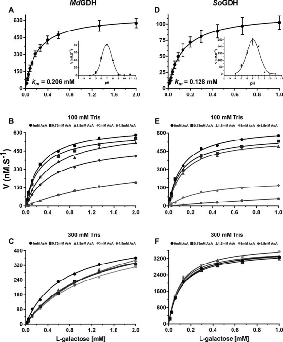
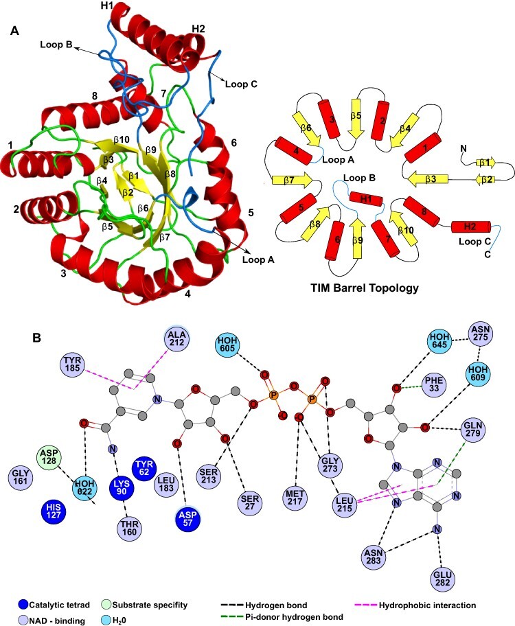
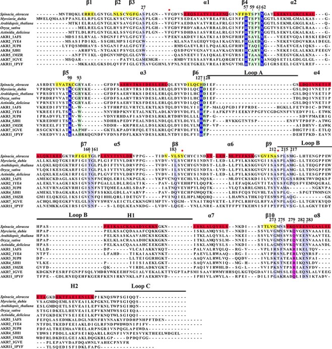
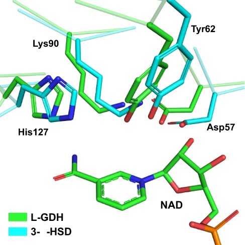
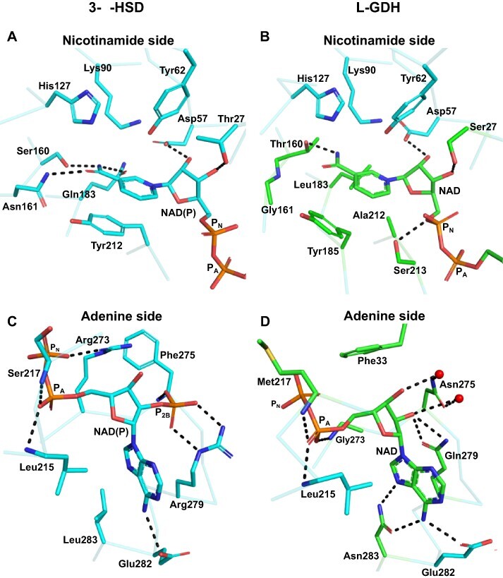
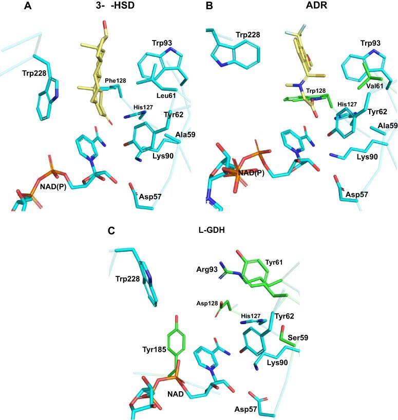
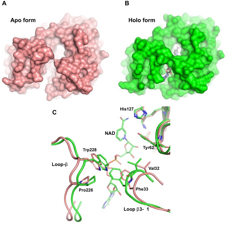
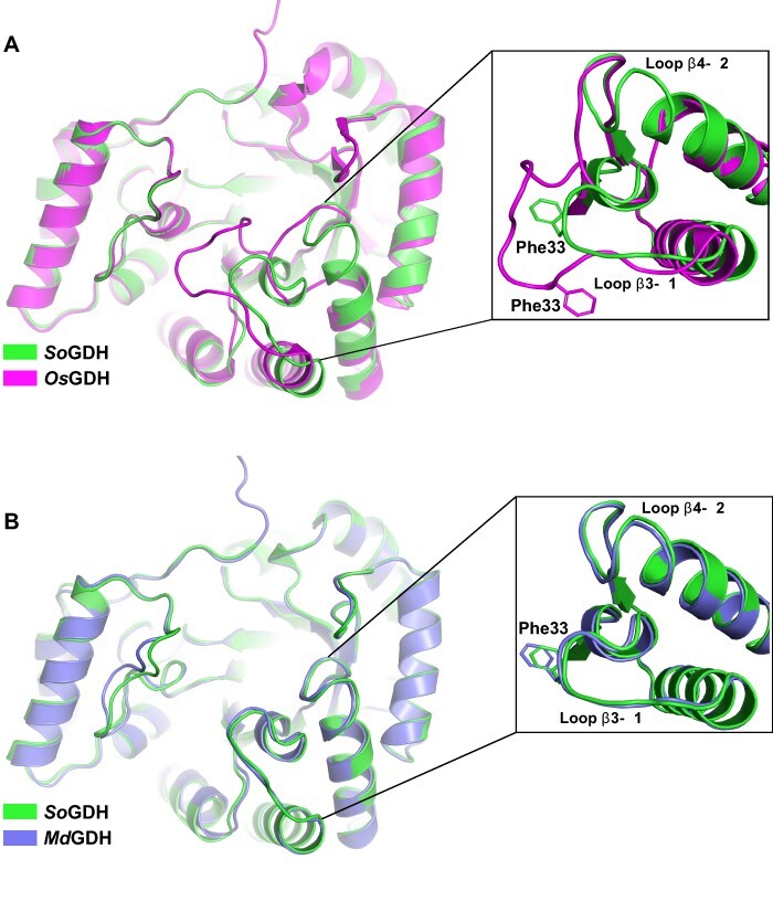
In plants, it is well-known that ascorbic acid (vitamin C) can be synthesized via multiple metabolic pathways but there is still much to be learned concerning their integration and control mechanisms. Furthermore, the structural biology of the component enzymes has been poorly exploited. Here we describe the first crystal structure for an L-galactose dehydrogenase [Spinacia oleracea GDH (SoGDH) from spinach], from the D-mannose/L-galactose (Smirnoff-Wheeler) pathway which converts L-galactose into L-galactono-1,4-lactone. The kinetic parameters for the enzyme are similar to those from its homolog from camu camu, a super-accumulator of vitamin C found in the Peruvian Amazon. Both enzymes are monomers in solution and have a pH optimum of 7, and their activity is largely unaffected by high concentrations of ascorbic acid, suggesting the absence of a feedback mechanism acting via GDH. Previous reports may have been influenced by changes of the pH of the reaction medium as a function of ascorbic acid concentration. The structure of SoGDH is dominated by a (β/α)8 barrel closely related to aldehyde-keto reductases (AKRs). The structure bound to NAD+ shows that the lack of Arg279 justifies its preference for NAD+ over NADP+, as employed by many AKRs. This favors the oxidation reaction that ultimately leads to ascorbic acid accumulation. When compared with other AKRs, residue substitutions at the C-terminal end of the barrel (Tyr185, Tyr61, Ser59 and Asp128) can be identified to be likely determinants of substrate specificity. The present work contributes toward a more comprehensive understanding of structure-function relationships in the enzymes involved in vitamin C synthesis.

Keywords: Myrciaria dubia ‘camu-camu’; Crystal structure; Enzyme kinetics; L-galactose dehydrogenase; Spinach; Vitamin C biosynthesis.


Figures

Fig. 1
Fig. 1
Kinetic properties of camu camu MdGDH and spinach SoGDH in the presence of excess NAD+ (200 μM). Michaelis–Menten-like type kinetics were observed for MdGDH (A) and SoGDH (D) with Km values of 0.206 and 0.128 mM, respectively (mean values of five independent measurements with corresponding error bars). Optimum pH curves are shown as insets. The inhibition of MdGDH and SoGDH by AsA in either 100 mM Tris–HCl buffer (B and E) or 300 mM Tris–HCl (C and F) are shown. The inhibitory effect of AsA drops off drastically in the higher concentration buffer.
Fig. 2
Fig. 2
SoGDH fold and NAD+-binding interactions. (A) Cartoon representation (left) and topology diagram (right) for the structure of SoGDH, which shows a (β/α)8-barrel (TIM-barrel) fold, characteristic of the AKR superfamily. This fold has eight β-strands interspersed by eight α-helices. At the bottom of the barrel, at the N-terminus of the polypeptide chain, there is a β-hairpin composed of strands β1 and β2. In addition, SoGDH presents two helices (H1 and H2) external to the barrel, which together with the loops loop-A, loop-B and loop-C are important for the classification of the members of AKRs. (B) The network of interactions with the NAD+ cofactor. Figures were generated with PyMol v2.05 (Schrödinger, LLC, San Diego, CA, USA) and Discovery Studio Visualizer V21.1.0 (BIOVIA, Dassault Systèmes, Waltham, MA, USA).
Fig. 3
Fig. 3
Multiple sequence alignment between L-GDHs and AKRs. Five L-GDH sequences from different species were aligned with seven representatives of different families of AKRs. Using the spinach SoGDH sequence, the position of secondary structure elements are indicated. Likewise, the numbering adopted for the description of the amino acid positions was based on SoGDH. Highlights: in dark blue the amino acids of the catalytic tetrad, in light blue those responsible for binding to NAD+, in light green the amino acids important in determining substrate specificity and in purple a conserved position in ARKs that confers a preference for the cofactor (NAD+ or NADP+). Positions with a red ball represent residues that interact with NAD+, which are only observed in the L-GDH structures presented here.
Fig. 4
Fig. 4
The active site in SoGDH. A superposition between the active site of SoGDH and 3-α-hydroxysteroid dehydrogenase (a representative of the AKR1 family) shows the catalytic tetrad (Asp27, Tyr52, Lys90 and His127) conserved in L-GDH. 3-α-HSD. PDB code: 1AFS.
Fig. 5
Fig. 5
Comparison between the NADP+-binding interactions in AKRs and NAD+ binding in SoGDH. For a better understanding, the interactions have been divided into the nicotinamide and adenine sides which are treated separately. (A and C) The interactions made with NADP+ in 3-α-HSD (PDB identifier: 1AFS). On the nicotinamide side (A), three hydrogen bonds are observed between Ser160, Asn161 and Gln183, which together with the pi-stacking formed by Tyr212, correctly orient the nicotinamide ring of NADP+ for catalysis. On the adenine side (C), the interaction between Arg279 (conserved in AKRs) and the P2B phosphate stands out and confers specificity for NADP+. On the nicotinamide side of SoGDH (B), the hydrogen bonds are lost, as well as the pi-stack at position 212. However, the orientation of the nicotinamide ring is maintained by the hydrophobic interaction of Leu183 and the pi-stacking of Tyr18. In addition, a hydrogen bond is maintained between Thr160 and the nicotinamide ring. On the adenine side (D), we observe that the absence of P2B allows for interaction via water molecules between Asn275 and the ribose. Likewise, Gln279 interacts directly with the ribose. A hydrophobic interaction involving Phe33 and two new hydrogen bonds between Asn283 and the adenine appear in SoGDH. PN: phosphate on nicotinamide side of the cofactor; PA: phosphate on adenine side.
Fig. 6
Fig. 6
Substrate specificity in AKRs and SoGDH. (A) 3-α-HSD bound to testosterone, showing amino acids important for substrate binding. Leu61 and Phe128 are specific for AKRs that bind steroids. (B) Human ADR bound to the inhibitor tolrestat. Val61 and Trp128 are specific for AKRs that bind sugars. (C) The empty active site as observed in the SoGDH–NAD+ complex. Despite SoGDH having a sugar as its natural substrate, it presents a Tyr at position 61 and an Asp at 128. It also has differences in positions 59 (Ser) and 93 (Arg). Tyr185 appears to be specific for L-GDH, since galactose-binding proteins use an aromatic amino acid to orient the ligand to the active site. 3-α-HSD PDB code: 1AFS; ADR PDB code: 2FZD.
Fig. 7
Fig. 7
The open and close states of SoGDH. (A) The apo form of SoGDH shows the open state, with little contact between loop-B and loop-β3–α1. (B) The holo form of SoGDH–NAD+ shows the closed state, forming a tunnel generated by the contact made between loop- B and loop-β3–α1 (NAD+ is shown within the tunnel at the bottom). (C) A superposition between SoGDH apo and holo forms shows a close-up of the loop-B and loop-β3–α1 regions. NAD+-binding results in hydrophobic contacts between Val32, Phe33, Pro226 and Trp228, as well as hydrogen bonding between the indole nitrogen of Trp228 and the Val32 main chain.
Fig. 8
Fig. 8
L-GDH structure comparisons. (A) A superposition between spinach SoGDH and rice OsGDH crystal structures in the apo form. Despite having an RMSD of 0.45 Å for Cα atoms, the orientations of loop-β3–α1 and loop-β4–α2 are very different (with a displacement of approximately 6 Å). (B) Superposition between the crystal structure of spinach SoGDH and the Alphafold2 model for camu camu MdGDH. loop-β3–α1 and loop-β4–α2 show the same orientation. Rice OsGDH PDB code: 7EZI.

References

    1. Adams P.D., Afonine P.V., Bunkóczi G., Chen V.B., Davis I.W., Echols N., et al. (2010) PHENIX: a comprehensive Python-based system for macromolecular structure solution. Acta Crystallogr. Sect. D Biol. Crystallogr. 66: 213–221. - PMC - PubMed
    1. Barski O.A., Tipparaju S.M. and Bhatnagar A. (2008) The aldo-keto reductase superfamily and its role in drug metabolism and detoxification. Drug Metab. Rev. 40: 553–624. - PMC - PubMed
    1. Bennett M.J., Albert R.H., Jez J.M., Ma H., Penning T.M. and Lewis M. (1997) Steroid recognition and regulation of hormone action: crystal structure of testosterone and NADP+ bound to 3α-hydroxysteroid/dihydrodiol dehydrogenase. Structure 5: 799–812. - PubMed
    1. Caruso P., Russo M.P., Caruso M., Guardo M.D., Russo G., Fabroni S., et al.(2021) Plants. - PMC - PubMed
    1. Castro J.C., Maddox J.D., Cobos M., Requena D., Zimic M., Bombarely A., et al. (2015) De novo assembly and functional annotation of Myrciaria dubia fruit transcriptome reveals multiple metabolic pathways for L-ascorbic acid biosynthesis. BMC Genomics 16: 997. - PMC - PubMed