. 2022 Jun 29;7(3):e0002922.
doi: 10.1128/msphere.00029-22. Epub 2022 May 9.

The Pathogenic Yeast Candida parapsilosis Forms Pseudohyphae through Different Signaling Pathways Depending on the Available Carbon Source

Affiliations

The Pathogenic Yeast Candida parapsilosis Forms Pseudohyphae through Different Signaling Pathways Depending on the Available Carbon Source

Christopher B Rupert et al. mSphere. .

Abstract

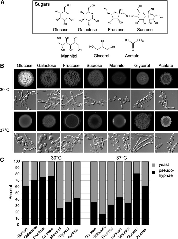
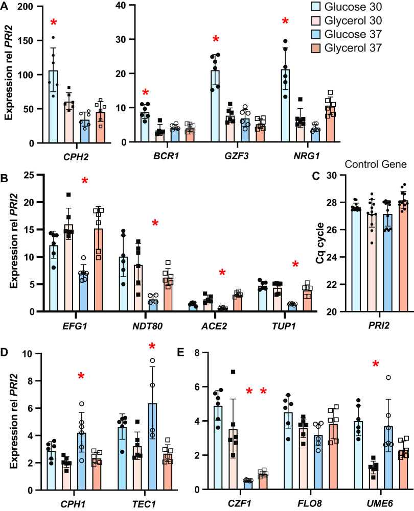
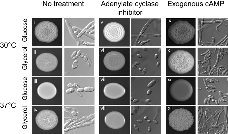
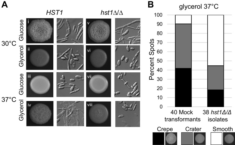
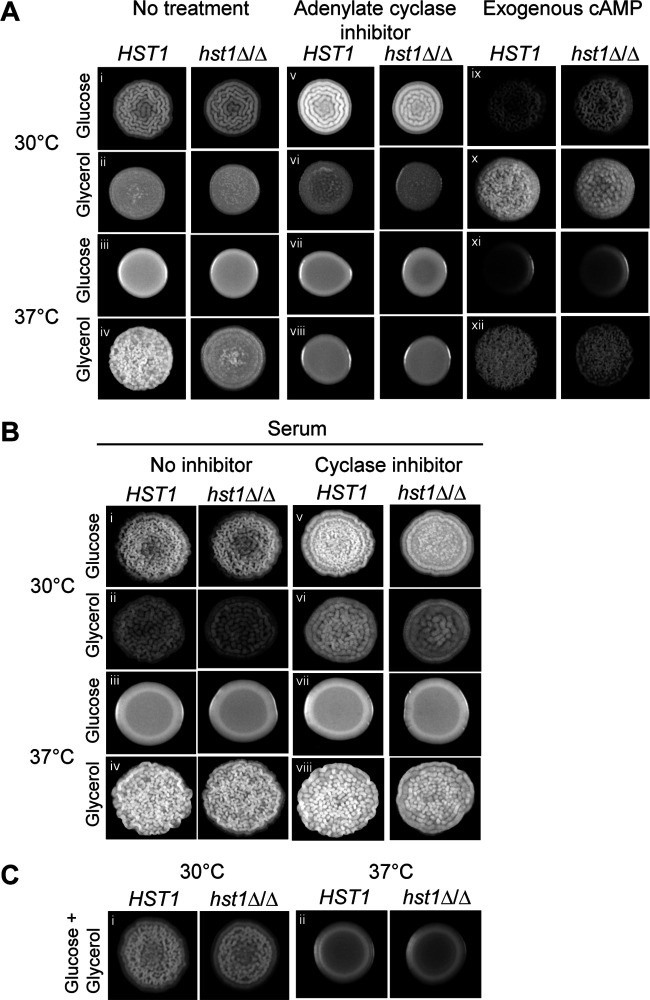
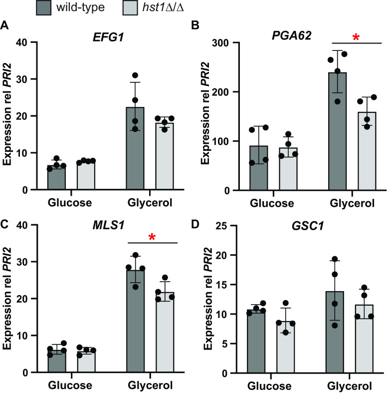
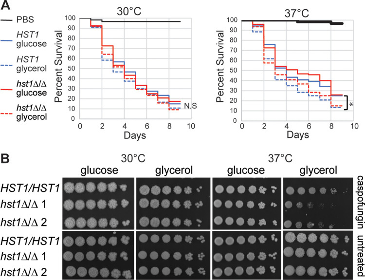
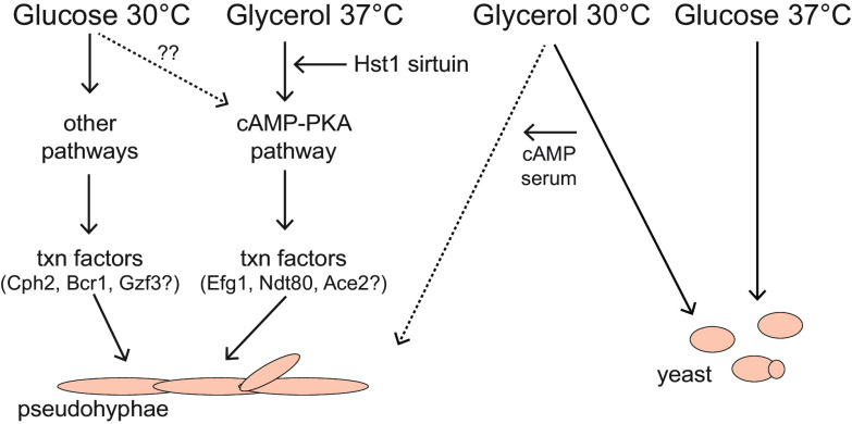
Candida parapsilosis is an emerging fungal pathogen that primarily affects immunocompromised patients in hospitals. A significant risk factor is the use of implanted medical devices, which support the growth of biofilms composed of a mixture of individual yeast cells and chains of elongated pseudohyphal cells. The morphological switch between these two forms is triggered by cues from the environment, including nutrient availability and temperature. We examined how different nutrient sources affect the balance between yeast and pseudohyphae and found that cells grown in the presence of five- or six-carbon sugars form more pseudohyphae at 30°C than at 37°C. Conversely, cells grown on glycerol, a three-carbon polyalcohol, form more pseudohyphae at 37°C. Furthermore, we found that different regulators influence pseudohyphal growth on glucose at 30°C compared with those on glycerol at 37°C. In particular, cAMP signaling and the sirtuin deacetylase Hst1 were required for pseudohyphal growth on glycerol at 37°C but not on glucose at 30°C. Finally, we found that the carbon source on which C. parapsilosis is grown can influence its ability to establish an infection in a wax moth model. Overall, this study reveals that environmental conditions affect not only the extent of pseudohyphal growth but also which pathways and regulators govern pseudohyphal formation. IMPORTANCE Candida parapsilosis is one of the leading causes of hospital-acquired yeast infections and poses a significant risk to immunocompromised people. Two of its properties that contribute to infection are metabolic flexibility, to use a range of nutrients available in the host, and cellular dimorphism, to switch between round yeast cells and chains of elongated pseudohyphal cells. Uncovering the molecular mechanisms that regulate these processes could reveal new targets for antifungal drugs. We found that for C. parapsilosis, the balance between yeast and pseudohyphal cells depends on the nutrients available and the growth temperature. Moreover, these environmental changes can affect its ability to cause infections. Finally, we found that a potential sensor of the cell's metabolic state, the sirtuin Hst1, contributes to pseudohyphal growth for cells grown on glycerol. These findings indicate that the shape and virulence of C. parapsilosis likely vary depending on its location in the host.

Keywords: Candida; cyclic AMP; filamentation; glycerol; sirtuin.


Conflict of interest statement

The authors declare no conflict of interest.

Figures

FIG 1
FIG 1
Carbon source and temperature influenced the growth pattern of C. parapsilosis. (A) Molecular structures of the carbon sources used in this study. (B) Colony and cell morphology of wild-type C. parapsilosis cells (CLIB214) grown on rich medium (yeast extract and peptone; YP) containing the indicated carbon sources at 30°C (top) and 37°C (bottom). Spots were inoculated with 105 cells in 10 μL H2O. Spots and cells from the pictured spots were imaged after 3 days of growth and were representative of two separate experiments done in duplicate. (C) The proportions of yeast and pseudohyphae were determined for spots grown on each carbon source. Each bar represents the average of two separate experiments. For each one, approximately 150 cells were scored from 8 to 12 fields of view.
FIG 2
FIG 2
Transcription factor genes were expressed differently under the two filament-promoting conditions. RNA was extracted from wild-type CLIB214 cells grown on solid medium (YP) containing glucose (blue bars) or glycerol (orange bars) at 30°C (lighter bars) or 37°C (darker bars). Each bar represents the average and standard deviation of six independent extractions, with expression normalized to the control gene PRI2. Samples that were statistically different (P < 0.05) based on two-way ANOVA with Tukey post hoc analysis are indicated by an asterisk (*). (C) For the control gene PRI2, the threshold cycle (Cq) did not vary across conditions, indicating that PRI2 is expressed consistently.
FIG 3
FIG 3
Pseudohyphal growth on glycerol at 37°C was dependent on adenylate cyclase. Wild-type cells (CLIB214) were spotted onto solid medium containing glucose or glycerol, with or without 10 mM cAMP or 50 μM MD-12,330A, an inhibitor of adenylate cyclase. Spots and cells were imaged after 3 days of growth. The experiment was performed in duplicate two separate times, with similar results.
FIG 4
FIG 4
The deacetylase Hst1 was required for pseudohyphal growth on glycerol at 37°C. (A) Communities of wild-type (CLIB214) or hst1Δ/Δ (LRY3263) cells grown on glucose or glycerol. Spots and cells were imaged after 3 days of growth and were performed in duplicate in two separate experiments. Similar results were obtained for a second hst1Δ/Δ isolate (LRY3296). (B) Growth phenotypes on glycerol at 37°C for 38 independent hst1Δ/Δ isolates (LRY3259 to 3296) and 40 mock-transformed isolates (LRY3297 to 3331) exposed to a CRISPR plasmid lacking the HST1-directed guide RNA. Phenotypes were classified as crepe, crater, or smooth following published categories (19) and were scored in a blind manner by three observers evaluating two separate rounds of growth.
FIG 5
FIG 5
The pseudohyphal defect in hst1Δ/Δ cells could be rescued by cAMP or serum. (A) Wild-type (CLIB214) and hst1Δ/Δ cells (LRY3263) were spotted onto plates containing glucose or glycerol, with or without 50 μM MDL-12,330A or 10 mM cAMP. (B) Wild-type and hst1Δ/Δ cells were spotted onto plates containing glucose or glycerol with 10% fetal bovine serum and with or without MDL-12,330A. (C) Cells were spotted onto plates containing both glucose and glycerol. All spot pictures were taken after 3 days of growth and were performed in duplicate on two separate days. Similar results were obtained for another hst1Δ/Δ isolate (LRY3296).
FIG 6
FIG 6
On glycerol at 37°C, hst1Δ/Δ cells had a lower expression of EFG1 and Efg1-target genes than wild-type cells. RNA was extracted from wild-type (CLIB214) or hst1Δ/Δ (LRY3263 and 3296) spots grown 3 days on glucose or glycerol at 37°C. mRNAs were quantified by RT-PCR and normalized to the PRI2 gene. Expression for the wild type represents four wild type spots harvested on two separate days, whereas expression for hst1Δ/Δ cells represents two spots each of LRY3263 and LRY3296 harvested on 2 days. A significant difference between wild-type and hst1Δ/Δ cells grown on glycerol was evaluated using Student’s t-test; *, P < 0.05.
FIG 7
FIG 7
Effect of growth condition and hst1Δ/Δ mutation on virulence traits of C. parapsilosis. (A) Virulence assay using G. mellonella larvae. Wild-type (CLIB214) and hst1Δ/Δ cells (LRY3263 and LRY3296) were grown on solid medium containing glucose or glycerol at 30°C or 37°C for 3 days. Approximately 4 × 106 cells were injected into each of 30 larvae, which were incubated at 30°C (left) or 37°C (right) for 8 days. Live larvae were counted every day. Lines represent the percent survival of 120 larvae from four different experiments. *, P < 0.05; using log rank test. (B) Caspofungin sensitivity assay. Wild-type or hst1Δ/Δ cells (LRY3263 and 3296) were grown overnight in YPD at 30°C and then subcultured to an OD600 of 0.2 and grown until an OD600 of 2.5. Five-fold serial dilutions were spotted on plates with or without 0.5 μg/mL caspofungin and imaged after 2 days of growth.
FIG 8
FIG 8
Model depicting pathways contributing to pseudohyphal growth depending on the available carbon source and the incubation temperature.

Similar articles

Cited by

References

    1. Almirante B, Rodriguez D, Cuenca-Estrella M, Almela M, Sanchez F, Ayats J, Alonso-Tarres C, Rodriguez-Tudela JL, Pahissa A, the Barcelona Candidemia Project Study Group . 2006. Epidemiology, risk factors, and prognosis of Candida parapsilosis bloodstream infections: case-control population-based surveillance study of patients in Barcelona, Spain, from 2002 to 2003. J Clin Microbiol 44:1681–1685. doi:10.1128/JCM.44.5.1681-1685.2006. - DOI - PMC - PubMed
    1. Brito LR, Guimaraes T, Nucci M, Rosas RC, Paula Almeida L, Da Matta DA, Colombo AL. 2006. Clinical and microbiological aspects of candidemia due to Candida parapsilosis in Brazilian tertiary care hospitals. Med Mycol 44:261–266. doi:10.1080/13693780500421476. - DOI - PubMed
    1. Costa-de-Oliveira S, Pina-Vaz C, Mendonca D, Goncalves Rodrigues A. 2008. A first Portuguese epidemiological survey of fungaemia in a university hospital. Eur J Clin Microbiol Infect Dis 27:365–374. doi:10.1007/s10096-007-0448-4. - DOI - PubMed
    1. Fridkin SK, Kaufman D, Edwards JR, Shetty S, Horan T, the National Nosocomial Infections Surveillance System Hospitals . 2006. Changing incidence of Candida bloodstream infections among NICU patients in the United States: 1995–2004. Pediatrics 117:1680–1687. doi:10.1542/peds.2005-1996. - DOI - PubMed
    1. Messer SA, Jones RN, Fritsche TR. 2006. International surveillance of Candida spp. and Aspergillus spp.: report from the SENTRY Antimicrobial Surveillance Program (2003). J Clin Microbiol 44:1782–1787. doi:10.1128/JCM.44.5.1782-1787.2006. - DOI - PMC - PubMed

Publication types

LinkOut - more resources