. 2022 Jul 19;66(7):e0046322.
doi: 10.1128/aac.00463-22. Epub 2022 Jun 29.

MBZM-N-IBT, a Novel Small Molecule, Restricts Chikungunya Virus Infection by Targeting nsP2 Protease Activity In Vitro, In Vivo, and Ex Vivo

Affiliations

MBZM-N-IBT, a Novel Small Molecule, Restricts Chikungunya Virus Infection by Targeting nsP2 Protease Activity In Vitro, In Vivo, and Ex Vivo

Saikat De et al. Antimicrob Agents Chemother. .

Abstract

The increase in disease incidences and persistent Chikungunya virus (CHIKV)-induced arthritis have been a huge burden on public health globally. In the absence of specific antivirals or vaccines, it is essential to continue efforts to develop effective anti-CHIKV strategies. Our previous study showing the in vitro anti-CHIKV potential of a novel molecule 1-[(2-methylbenzimidazol-1-yl) methyl]-2-oxo-indolin-3-ylidene] amino] thiourea (MBZM-N-IBT) encouraged us to further validate its efficacy. Here, the effect of MBZM-N-IBT was evaluated in vitro in RAW 264.7 cells, in vivo in C57BL/6 mice, and ex vivo in human peripheral blood mononuclear cells (hPBMCs). The study demonstrated that CHIKV infection was efficiently abrogated in RAW 264.7 cells (IC50 = 22.34 μM) with significant inhibition in viral proteins. The inhibition was effective in the postentry step, and MBZM-N-IBT predominately interfered in the early stages of CHIKV life cycle. It was further supported when the protease activity of CHIKV-nsP2 was hindered by the compound. Moreover, it diminished the CHIKV-induced inflammatory responses in vitro through significant downregulation of all the major mitogen-activated protein kinases (MAPKs), NF-κB, cyclooxygenase (COX)-2, and cytokines. Furthermore, MBZM-N-IBT restricted CHIKV infection and inflammation in vivo, leading to reduced clinical scores and complete survival of C57BL/6 mice. Additionally, it has been noticed that the CHIKV infection was reduced remarkably in hPBMC-derived monocyte-macrophage populations ex vivo by the compound. In conclusion, it can be suggested that this novel compound MBZM-N-IBT has been demonstrated to be a potential anti-CHIKV molecule in vitro, in vivo, and ex vivo and fulfilled all the criteria to investigate further for successful treatment of CHIKV infection.

Keywords: Chikungunya; anti-viral; infection; inflammation; replication.


Conflict of interest statement

The authors declare no conflict of interest.

Figures
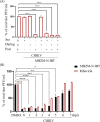
FIG 1
FIG 1
Post-treatment of 1-[(2-methylbenzimidazol-1-yl) methyl]-2-oxo-indolin-3-ylidene] amino] thiourea (MBZM-N-IBT) reduces Chikungunya virus (CHIKV) infection significantly. (A) The RAW 264.7 cells were treated with the compound (100 μM) separately (3 h before infection), during (1.5 h during infection), and after infection (8 h after infection). Compound was present before and during for “pre + during” conditions. Compound was present before, during, and after infection for “pre + during + post” infection. Supernatants collected at 8 h postinfection (hpi) were subjected to plaque assay. The bar diagram shows the percentage of virus particle. The data are presented as means ± standard error of the mean (SEM) (n = 3, P ≤ 0.05 was considered statistically significant). (B) The RAW 264.7 cells were infected by CHIKV with a multiplicity of infection (MOI) of 5, and 100 μM compound was added to each sample, every 1-h interval up to 7 hpi. The open bars represent the viral titer obtained using ribavirin (10 μM) as a control. The bar diagram represents the percentage of virus titers of the supernatants collected at 8 hpi for all the samples. DMSO, dimethyl sulfoxide. *, P ≤ 0.05; **, P ≤ 0.01; ***, P ≤ 0.001; and ****, P ≤ 0.0001 were considered statistically significant.
FIG 2
FIG 2
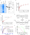
MBZM-N-IBT interacts with CHIKV-nsP2-CT and reduces its protease activity. (A) Crude and purified nsP2-CT (47 kDa) was resolved with SDS-PAGE under reducing conditions and subjected to Coomassie blue staining (left panel) and Western blot analysis (right panel) by nsP2-CT-specific antibody. (B) 1 μM nsP2-CT was incubated with or without increasing concentration (1, 2, 4, 6, 10, 20, 30, 50, 75, and 100 μM) of compound. The intrinsic tryptophan emission intensity was measured at 335 nm, and a gradual reduction in fluorescent intensity was observed. The graph shows binding of MBZM-N-IBT to nsP2-CT. (C) The dissociation constant (Kd) for the interaction between the compound and nsP2-CT was derived by the double reciprocal plot. (D) Schematic diagram illustrating the principle of the fluorescence resonance energy transfer (FRET) assay. The cleavage site present between nonstructural proteins (nsP3-nsP4) of CHIKV (RAGGYIFS) is inserted between the fluorescence quenching pair (4-(dimethylaminoazo) benzene-4-carboxylic acid [DABCYL] and 5-((2-aminoethyl) amino) naphthalene-1-sulfonic acid [EDANS]. Cleavage of the peptide bond at RAGG*YIFS by the nsP2-CT proteins separates DABCYL and EDANS. Enhanced fluorescence can be monitored at 460 nm with excitation at 355 nm. (E) Kinetic parameters of nsP2-CT with or without increasing concentrations of compound were determined by fitting untransformed data into the Michaelis-Menten equation using GraphPad Prism software. (F) The percentage of enzymatic activity was calculated as a function of inhibitor concentration, and the IC50 value of the compound is represented in this graph obtained through GraphPad Prism software. Telmisartan was used as a positive control. All the experiments were performed in duplicate. Values from three independent experiments were used to obtain all the graphs.
FIG 3
FIG 3
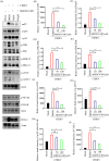
MBZM-N-IBT reduces the (CHIKV-induced) upregulated of mitogen-activated protein kinases (MAPKs), NF-κB, and COX-2 in RAW 264.7 cells. The RAW 264.7 cells were infected with CHIKV, treated with two different concentrations of MBZM-N-IBT (50 and 100 μM), and harvested at 8 hpi. (A) Western blot showing the nsP2 protein level along with phosphorylation status of p38, JNK, extracellular signal-regulated kinase (ERK), cJUN, ATF2/7, NF-κB, and total expression levels of COX-2 in RAW 264.7 cell lysates. Actin was used as a loading control. (B to H) Bar diagrams indicating relative band intensities of nsP2, phosphorylated (p)-p38, p-JNK, p-ERK, p-cJUN, p-ATF2/7, and p-NF-κB. (I) Bar diagram depicting relative band intensities of COX-2 protein. The data represent the means ± SEM (n = 3). P ≤ 0.05 was considered statistically significant.
FIG 4
FIG 4
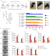
MBZM-N-IBT protects mice from CHIKV infection by diminishing the overall viral burden, viral RNA, and protein levels. (A) C57BL/6 mice were infected subcutaneously with 106 PFU of CHIKV and treated with 15 mg/kg MBZM-N-IBT at 24-h intervals up to 4 days postinfection (dpi). The mice were sacrificed at 5 dpi. Sera and different tissues were also collected for further downstream experiments. (B) Image of mock, CHIKV-infected, and compound-treated mice. (C) Equal amount of muscle tissue was taken and homogenized followed by filtration (0.22 μm). Homogeneous sample was subjected to plaque assay. The bar diagram shows CHIKV percentage of PFU/mL in virus-infected and compound-treated mice serum. (D) Whole RNA was isolated from the CHIKV-infected and compound-treated samples, and the CHIKV E1 gene was amplified by reverse transcription-quantitative PCR (qRT-PCR). The bar diagram shows the fold change of viral RNA in infected and compound-treated samples. (E) Western blot showing the viral E2 and nsP2 protein in different tissue samples. Actin was used as a loading control. (F to J) Bar diagrams showing the relative band intensities of E2 in infected and compound-treated samples of different tissues. All bar diagrams were obtained through GraphPad Prism software (n = 3). The data are presented as means ± SEM (P ≤ 0.05 was considered statistically significant). PBS, phosphate-buffered saline; plaque-forming units per milliliter (PFU/mL).
FIG 5
FIG 5
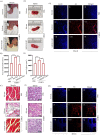
MBZM-N-IBT decreases the swelling of muscle and spleen and improves their histopathology in CHIKV-infected mice. C57BL/6 mice were infected subcutaneously with 106 PFU of CHIKV and treated with 15 mg/kg MBZM-N-IBT at 24-h intervals up to 4 dpi. The mice were sacrificed at 5 dpi. Muscle and spleen tissues were collected for morphological and histopathological analysis. (A) Image panels indicating evident hind limb muscle swelling of mock, infected, and treated groups of mice. (B) Bar diagram showing the quantitation of limb swelling. (C) Image panels showing the hematoxylin and eosin (H&E)-stained muscle section. (D) Image panels depicting gross swelling of spleen (splenomegaly) of mock, infected, and treated groups of mice. (E) Bar diagram showing the quantitation of splenomegaly. (F) Image panels showing the H&E-stained spleen section. (G) Image panels showing the CHIKV-E2 stained muscle sections. (H) Image panels showing the CHIKV-E2 stained spleen tissue sections. All bar diagrams were obtained through GraphPad Prism software (n = 3). The data are presented as the means ± SEM (P ≤ 0.05 was considered statistically significant). DAPI, 4′,6-diamidino-2-phenylindole.
FIG 6
FIG 6
MBZM-N-IBT treatment ameliorates CHIKV-induced disease score and provides better survival. (A) C57BL/6 mice were infected subcutaneously with 106 PFU of CHIKV and treated with 15 mg/kg MBZM-N-IBT at 24- or 12-h intervals up to 12 dpi. Six mice were taken in each group. (B) Graph showing cumulative body weight of each group of mice throughout the study. (C) Graph describing mean body weight of animals in each group at each dpi. (D) Line diagram showing the survivability of infected mice along with the treated groups. The percentage survivability data were obtained through GraphPad Prism software.
FIG 7
FIG 7
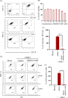
MBZM-N-IBT reduces CHIKV infection in human peripheral blood mononuclear cell (hPBMC)-derived monocyte-macrophage populations ex vivo. (A) Dot plot showing the percentages of CD14+CD11b+ monocyte-macrophage lineage of adherent hPBMCs, B cells (CD19), and T cells (CD3) by flow cytometry. (B) Bar diagram referring the cytotoxicity of MBZM-N-IBT in hPBMC-derived adherent population by 3-(4,5-dimethyl-2-thiazolyl)-2,5-diphenyl-2H-tetrazolium bromide (MTT) assay. (C) Bar diagram depicting percentage of the viral particle formation by plaque assay. (D) Dot plot showing the percentage of viral E2 positive monocyte-macrophage population in mock, CHIKV-infected, and compound-treated samples from three healthy donors’ hPBMCs by flow cytometry. (E) Bar diagram showing the percentage of positive cells for the CHIKV-E2 protein. The data shown are represented as means ± SEM of three independent experiments. *, P < 0.05. Side scatter (SSC), 0.15 M NaCl plus 0.015 M sodium citrate.

References

    1. Caglioti C, Lalle E, Castilletti C, Carletti F, Capobianchi MR, Bordi L. 2013. Chikungunya virus infection: an overview. New Microbiol 36:211–227. - PubMed
    1. Burt FJ, Chen W, Miner JJ, Lenschow DJ, Merits A, Schnettler E, Kohl A, Rudd PA, Taylor A, Herrero LJ, Zaid A, Ng LFP, Mahalingam S. 2017. Chikungunya virus: an update on the biology and pathogenesis of this emerging pathogen. Lancet Infect Dis 17:e107–e117. 10.1016/S1473-3099(16)30385-1. - DOI - PubMed
    1. Mbaika S, Lutomiah J, Chepkorir E, Mulwa F, Khayeka-Wandabwa C, Tigoi C, Oyoo-Okoth E, Mutisya J, Ng'ang'a Z, Sang R. 2016. Vector competence of Aedes aegypti in transmitting chikungunya virus: effects and implications of extrinsic incubation temperature on dissemination and infection rates. Virol J 13:114. 10.1186/s12985-016-0566-7. - DOI - PMC - PubMed
    1. Kril V, Aiqui-Reboul-Paviet O, Briant L, Amara A. 2021. New insights into chikungunya virus infection and pathogenesis. Annu Rev Virol 8:327–347. 10.1146/annurev-virology-091919-102021. - DOI - PubMed
    1. Ganesan VK, Duan B, Reid SP. 2017. Chikungunya virus: pathophysiology, mechanism, and modeling. Viruses 9:368. 10.3390/v9120368. - DOI - PMC - PubMed

Publication types

Substances