Review
. 2022 Jun 29;13(1):108.
doi: 10.1186/s13244-022-01243-2.

Clinical and radiological features of lung disorders related to connective-tissue diseases: a pictorial essay

Affiliations
Review

Clinical and radiological features of lung disorders related to connective-tissue diseases: a pictorial essay

Stefano Palmucci et al. Insights Imaging. .

Abstract

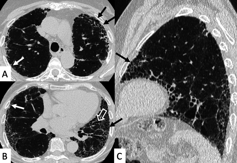
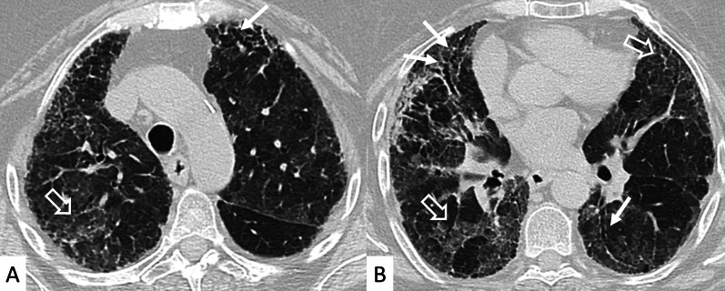
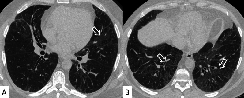
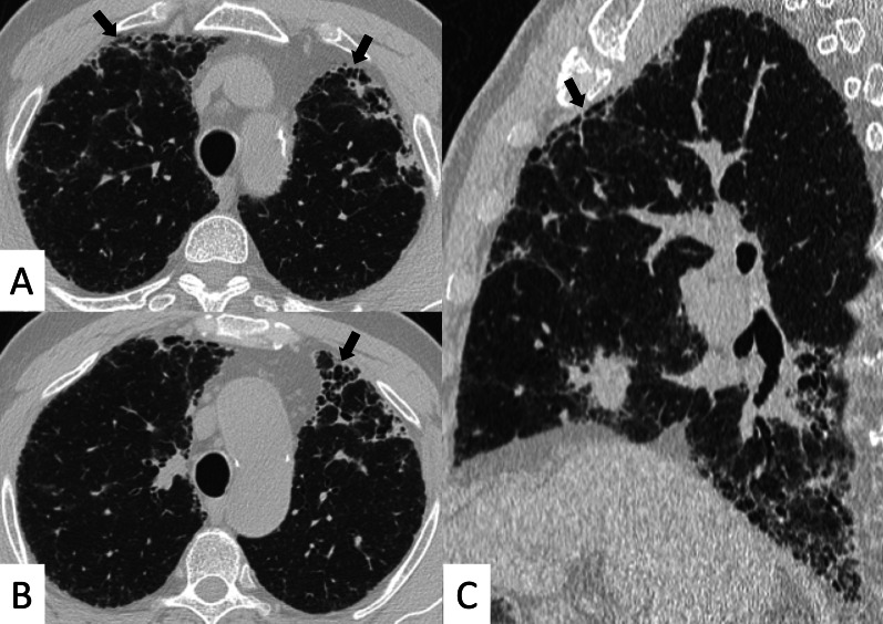
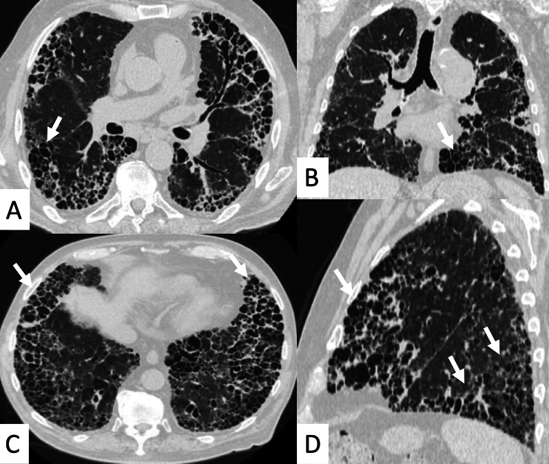
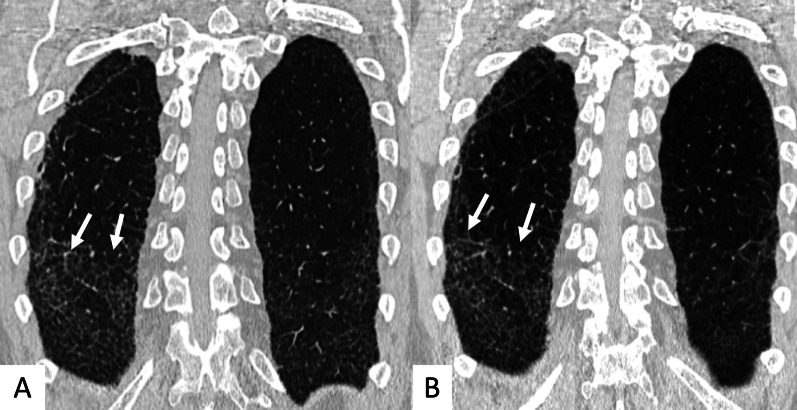
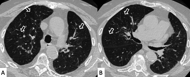
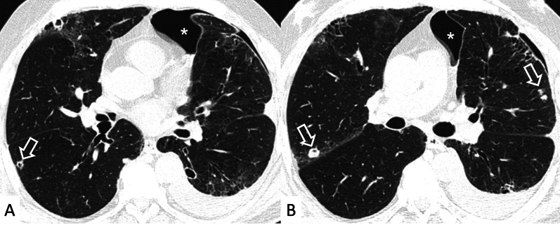
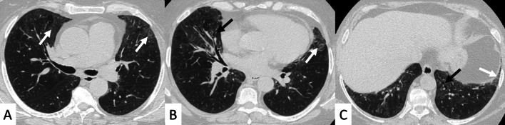
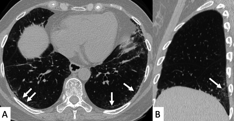
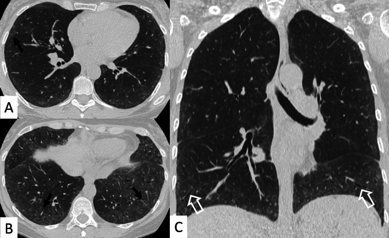
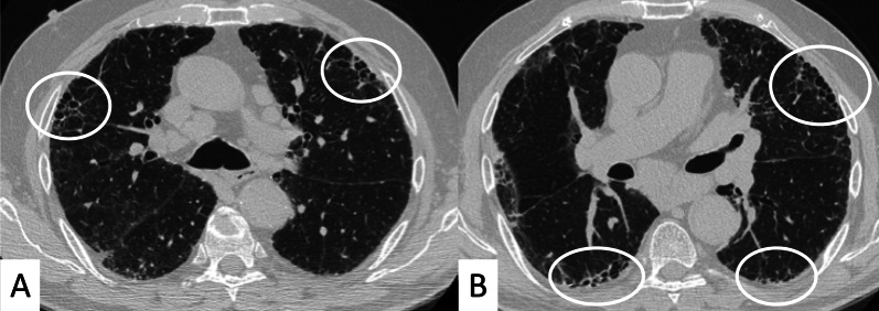
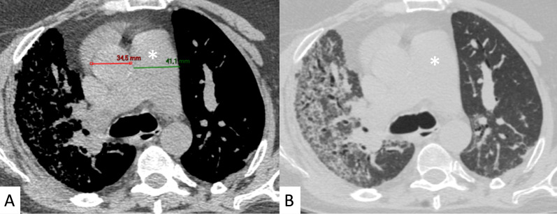
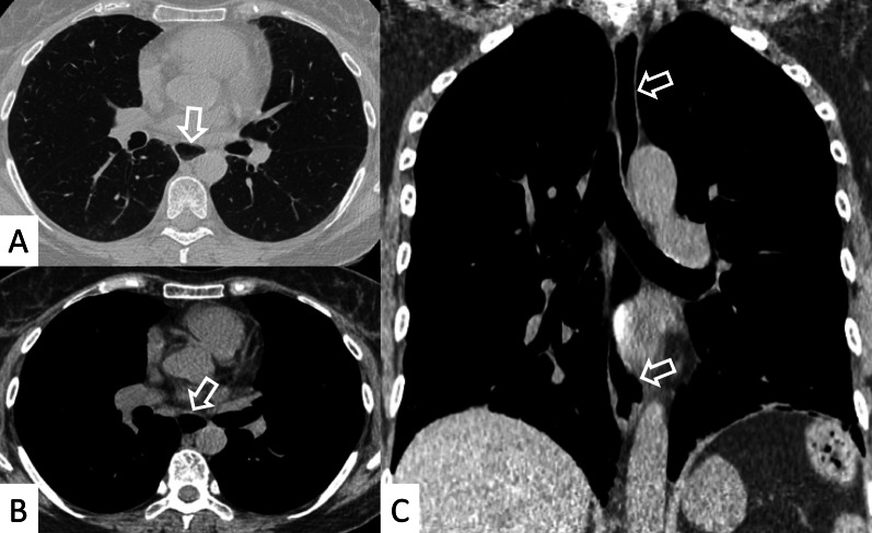
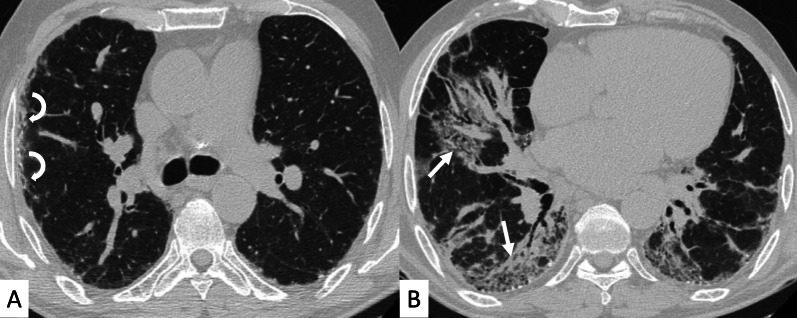
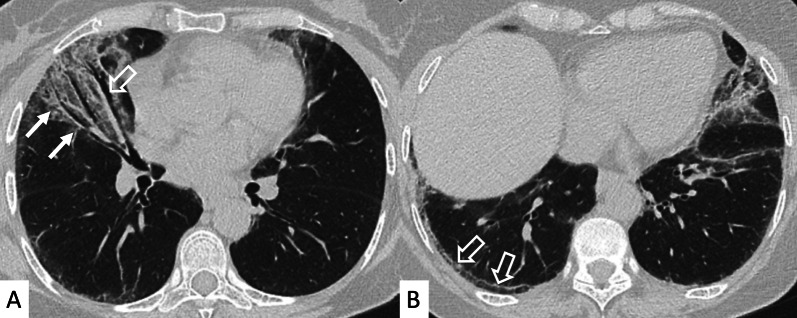
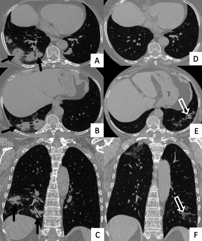
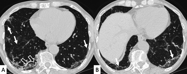
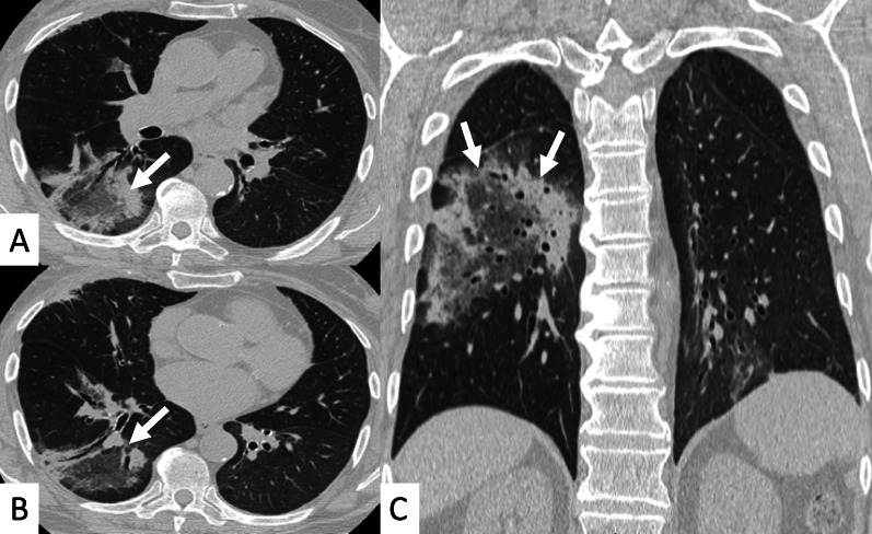
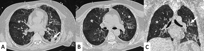
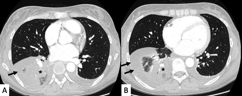
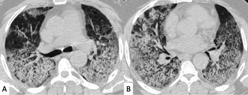
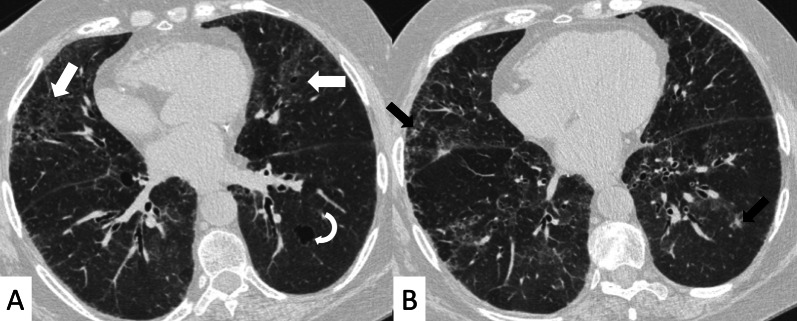
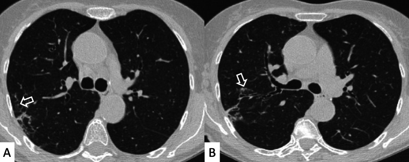
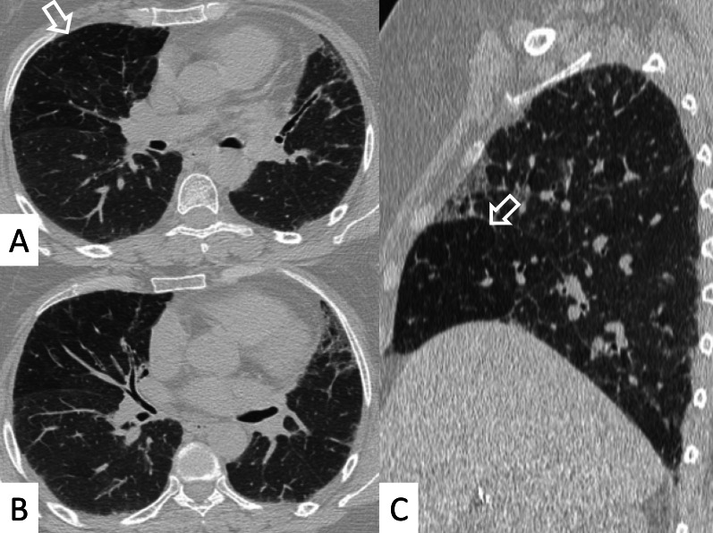
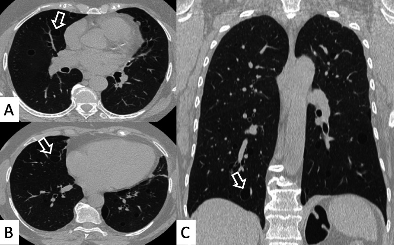
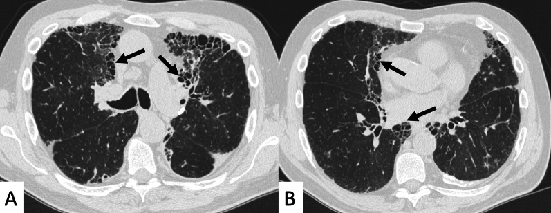
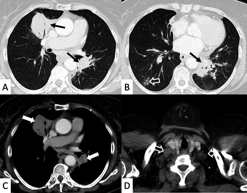
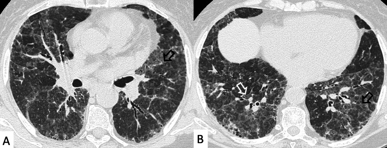
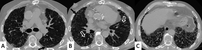
Connective tissue diseases (CTDs) include a spectrum of disorders that affect the connective tissue of the human body; they include autoimmune disorders characterized by immune-mediated chronic inflammation and the development of fibrosis. Lung involvement can be misdiagnosed, since pulmonary alterations preceded osteo-articular manifestations only in 20% of cases and they have no clear clinical findings in the early phases. All pulmonary structures may be interested: pulmonary interstitium, airways, pleura and respiratory muscles. Among these autoimmune disorders, rheumatoid arthritis (RA) is characterized by usual interstitial pneumonia (UIP), pulmonary nodules and airway disease with air-trapping, whereas non-specific interstitial pneumonia (NSIP), pulmonary hypertension and esophageal dilatation are frequently revealed in systemic sclerosis (SSc). NSIP and organizing pneumonia (OP) may be found in patients having polymyositis (PM) and dermatomyositis (DM); in some cases, perilobular consolidations and reverse halo-sign areas may be observed. Systemic lupus erythematosus (SLE) is characterized by serositis, acute lupus pneumonitis and alveolar hemorrhage. In the Sjögren syndrome (SS), the most frequent pattern encountered on HRCT images is represented by NSIP; UIP and lymphocytic interstitial pneumonia (LIP) are reported with a lower frequency. Finally, fibrotic NSIP may be the interstitial disease observed in patients having mixed connective tissue diseases (MCTD). This pictorial review therefore aims to provide clinical features and imaging findings associated with autoimmune CTDs, in order to help radiologists, pneumologists and rheumatologists in their diagnoses and management.

Keywords: Autoimmune diseases; Connective tissue disease; Lung disease (Interstitial); Multidetector computed tomography; Pulmonary fibrosis.


Conflict of interest statement

SP reports personal consulting fees and/or speaker fees from Boehringer Ingelheim and F. HoffmannLa Roche Ltd. outside the submitted work. GS reports personal fees from Boehringer Ingelheim outside the submitted work. CV is part of the F. Hoffmann-La Roche Ltd. and Boehringer Ingelheim Scientific board. He has received consulting fees and/or speaker fees from AstraZeneca, Boehringer Ingelheim, Chiesi, F. Hoffmann-La Roche Ltd and Menarini.

Figures

Fig. 1
Fig. 1
Axial (A, B) and sagittal (C) HRCT images of a UIP pattern in a 72-year-old patient with rheumatoid arthritis. The images show the typical UIP pattern, predominantly located on basal and subpleural regions, and represented by honeycombing areas (black arrows in AC), reticulations (white arrows in A, B) and traction bronchiectasis (white empty arrow in B)
Fig. 2
Fig. 2
Axial HRCT images of a patient with rheumatoid arthritis. A, B clearly show ground-glass opacities (empty white arrows) and bronchiectasis (white arrows), resembling an NSIP pattern
Fig. 3
Fig. 3
Axial HRCT images of an NSIP pattern in a patient with rheumatoid arthritis. A, B Ground-glass opacities (empty white arrows) can be appreciated through the pulmonary parenchyma. The heterogeneous distribution of ground glass regions reflects areas of inflammation—alternated to normal lung regions
Fig. 4
Fig. 4
Anterior upper lobe sign in a patient with RA. It consists of a particular fibrosis distribution, most concentrated along the anterior side of the upper lobes; it can be appreciated on axial (black arrows in A, B) and sagittal MPR image (black arrow in C)
Fig. 5
Fig. 5
Axial (A, C), coronal (B) and sagittal (D) HRCT images demonstrate—in an RA patient—the presence of a florid honeycombing, involving more than 50% of the lung. This finding is typically due to macrocystic spaces (white arrows) and has been defined as “exuberant honeycombing sign”
Fig. 6
Fig. 6
Coronal multiplanar reformatted images. A, B demonstrate the presence of straight-edge sign in female RA patients showing interstitial lung fibrosis. This sign consists of a more pronounced fibrosis in the lung bases—with sharp demarcation in the cranio-caudal plane (white arrows in A, B)
Fig. 7
Fig. 7
Airway disease in RA patient, clearly depicted in A, B. Lobular decreased attenuation areas (white empty arrows) are caused by follicular bronchiolitis and obliterative bronchiolitis. This finding represents an early manifestation of disease, due to airway involvement—typically found when no fibrosing alterations are observed
Fig. 8
Fig. 8
Cavitate nodular lesions (white empty arrows in A, B) in a male RA patient. Rheumatoid nodules represent a rare manifestation of disease; generally, they have bilateral and subpleural location, with rounded shape. The development of cavitation has been reported in literature related to vasculitis. Left pneumothorax (white asterisk) is also evident along the anterior part of the lung
Fig. 9
Fig. 9
Typical NSIP pattern in an SSc patient. A Ground-glass opacities and fine reticulations (white arrows), predominantly located in peripheral regions; in B, C traction bronchiectasis are also depicted (black arrows)
Fig. 10
Fig. 10
NSIP pattern with subpleural sparing in a female SSc patient. Axial (A) and sagittal (B) HRCT images demonstrate ground-glass opacities predominantly located in the basal pulmonary regions, with peripheral and subpleural distribution (white arrows). Pulmonary alterations may spare subpleural regions, as well demonstrated in A—namely in the left lower lobe
Fig. 11
Fig. 11
Axial (A, B) and coronal (C) HRCT images of an SSc patient demonstrate NSIP pattern with ground-glass opacities (black arrows), well depicted in the subpleural and basal regions of the lung; fine reticulations may be also observed. Coronal MPR image is useful to better delineate the presence of small bronchiectasis, which may be seen in the peripheral basal regions (white empty arrows)
Fig. 12
Fig. 12
The Four Corner Sign. This sign is defined by the presence of pulmonary fibrosis in the “4 corners regions of the thorax”—represented by anterolateral regions of mid-upper lobes and the posterosuperior regions of lower lobes. In this SSc patient, honeycomb areas are clearly recognizable in the upper lobes (white circles in A, B) and in the upper parts of lower lobes (white circles in B)
Fig. 13
Fig. 13
A patient with pulmonary hypertension (PH) assessed by chest CT. A Mediastinal window preset of visualization and (B) lung windowing preset of visualization clearly demonstrate the enlargement of the pulmonary trunk (white asterisk); commonly, the main pulmonary artery/ascending aortic/ratio is calculated. Generally, normal values are less than 1.0, whereas in cases of superior values, PH may be suspected
Fig. 14
Fig. 14
Axial (A, B) and coronal (C) images of an SSc patient. Dilatation of the esophagus is clearly recognizable, being depicted (white empty arrows) in the middle and distal parts of the mediastinum
Fig. 15
Fig. 15
NSIP pattern in a patient with PM. Subpleural reticulations are located in the subpleural regions, namely located in the right lung (white curved arrows in A. Ground-glass opacifications and reticulations are demonstrated through the lung, predominantly located in the basal right lung and in the middle lobe (white arrows in B), with small bronchiectasis included
Fig. 16
Fig. 16
NSIP pattern in a patient with PM. HRCT features include reticulations superimposed on ground-glass opacifications, as clearly depicted in the middle lobe (white arrows); traction bronchiectasis (empty white arrow) can also be appreciated in the same area. Subpleural reticulations are also evident in B, in the basal part of the right lung (empty white arrows)
Fig. 17
Fig. 17
Axial (A, B) and coronal (C) HRCT of a PM-DM patient, showing OP pattern. AC Peripheral and bronchocentric consolidations (black arrows); the “air bronchogram sign” is clearly recognizable. Follow-up HRCT images (DF) demonstrate the typical evolution of organizing form, with tendency to migration and location changing (empty white arrows)
Fig. 18
Fig. 18
OP and NSIP pattern in a clinical case of antisynthetase syndrome. Bronchiectasis are clearly seen in the dorsal region of the right lung (white empty arrows in A); small linear opacities and lines (white arrows) are also appreciable in A. Subtle consolidations resembling a perilobular distribution are well depicted in B (white arrows)
Fig. 19
Fig. 19
Reverse halo sign or “atoll sign” in a patient with OP. HCRT images show circular consolidations surrounding normal parenchyma (white arrows in AC)
Fig. 20
Fig. 20
A 62-year-old man with acute respiratory failure. He referred Gottron papules and acral cutaneous ulcerations one month previously; laboratory exams revealed increased Creatine-Phosphokinase levels (800 u/l) and presence of Anti-melanoma differentiation-associated gene 5 (MDA5) antibodies. Diffuse ground-glass opacifications (white asterisks in AC) and small consolidations (white arrows in A, C) are depicted, suggesting HRCT features of diffuse alveolar damage
Fig. 21
Fig. 21
A 37-year-old patient with ALP, characterized by dyspnea, fever and pleuritic pain; on HRCT images (A, B), bilateral pleural effusions (black arrows) and right lower lobe consolidation (black asterisk) are clearly recognizable
Fig. 22
Fig. 22
DAH in a patient with LSD. Images (A, B) show diffuse bilateral consolidations and ground-glass opacities
Fig. 23
Fig. 23
LIP pattern in a 77-year-old female patient. A, B Ground-glass opacifications superimposed on a reticular pattern (white arrows in A)—reflecting the lymphocytic infiltration; there are also thin-walled peribronchovascular cysts (white curved arrow in A) and centrilobular and subpleural nodules (black arrows in B)
Fig. 24
Fig. 24
Pulmonary nodules and reticulations in a pattern LIP. Nodules (white empty arrows in A, B) may be reproduced in LIP patients, due to the presence of lymphocytic bronchiolitis, with thickening of the bronchovascular bundles
Fig. 25
Fig. 25
LIP pattern with inhomogeneous attenuation of pulmonary parenchyma. Nodules, due to the peribronchovascular locations, may reproduce inhomogeneous lung attenuation, due to air-trapping mechanisms (white empty arrows in A, C)
Fig. 26
Fig. 26
Typical peribronchovascular locations of cysts in a female patient with SS. Cysts—sharply demarcated and thin-walled—are distributed along peribronchovascular structures (white empty arrows in AC); they may be not associated with other pulmonary findings
Fig. 27
Fig. 27
A 77-year-old man with diagnosis of Sjöegren, achieved from histological samples of parotid glands. Cystic spaces are clearly depicted through the lung regions, with anterior and subpleural mediastinal location (black arrows in A, B). Peripheral cystic areas have been associated with perilymphatic distribution of disease
Fig. 28
Fig. 28
A 77-old-year female patient affected by SS. After a chest radiograph—which showed pulmonary consolidations—she underwent a CT examination. A, B Right and left pulmonary consolidations are depicted (black arrows in the middle and left lower lobes); B a ground-glass attenuation area, with small nodules, is also demonstrated (empty white arrow). On mediastinal windowing level, the consolidations have homogenous density, appearing as soft attenuation masses (white arrows in C). A CT scan acquired at the cervical level, clearly demonstrate enlarged nodes in the supraclavicular spaces (white empty arrows in D); histological samples confirmed the development of a lymphoma in this SS patient. Consolidations, large nodules (> 1 cm) or pleural effusions; all these signs have been associated with lymphoma
Fig. 29
Fig. 29
A NSIP pattern in a female patient with diagnosis of MCTD. HRCT show ground-glass areas (white and black empty arrows) in figures A and B. Reticulations and small cystic areas are also evident, in this fibrosing pattern
Fig. 30
Fig. 30
A 70-year-old man with MCTD, positive for ANCA antibodies. HRCT images show reticulations (black arrows in A, B) and bronchiectasis, with irregular course through the lung (white empty arrows), and cystic appearance, clearly recognizable in the basal regions in C

References

    1. Spagnolo P, Cordier JF, Cottin V. Connective tissue diseases, multimorbidity and the ageing lung. Eur Respir J. 2016;47:1535–1558. doi: 10.1183/13993003.00829-2015. - DOI - PubMed
    1. Wells AU, Denton CP. Interstitial lung disease in connective tissue disease—mechanisms and management. Nat Rev Rheumatol. 2014;10:728–739. doi: 10.1038/nrrheum.2014.149. - DOI - PubMed
    1. Fischer A, du Bois RM. Interstitial lung disease in connective tissue disorders. Lancet. 2012;380:689–698. doi: 10.1016/S0140-6736(12)61079-4. - DOI - PubMed
    1. Mira-Avendano I, Abril A, Burger CD, et al. Interstitial lung disease and other pulmonary manifestations in connective tissue diseases. Mayo Clin Proc. 2019;94:309–325. doi: 10.1016/j.mayocp.2018.09.002. - DOI - PubMed
    1. Strand MJ, Sprunger D, Cosgrove GP, et al. Pulmonary function and survival in idiopathic vs secondary usual interstitial pneumonia. Chest. 2014;146:775–785. doi: 10.1378/chest.13-2388. - DOI - PMC - PubMed

LinkOut - more resources