. 2022 Jun 29;17(6):e0270689.
doi: 10.1371/journal.pone.0270689. eCollection 2022.

Effects of image homogeneity on stenosis visualization at 7 T in a coronary artery phantom study: With and without B1-shimming and parallel transmission

Affiliations

Effects of image homogeneity on stenosis visualization at 7 T in a coronary artery phantom study: With and without B1-shimming and parallel transmission

Stefan Herz et al. PLoS One. .

Abstract

Background: To investigate the effects of B1-shimming and radiofrequency (RF) parallel transmission (pTX) on the visualization and quantification of the degree of stenosis in a coronary artery phantom using 7 Tesla (7 T) magnetic resonance imaging (MRI).

Methods: Stenosis phantoms with different grades of stenosis (0%, 20%, 40%, 60%, 80%, and 100%; 5 mm inner vessel diameter) were produced using 3D printing (clear resin). Phantoms were imaged with four different concentrations of diluted Gd-DOTA representing established arterial concentrations after intravenous injection in humans. Samples were centrally positioned in a thorax phantom of 30 cm diameter filled with a custom-made liquid featuring dielectric properties of muscle tissue. MRI was performed on a 7 T whole-body system. 2D-gradient-echo sequences were acquired with an 8-channel transmit 16-channel receive (8 Tx / 16 Rx) cardiac array prototype coil with and without pTX mode. Measurements were compared to those obtained with identical scan parameters using a commercially available 1 Tx / 16 Rx single transmit coil (sTX). To assess reproducibility, measurements (n = 15) were repeated at different horizontal angles with respect to the B0-field.

Results: B1-shimming and pTX markedly improved flip angle homogeneity across the thorax phantom yielding a distinctly increased signal-to-noise ratio (SNR) averaged over a whole slice relative to non-manipulated RF fields. Images without B1-shimming showed shading artifacts due to local B1+-field inhomogeneities, which hampered stenosis quantification in severe cases. In contrast, B1-shimming and pTX provided superior image homogeneity. Compared with a conventional sTX coil higher grade stenoses (60% and 80%) were graded significantly (p<0.01) more precise. Mild to moderate grade stenoses did not show significant differences. Overall, SNR was distinctly higher with B1-shimming and pTX than with the conventional sTX coil (inside the stenosis phantoms 14%, outside the phantoms 32%). Both full and half concentration (10.2 mM and 5.1 mM) of a conventional Gd-DOTA dose for humans were equally suitable for stenosis evaluation in this phantom study.

Conclusions: B1-shimming and pTX at 7 T can distinctly improve image homogeneity and therefore provide considerably more accurate MR image analysis, which is beneficial for imaging of small vessel structures.


Conflict of interest statement

The authors have declared that no competing interests exist.

Figures

Fig 1
Fig 1. Side view of the MRI scanner showing the position of the stenosis phantoms in the thorax phantom and the cardiac array transmit-receive coil.
Magnified out in the red box is the cross-section of a stenosis phantom (computer-aided design (CAD)-Model for 3D-printing) with a stenosis of 40% in the center of the vessel phantom.
Fig 2
Fig 2. Signal-to-noise ratio (SNR) evaluation.
Mean signal intensity was calculated by averaging the region of interest’s (ROI) pixels’ intensities (Gd-DOTA concentration 5.1 mM). Square ROIs were fitted into each of the contrast filled phantoms (1–6), circular ROIs were horizontally positioned in between the phantoms (a-g). Noise signal was drawn from the non-signal producing regions outside the phantom. B1-shimming and parallel transmission yielded an 18% higher SNR inside the stenosis phantoms and 47% for outside compared to the conventional single transmit coil.
Fig 3
Fig 3. Flip angle maps of stenosis phantoms with and without B1-shimming.
Displayed values are limited to 90°. Distinct flip angle inhomogeneities in parallel transmission (pTX) without B1-modulation (1) are displayed as prominent dark blue shadows in the upper-right quadrant. In contrast, a much more homogenous flip angle distribution was observed using pTX with automated B1-shimming (2).
Fig 4
Fig 4. Imaging of stenosis phantoms (Gd-DOTA concentration 5.1 mM).
First row: stenosis grade from left to right 0%, 20%, 40%, 60%, 80%, 100%. Second row: Magnified views of stenosis phantoms (60%, 80%, and 100%). To better visualize the stenosis area against the background, strongly windowed images were chosen for illustration. (1) Image acquired with a single transmit (sTX) coil shows strong shading artifacts only occurring at the edge of the lower-left quadrant (red arrow) and, thus, not affecting stenosis quantification. Minor field inhomogeneities in the center partly involve stenosis phantoms 0%, 60%, and 80% (yellow arrows). (2) Image from a parallel transmission (pTX) coil without B1-shimming depicts strong shading artifacts in the upper-right quadrant overlaying the 80% stenosis phantom (red arrow). In this case, reasonable stenosis quantification is not possible. (3) Images acquired with a pTX coil using automated B1-shimming provide superior image homogeneity and do not show relevant shading artifacts. High-grade stenoses (60% and 80%, cyan arrows) were graded significantly (p<0.01) more precise compared to the conventional coil. No significant differences in stenosis grade between pTX and sTX coils were shown for 0%, 20%, and 40%.
Fig 5
Fig 5. Quantification of stenosis grades in 3D-printed phantom models using a conventional single transmit coil (sTX, red dots) and a parallel transmission (pTX) coil with B1-shimming (black dots).
Stenosis grades were determined by comparing the minimal luminal diameter at the site of maximal stenosis with normal reference diameters proximal or distal. Error bars denote single standard deviation. In images acquired with the conventional coil, higher grade stenoses were slightly overestimated, whereas pTX with B1-shimming enabled significantly (p < 0.01) more precise stenosis quantification. Quantification of stenoses in images of pTX coil without B1-shimming was not reasonably achievable in high-grade stenoses due to severe B1+-inhomogeneities.
Fig 6
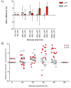
Fig 6. Measurement accuracy in stenosis quantification using a conventional single transmit coil (sTX) and a parallel transmission (pTX) coil with B1-shimming.
(1) For higher-grade stenoses, the mean differences obtained by subtracting real measures of the 3D-printed phantoms from MRI measurements were significantly lower (60% and 80%: p<0.01) using pTX with B1-shimming. Error bars indicate the range of the differences across the phantoms. (2) Deviation from actual stenosis determined with pTX (white squares) and sTX (red circles). To visualize the individual data points, the values on the x-axis (true degree of stenosis) are not fixed to a single value (e.g. 20%), but have a range of +/-5% (e.g. 15% to 25%). The y-axis indicates the difference between the stenosis measured with the respective technique and the true degree of stenosis.

Similar articles

Cited by

References

    1. Townsend N, Wilson L, Bhatnagar P, Wickramasinghe K, Rayner M, Nichols M. Cardiovascular disease in Europe: epidemiological update 2016. European Heart Journal. 2016;37:3232–45. doi: 10.1093/eurheartj/ehw334 - DOI - PubMed
    1. Adamson PD, Newby DE. Non-invasive imaging of the coronary arteries. European Heart Journal. 2019;40:2444–54. doi: 10.1093/eurheartj/ehy670 - DOI - PMC - PubMed
    1. Task Force Members, Montalescot G, Sechtem U, Achenbach S, Andreotti F, Arden C, et al.. 2013 ESC guidelines on the management of stable coronary artery disease: the Task Force on the management of stable coronary artery disease of the European Society of Cardiology. European Heart Journal. 2013. pp. 2949–3003. doi: 10.1093/eurheartj/eht296 - DOI - PubMed
    1. Fihn SD, Gardin JM, Abrams J, Berra K, Blankenship JC, Dallas AP, et al.. 2012 ACCF/AHA/ACP/AATS/PCNA/SCAI/STS guideline for the diagnosis and management of patients with stable ischemic heart disease: a report of the American College of Cardiology Foundation/American Heart Association task force on practice guidelines, and the American College of Physicians, American Association for Thoracic Surgery, Preventive Cardiovascular Nurses Association, Society for Cardiovascular Angiography and Interventions, and Society of Thoracic Surgeons. 11 ed. Circulation. 2012. pp. e354–471. doi: 10.1161/CIR.0b013e318277d6a0 - DOI - PubMed
    1. Liu X, Zhao X, Huang J, Francois CJ, Tuite D, Bi X, et al.. Comparison of 3D free-breathing coronary MR angiography and 64-MDCT angiography for detection of coronary stenosis in patients with high calcium scores. AJR Am J Roentgenol. American Roentgen Ray Society; 2007;189:1326–32. doi: 10.2214/AJR.07.2805 - DOI - PMC - PubMed

Publication types