. 2022 Jun 29;18(6):e1010435.
doi: 10.1371/journal.ppat.1010435. eCollection 2022 Jun.

A surface pocket in the cytoplasmic domain of the herpes simplex virus fusogen gB controls membrane fusion

Affiliations

A surface pocket in the cytoplasmic domain of the herpes simplex virus fusogen gB controls membrane fusion

Zemplen Pataki et al. PLoS Pathog. .

Abstract

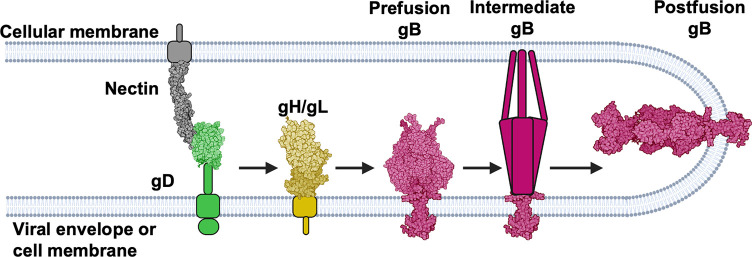
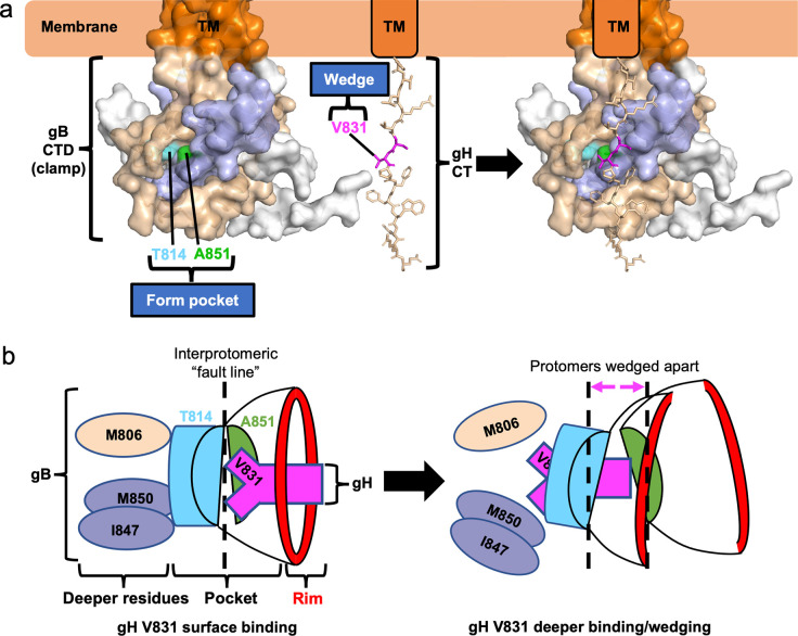
Membrane fusion during the entry of herpesviruses is carried out by the viral fusogen gB that is activated by its partner protein gH in some manner. The fusogenic activity of gB is controlled by its cytoplasmic (or intraviral) domain (gBCTD) and, according to the current model, the gBCTD is a trimeric, inhibitory clamp that restrains gB in the prefusion conformation. But how the gBCTD clamp is released by gH is unclear. Here, we identified two new regulatory elements within gB and gH from the prototypical herpes simplex virus 1: a surface pocket within the gBCTD and residue V831 within the gH cytoplasmic tail. Mutagenesis and structural modeling suggest that gH V831 interacts with the gB pocket. The gB pocket is located above the interface between adjacent protomers, and we hypothesize that insertion of the gH V831 wedge into the pocket serves to push the protomers apart, which releases the inhibitory clamp. In this manner, gH activates the fusogenic activity of gB. Both gB and gH are conserved across all herpesviruses, and this activation mechanism could be used by other gB homologs. Our proposed mechanism emphasizes a central role for the cytoplasmic regions in regulating the activity of a viral fusogen.


Conflict of interest statement

The authors have declared that no competing interests exist.

Figures

Fig 1
Fig 1. HSV-1 fusion pathway model.
gD (2C36 [28]) binds a receptor (3U83 [29]) on the target cell and activates gH/gL; gH/gL (3M1C [27]) triggers gB (6Z9M [2] and 5V2S [5]) to refold and cause fusion. gD has been suggested to be a dimer [28] but is shown here as a monomer for clarity. Figure created with BioRender.com.
Fig 2
Fig 2. The gBCTD mutant A851V reduced the rate and extent of fusion.
a) gB A851V and gB868 cell surface expression measured by flow cytometry. R68 primary antibody. Columns show mean. Error bars are SEM. b) Split-luciferase cell-cell fusion assay experimental setup. Cells expressing HSV-1 glycoproteins gD (2C36 [28]), gH/gL (3M1C [27]), and gB (6Z9M [2], and 5V2S [5]) fuse with cells expressing a nectin-1 receptor (3U83 [29]). Reconstitution of luciferase reports on fusion. Created with BioRender.com. c) Fusion of cells transfected with WT HSV-1 gB, gH, gL, gD compared to pCAGGS. The initiation of fusion is defined as the first reading at which luminescence is greater than twice that of the pCAGGS negative control. Early and late rates of fusion are the slopes of the fusion curves between 20–120 minutes and 3–8 hours post addition of target cells to effector cells, respectively. Early and late extent of fusion is defined as luminescence at 2 and 8 hours post addition of target cells to effector cells, respectively. This represents the total amount of fusion that has occurred over that time. d) Fusion of A851V over 8 hr by the split-luciferase fusion assay. *: p < 0.05 compared to WT gB at 8 hr. gB868 was used as a hyperfusogenic positive control [48]. Curve indicates mean values. Shaded area represents SEM. e-i) Initiation of fusion, early and late rates and extents of fusion of A851V. Columns show mean. Error bars are SEM. ns: not significant, *: p < 0.05, **: p < 0.01, ****: p <0.0001 in all panels. Data in all panels are from 3–6 independent experiments.
Fig 3
Fig 3. Fusogenicity and structural effects of mutants in the newly identified gBCTD pocket and rim.
a) gBCTD crystal structure and the structure of the pocket on the gBCTD. The pocket is formed at the junction of opposing gBCTD protomers, which are colored in wheat and light blue, with the third protomer in white. The residues that form the pocket on the gBCTD are highlighted in colors (left panel). The residues of the outer rim of the pocket are indicated in red (right panel). b) The bottom of the pocket is made up of T814 and A851, in sky blue and green, respectively. They do not contact each other, leaving a space between them at the bottom of the pocket. c-e) Pocket mutations of T814 and A851 are hypofusogenic and were modeled in PyMOL. All three hypofusogenic mutations of T814 and A851 were predicted to fill the gBCTD pocket as well as the space between T814 and A851 at the bottom of the pocket. f) Fusogenicity of T814 and A851 pocket mutants at 2 hr. A851V data is the same as shown in Fig 2. Fusion trends were the same at 8 hr. g) Cell surface expression of pocket mutants by flow cytometry. A851V data is the same as shown in Fig 2. h) Fusogenicity of mutations of the pocket rim at 2 hr. Fusion trends were the same at 8 hr. i) Cell surface expression of rim mutants. Columns show mean and error bars are SEM in all panels. *: p < 0.05, **: p < 0.01, ****: p < 0.0001 in all panels. Data in all panels are from three to six independent experiments.
Fig 4
Fig 4. gH V831 is the most important gHCT residue for fusion.
a) Structural modeling of the gHCT. b) Summary of gHCT truncations and mutations tested to determine which gHCT residues are the most important for fusion and probe their mechanism of action. c) Kinetics of fusion of gHCT truncations over 8 hr. Statistical significance shown is based on comparisons to WT gH fusion at 8 hr. Curves are the mean. Shaded area is SEM. d) Fusion of gHCT truncations at 2 hr. e) Fusion of mutations of gH829-832 residues at 2 hr to probe the function of the residues. Fusion trends were the same at 8 hr. f) Fusion of V831 mutations designed to make the putative gH wedge smaller (V831A gH832) or larger (V831L gH832), at 2 hr. Fusion trends were the same at 8 hr. g) Fusion of mutations creating a smaller wedge (V831A gH832) combined with mutations creating smaller pockets (gB T814L, gB A851V), at 2 hr. Fusion trends were the same at 8 hr. h) Cell surface expression of the gHCT truncations and mutations. Expression of gH829 and gH832 was determined previously to be the same as WT gH expression [44]. LP11 primary antibody. *: p < 0.05, **: p < 0.01, ***: p < 0.001, ****: p < 0.0001 in all panels. Bars indicate the mean and error bars are SEM. Statistical comparisons are to WT gH and gB. All panels represent averages of three independent experiments.
Fig 5
Fig 5. A model for gBCTD/gHCT interactions and fusion triggering.
a) gBCTD residues T814 (sky blue) and A851 (green) form a gH-binding pocket on the surface of the gBCTD trimer. gH V831 (magenta) acts as a wedge. Modeling shows that the gH V831 wedge and the gBCTD pocket are equidistant from the membrane. The V831 wedge binds between gB T814 and A851 in the gB pocket and pushes the gBCTD protomers (wheat and light blue) apart to destabilize gB and trigger fusogenic refolding of gB into the postfusion conformation. b) gH V831 (magenta) acts as a wedge that initially binds between gBCTD residues T814 (sky blue) and A851 (green) in the pocket on the surface of the gBCTD trimer. gH V831 then binds to deeper hydrophobic residues of the gBCTD (colored by protomer), forming favorable hydrophobic interactions. This causes the protomers of gB to be pushed apart as gH V831 enters deeper into the gBCTD, destabilizing the gBCTD clamp and triggering gB to refold and cause fusion. The interprotomeric “fault line” refers to the boundary between the wheat and blue protomers, which widens as the protomers are wedged apart.
Fig 6
Fig 6. Conservation of gBCTD and gHCT sequences across selected herpesviruses.
a) Alignment of gBCTD sequences of selected herpesviruses in the region that includes all HSV-1 pocket and rim residues [60]. Chosen sequences include all human alphaherpesviruses (HSV-1, HSV-2, VZV), a closely related non-human alphaherpesvirus (PRV), and important human beta- and gammaherpesviruses (HCMV, EBV). Equivalent residues to the HSV-1 pocket and rim residues are bolded and labeled with HSV-1 residue numbers. Identical and similar residues across herpesviruses are colored. b) Alignment of gHCT sequences of selected herpesviruses [60]. Equivalent residues to the HSV-1 V831 residue is bolded and colored. Pink indicates similar to HSV-1. Green is different from HSV-1.

Similar articles

Cited by

References

    1. Harrison SC. Viral membrane fusion. Nat Struct Mol Biol. 2008;15(7):690–8. Epub 2008/07/04. nsmb.1456 [pii] doi: 10.1038/nsmb.1456 ; PubMed Central PMCID: PMC2517140. - DOI - PMC - PubMed
    1. Vollmer B, Prazak V, Vasishtan D, Jefferys EE, Hernandez-Duran A, Vallbracht M, et al.. The prefusion structure of herpes simplex virus glycoprotein B. Sci Adv. 2020;6(39). Epub 2020/09/27. doi: 10.1126/sciadv.abc1726 ; PubMed Central PMCID: PMC7518877. - DOI - PMC - PubMed
    1. Liu Y, Heim KP, Che Y, Chi X, Qiu X, Han S, et al.. Prefusion structure of human cytomegalovirus glycoprotein B and structural basis for membrane fusion. Sci Adv. 2021;7(10). Epub 2021/03/07. doi: 10.1126/sciadv.abf3178 ; PubMed Central PMCID: PMC7935361. - DOI - PMC - PubMed
    1. Heldwein EE, Lou H, Bender FC, Cohen GH, Eisenberg RJ, Harrison SC. Crystal structure of glycoprotein B from herpes simplex virus 1. Science. 2006;313(5784):217–20. doi: 10.1126/science.1126548 . - DOI - PubMed
    1. Cooper RS, Georgieva ER, Borbat PP, Freed JH, Heldwein EE. Structural basis for membrane anchoring and fusion regulation of the herpes simplex virus fusogen gB. Nat Struct Mol Biol. 2018;25(5):416–24. Epub 2018/05/08. doi: 10.1038/s41594-018-0060-6 ; PubMed Central PMCID: PMC5942590. - DOI - PMC - PubMed

Publication types