. 2022 Jun 29;14(651):eabm5868.
doi: 10.1126/scitranslmed.abm5868. Epub 2022 Jun 29.

Closed-loop stimulation using a multiregion brain-machine interface has analgesic effects in rodents

Affiliations

Closed-loop stimulation using a multiregion brain-machine interface has analgesic effects in rodents

Guanghao Sun et al. Sci Transl Med. .

Abstract

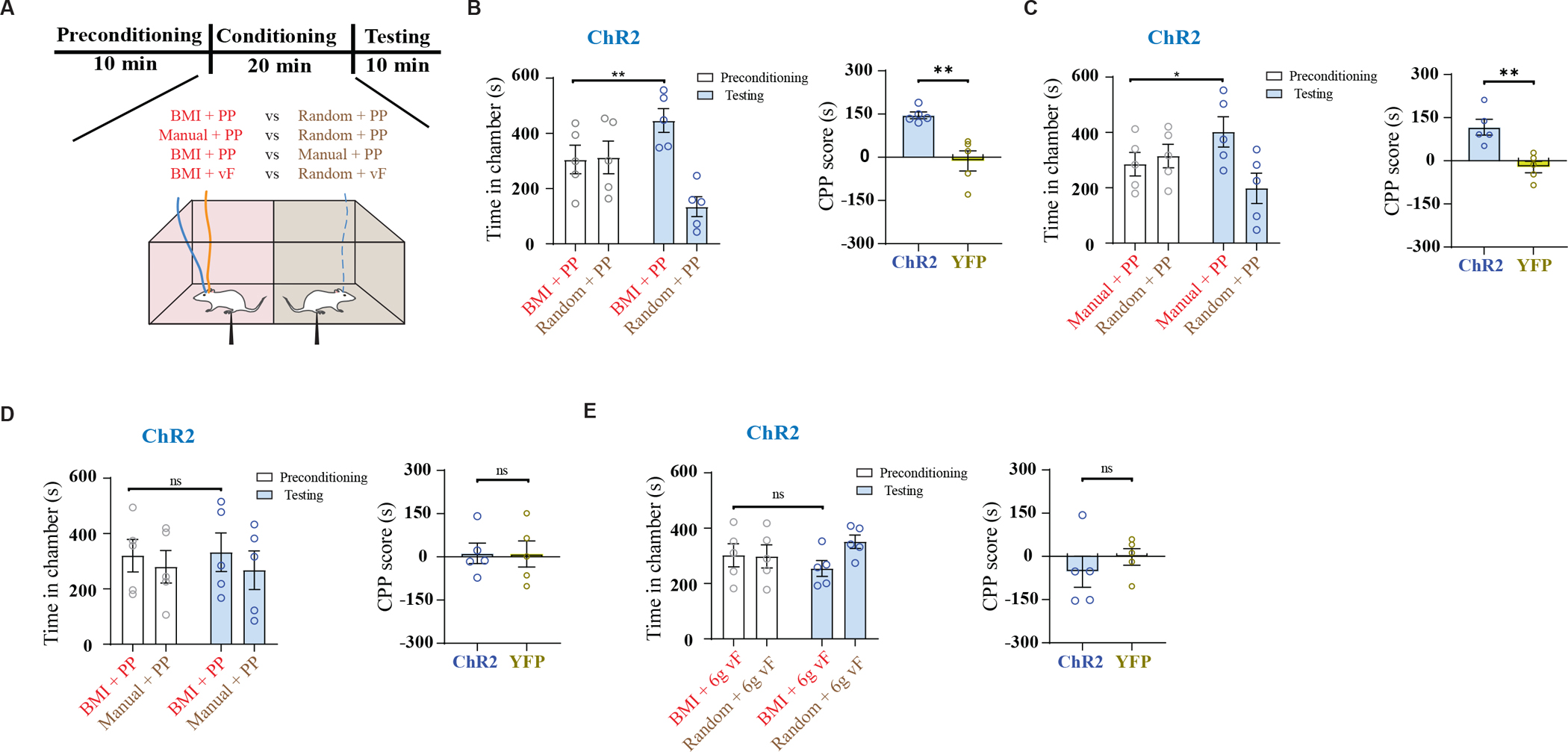
Effective treatments for chronic pain remain limited. Conceptually, a closed-loop neural interface combining sensory signal detection with therapeutic delivery could produce timely and effective pain relief. Such systems are challenging to develop because of difficulties in accurate pain detection and ultrafast analgesic delivery. Pain has sensory and affective components, encoded in large part by neural activities in the primary somatosensory cortex (S1) and anterior cingulate cortex (ACC), respectively. Meanwhile, studies show that stimulation of the prefrontal cortex (PFC) produces descending pain control. Here, we designed and tested a brain-machine interface (BMI) combining an automated pain detection arm, based on simultaneously recorded local field potential (LFP) signals from the S1 and ACC, with a treatment arm, based on optogenetic activation or electrical deep brain stimulation (DBS) of the PFC in freely behaving rats. Our multiregion neural interface accurately detected and treated acute evoked pain and chronic pain. This neural interface is activated rapidly, and its efficacy remained stable over time. Given the clinical feasibility of LFP recordings and DBS, our findings suggest that BMI is a promising approach for pain treatment.


Conflict of interest statement

Competing interests: J.W. and Z.S.C. have a provisional patent based on this work (“Closed-loop neural interface for pain control,” USPTO #121587). The other authors declare no competing interests.

Figures

Fig. 1.
Fig. 1.. Design of a multi-region LFP-based neural interface for pain.
(A) Schematic of experiments. The online closed-loop brain-machine interface (BMI) consists of three steps. In step ①, silicon probe arrays are implanted in the rat anterior cingulate cortex (ACC) and primary somatosensory cortex (S1) to record local field potentials (LFPs) simultaneously. In step ②, LFP signals are processed and sent to an automated decoder based on a state space model (SSM) to detect the onset of pain. In step ③, detected pain onset triggers neurofeedback in the form of optogenetic or electrical activation of the prelimbic prefrontal cortex (PL-PFC) to deliver pain modulation. (B) Placement of optic fiber or deep brain stimulating (DBS) electrode in the PL-PFC and recording silicon probes in the ACC and S1. (C) Raw LFP signals were processed to compute three band-limited LFP power features for the ACC channel: {y1,kACC,y2,kACC,y3,kACC} and S1 channel: {y1,kS1,y2,kS1,y3,kS1}, where the index k denotes the k-th temporal window (bin size 100 ms). MUA: multi-unit activity (300–500 Hz). (D) Schematic of two SSMs used to independently infer the latent variables {zkACC}v and {zkS1} from the LFP features {YkACC} and {YkS1} of ACC and S1, respectively (see Supplementary Materials and Methods for details). The SSM is illustrated by a graphical model with a Markovian structure, in which each node denotes a random variable, and the arrow indicates statistical dependency between two random variables. (E) Illustration of a multi-region decoding strategy for pain onset. First, the Z-scores were derived from the latent variables {zkACC} and {zkS1} (horizontal dashed lines denote the 95% confidence intervals for statistical significance). Next, a moving average cross-correlation function (CCF) was used to compute the correlation between the two Z-score series. The area beyond statistical significance (horizontal dashed lines) was computed to determine the change point (Supplementary Materials and Methods). When pain onset was detected, the decoder automatically triggered optogenetic or DBS stimulation to activate the PL-PFC.
Fig. 2.
Fig. 2.. The multi-region LFP-based neural interface reduces acute mechanical and thermal pain.
(A) Schematic of pain experiments demonstrating peripheral stimulation with either pin prick (PP) or von Frey filament (vF). LFP signals were recorded from ACC and S1 for pain detection, and optogenetic stimulation was administered to the PL-PFC for pain control. (B) Withdrawal response to mechanical stimulation, n = 10 rats; ****P < 0.0001, Wilcoxon Signed Rank test. (C) Illustration of mechanical pain onset detection using an LFP-based strategy. LFP features were computed from the ACC, S1, or both ACC and S1. The top two panels show single-channel LFP traces (white) overlying the spectrogram. The vertical dotted line indicates the onset of noxious peripheral stimulus (PP), and the vertical solid line indicates the time of paw withdrawal. The third and fourth panels show Z-scores (shaded areas denote the 95% confidence intervals) derived from the SSM-based decoder using LFPs recorded from ACC and S1, respectively (Methods). The two horizontal lines indicate the Z-score threshold ± 3.38. The fifth panel shows the cross-correlation function (CCF) between ACC and S1 from the third and fourth panels. The two horizontal dashed lines indicate the significance threshold. The bold triangle indicates the detection point. (D) Similar to panel c, except that the stimulus given is non-noxious (vF). (E) Demonstration of continuous online pain onset detection in a sample recording session. The vertical dotted line indicates the stimulus onset, and the vertical solid line indicates paw withdrawal. ♦ denotes true pain detection, * denotes false detection. (F) Comparison of detection rates between various non-noxious stimuli and noxious PP based on LFP decoding strategies using the ACC, S1 and combined (ACC + S1) signals. Each circle indicates data from one rat, n = 5 rats; ns, P > 0.05, **P < 0.01, ***P < 0.001 and ****P < 0.0001, one-way ANOVA with repeated measures and post-hoc Tukey’s multiple comparison tests to compare decoding rates for 2g vF, 6g vF, PP stimulation using signals from the ACC, S1 or ACC+S1. (G) The false positive (FP) detection rate per minute. n = 5 rats; P = 0.9679 (ACC vs S1), **P = 0.0036 (ACC vs ACC+S1), **P = 0.0036 (S1 vs ACC+S1), one-way ANOVA with repeated measures and post-hoc Tukey’s multiple comparison tests. (H) Comparison of detection rates based on LFP signals recorded in two different sessions, 3 months apart. Session 2 was recorded 3 months after session 1. Each pair of circles connected by a line indicates data from the same rat. We used the first 1–3 trials of each recording session to train the parameters of the SSM. n = 5 rats; *P = 0.0148 (ACC), P = 0.4651 (S1), P = 0.8650 (ACC+S1), paired t-test. (I) Comparison of detection rates based on model parameters set 5 days apart. We used the first 3 trials on Day 1 to train the parameters of SSM, and then used these same parameters to detect pain on the subsequent 5 days. n = 5 rats; P = 0.2339, one-way ANOVA with repeated measures and post-hoc Tukey’s multiple comparison tests.
Fig. 3.
Fig. 3.. The multi-region LFP-based neural interface reduces acute mechanical pain
(A) Schematic of conditioned place preference (CPP) assays to assess pain aversion. In a two-chamber set up, during conditioning, one of the chambers was paired with treatment shown in red, and the opposite chamber was paired with control conditions shown in brown (see Supplementary Materials and Methods for details). (B) Left panel: Time spent in preconditioning and testing phases in BMI+PP vs random+PP paired chambers, n = 5 rats; **P = 0.0016, paired t-test. Right panel: comparison of CPP scores of ChR2-expressing and YFP-expressing (control) rats (n = 5 ChR2 rats and 5 YFP rats, **P = 0.0027, unpaired t-test). (C) Left panel: Time spent in preconditioning and testing phases in manual+PP vs random+PP paired chambers, n = 5 rats; *P = 0.013, paired t-test. Right panel: comparison of CPP scores of ChR2 and YFP rats (n = 5 ChR2 rats and 5 YFP rats, **P = 0.0036, unpaired t-test). (D) Left panel: Time spent in preconditioning and testing phases in BMI+PP vs manual+PP paired chambers, n = 5 rats; P = 0.74, paired t-test. Right panel: comparison of CPP scores of ChR2 and YFP rats (n = 5 ChR2 rats and 5 YFP rats, P = 0.969, unpaired t-test). (E) Left panel: Time spent in preconditioning and testing phases in BMI+6g vF vs random+6g vF paired chambers, n = 5 rats; P = 0.46, paired t-test. Right panel: comparison of CPP scores of ChR2 and YFP rats (n = 5 ChR2 rats and 5 YFP rats, P = 0.428, unpaired t-test).
Fig. 4.
Fig. 4.. The multi-region neural interface reduces acute thermal pain
(A) Schematic of thermal stimulation experiments, with infrared intensity (IR) set to either 70 (noxious) or 10 (non-noxious). (B) IR 70 elicited paw withdrawals. n = 10 rats; ****P < 0.0001, Wilcoxon Signed Rank test. (C, D) Illustration of thermal pain onset detection using an LFP-based strategy, similar to panels 2C and 2D. (E) Comparison of the detection rate based on LFP decoding strategies using the ACC, S1, and combined (ACC + S1) signals. Each circle indicates data from a single rat. Comparison of the detection rates for three LFP-based decoding strategies. n = 5 rats; ns, P > 0.05, ***P < 0.001, ****P < 0.0001, one-way ANOVA with repeated measures and post-hoc Tukey’s multiple comparison tests to compare decoding rates for IR 10, 20, 70 using signals from ACC, S1 or ACC+S1. (F) Comparison of paw withdrawal latency for ChR2 rats. n = 5 rats; **P = 0.0089 (No opto vs BMI opto), **P = 0.0044 (No opto vs Manual opto), P = 0.9176 (BMI opto vs Manual opto), one-way ANOVA with repeated measures and post-hoc Tukey’s multiple comparison tests. (G) Comparison of paw withdrawal latency for YFP control rats. n = 5 rats; P = 0.5385 (No opto vs BMI opto), P = 0.9741 (No opto vs Manual opto), P = 0.7909 (BMI opto vs Manual opto), one-way ANOVA with repeated measures and post-hoc Tukey’s multiple comparison tests.
Fig. 5.
Fig. 5.. The multi-region neural interface performance in a chronic inflammatory pain model
(A) Schematic of experiments in CFA-treated rats. (B) CFA injection caused mechanical allodynia, n = 10 rats (5 ChR2 rats and 5 YFP rats); ****P < 0.0001, one-way ANOVA with repeated measures and post-hoc Tukey’s multiple comparison tests. (C) Paw withdrawal rate with vF stimulation. n = 10 rats, ****P < 0.0001, paired t-test. (D) Illustration of the multi-region LFP-based strategy for detecting the onset of evoked pain signal in a CFA-treated rat. Similar to panels 2C and 2D. (E) A comparison of different LFP-based strategies for decoding the pain onset in the CFA model. Comparison of the detection rates for the noxious vs non-noxious stimulus, n = 10 rats, ****P < 0.0001, paired t-test. Comparison of the detection rates for three LFP-based decoding strategies. For the noxious stimulus: n = 10 rats; P = 0.8139 (ACC vs S1), P = 0.9993 (ACC vs ACC+S1), P = 0.8231 (S1 vs ACC+S1). For the noxious stimulus: n = 10 rats; P = 0.4975 (ACC vs S1), *P = 0.0250 (ACC vs ACC+S1), **P = 0.0023 (S1 vs ACC+S1), one-way ANOVA with repeated measures and post-hoc Tukey’s multiple comparison tests. (F) Multi-region LFP-based BMI inhibited mechanical allodynia in CFA-treated rats. n = 5 rats; **P = 0.0026, paired t-test.
Fig. 6.
Fig. 6.. The multi-region neural interface reduces chronic inflammatory pain
(A) Schematic of CPP assays in CFA-treated rats. 6g vF represents noxious stimulation, 0.4g vF represents non-noxious stimulation. (B) Time spent in preconditioning and testing phases in chambers paired with BMI+6g vF vs random+6g vF, n = 5 rats; *P = 0.028, paired t-test. (C) Comparison of CPP scores of ChR2 and YFP rats, n = 5 ChR2 rats and 5 YFP rats; *P = 0.015, unpaired t-test. (D) Time spent in preconditioning and testing phases in chambers paired with BMI+0.4g vF vs random+0.4g vF, n = 5 rats; P = 0.392, paired t-test. (E) Comparison of CPP scores of ChR2 and YFP rats, n = 5 ChR2 rats and 5 YFP rats; P = 0.527, unpaired t-test. (F) Schematic of the CPP experiment to test tonic pain in CFA-treated rats. No peripheral stimuli were given. One chamber was paired with closed-loop BMI treatment, and the opposite chamber was paired with random PL-PFC activation of matching duration and intensity. (G) Demonstration of continuous decoding for spontaneous pain detection in the absence of peripheral stimuli. The first and second panels show the Z-score (shaded area denotes the 95% confidence intervals) derived from the LFP-based SSM decoder, where two horizontal dotted lines indicate the Z-score threshold ± 3.38. The third panel shows the cross-correlation function (CCF) between the two Z-scores. Two horizontal dashed lines indicate the significance threshold. The two black triangles mark the detection onset of spontaneous pain. (H) Time spent in preconditioning and testing phases in chambers paired with BMI vs random stimulation, n = 5 rats; *P = 0.0266, paired t-test. (I) Comparison of CPP scores of ChR2 and YFP rats, n = 5 ChR2 rats and 5 YFP rats; **P = 0.0087, unpaired t-test.
Fig. 7.
Fig. 7.. A BMI-driven, closed-loop DBS reduces acute and chronic inflammatory pain.
(A) Placement of stimulating electrode in the PL-PFC and recording electrodes in the ACC and S1. (B) Schematic of pain experiments during mechanical stimulus delivery. (C) Schematic of CPP experiments to assess aversion to evoked pain using DBS. (D) Time spent in preconditioning and testing phase in chambers paired with BMI+PP vs random+PP, n = 7 rats; *P = 0.0207, paired t-test. (E) Schematic of thermal experiments in DBS rats. Top panel: schematic of the Hargreaves test (IR 70). Bottom panel: comparison of paw withdrawal latency during different experimental conditions. n = 7 rats; ***P = 0.001, paired t-test. (F) Top panel: schematic of the experiment. Bottom panel: 50% paw withdrawal threshold in the presence of BMI-driven DBS vs control (no DBS). n = 7 rats; ****P < 0.0001, paired t-test. (G) Schematic of the CPP assay to assess aversion to evoked pain. (H) Time spent in preconditioning and testing phases in chambers paired with BMI+vF vs random+vF, n = 7 rats; *P = 0.0194, paired t-test. (I) Schematic of the CPP experiment to test tonic pain in CFA-treated rats. (J) Time spent in preconditioning and testing phases in chambers paired with BMI vs random DBS, n = 7 rats; ****P < 0.0001, paired t-test.
Fig. 8.
Fig. 8.. Closed-loop BMI reduces chronic neuropathic pain.
(A) Schematic of pain experiments in SNI-treated rats. (B) SNI operation resulted in mechanical allodynia, n = 5 rats; ****P < 0.0001, one-way ANOVA with repeated measures and post-hoc Tukey’s multiple comparison tests. (C) Multi-region LFP-based BMI inhibited mechanical allodynia in SNI-treated rats. n = 5 rats; ***P = 0.0004, paired t-test. (D) Schematic of the CPP experiment to test tonic pain in SNI-treated rats.. (E) Time spent in preconditioning and testing phases in chambers paired with BMI vs random DBS, n = 5 rats; *P = 0.0231, paired t-test.

Comment in

References

    1. Andersen RA, Aflalo T, Kellis S, From thought to action: The brain–machine interface in posterior parietal cortex. PNAS 116, 26274–26279 (2019). - PMC - PubMed
    1. Grosenick L, Marshel JH, Deisseroth K, Closed-loop and activity-guided optogenetic control. Neuron 86, 106–139 (2015). - PMC - PubMed
    1. Shanechi MM, Brain–machine interfaces from motor to mood. Nat Neurosci 22, 1554–1564 (2019). - PubMed
    1. Bergey GK, Morrell MJ, Mizrahi EM, Goldman A, King-Stephens D, Nair D, Srinivasan S, Jobst B, Gross RE, Shields DC, Barkley G, Salanova V, Olejniczak P, Cole A, Cash SS, Noe K, Wharen R, Worrell G, Murro AM, Edwards J, Duchowny M, Spencer D, Smith M, Geller E, Gwinn R, Skidmore C, Eisenschenk S, Berg M, Heck C, Van Ness P, Fountain N, Rutecki P, Massey A, O’Donovan C, Labar D, Duckrow RB, Hirsch LJ, Courtney T, Sun FT, Seale CG, Long-term treatment with responsive brain stimulation in adults with refractory partial seizures. Neurology 84, 810–817 (2015). - PMC - PubMed
    1. Taylor DM, Tillery SI, Schwartz AB, Direct cortical control of 3D neuroprosthetic devices. Science 296, 1829–1832 (2002). - PubMed

Publication types