. 2022 Sep 5;23(17):e202200178.
doi: 10.1002/cbic.202200178. Epub 2022 Jul 19.

Fragment Screening Yields a Small-Molecule Stabilizer of 14-3-3 Dimers That Modulates Client Protein Interactions

Affiliations

Fragment Screening Yields a Small-Molecule Stabilizer of 14-3-3 Dimers That Modulates Client Protein Interactions

Hendrik J Brink et al. Chembiochem. .

Abstract

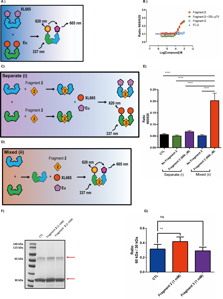
The development of protein-protein interaction (PPI) inhibitors has been a successful strategy in drug development. However, the identification of PPI stabilizers has proven much more challenging. Here we report a fragment-based drug screening approach using the regulatory hub-protein 14-3-3 as a platform for identifying PPI stabilizers. A homogenous time-resolved FRET assay was used to monitor stabilization of 14-3-3/peptide binding using the known interaction partner estrogen receptor alpha. Screening of an in-house fragment library identified fragment 2 (VUF15640) as a putative PPI stabilizer capable of cooperatively stabilizing 14-3-3 PPIs in a cooperative fashion with Fusicoccin-A. Mechanistically, fragment 2 appears to enhance 14-3-3 dimerization leading to increased client-protein binding. Functionally, fragment 2 enhanced potency of 14-3-3 in a cell-free system inhibiting the enzyme activity of the nitrate reductase. In conclusion, we identified a general PPI stabilizer targeting 14-3-3, which could be used as a tool compound for investigating 14-3-3 client protein interactions.

Keywords: drug discovery; estrogen receptor alpha; fragments; protein-protein interactions; stabilizers.


Conflict of interest statement

The authors declare no conflict of interest.

Figures

Figure 1
Figure 1
Fragment library screen for PPI stabilizers of the ERα/14‐3‐3 interaction complex. A) Cartoon illustrating our homogeneous time resolved FRET (HTRF) assay in which an N‐terminally GST tagged 14‐3‐3η is targeted with an anti‐GST europium (Eu) cryptate labelled monoclonal antibody and ERα peptides used are N‐terminally biotinylated and labelled with streptavidin conjugated to XL665. Europium is excited at a wavelength 337 nm resulting in emission at a wavelength of 620 nm. If the 14‐3‐3 interacts with the ERα peptide, resonance energy transfer can occur resulting in excitation and emission of XL665 at a wavelength of 665 nm. B) ERα‐pTV peptide binding to 10 nM GST14‐3‐3η in the absence (blue) or presence of 10 μM Fusicoccin (FC‐A, red). C) Overview of HTRF based fragment library screen. D) XY plots of fragments selected for further validation in which the effect on the 14‐3‐3/ERα‐pTV interaction complex is plotted against the effect on only the HTRF probes in absence of the protein or peptides. E) Structure of FC‐A. F) Structure of compound 13. Representative figures are shown of n=3 experiments; data are shown as mean±SD.
Figure 2
Figure 2
Validating fragment 2 in HTRF and orthogonal fluorescent polarization assay. A) Concentration‐response curves of FC‐A, fragment 2 and compound 1 generated with the HTRF assay with 50 nM ERα‐pTV peptide and 10 nM GST14‐3‐3η. B) Cartoon of fluorescent polarization assay. C) GST14‐3‐3η titration against fixed saturating concentrations of FC‐A (100 μM), fragment 2 (1 mM) or buffer control (CTL) in combination with 100 nM FAM‐ERα‐pTV in fluorescent polarization assay. D) Concentration‐response curves of FC‐A and fragment 2 generated in the fluorescent polarization assay using 100 nM of the FAM – ERα‐pTV peptide and 1.5 μM of GST14‐3‐3η with or without a fixed 100 μM of the non‐labelled 14‐3‐3 inhibitor peptide difopein. E) Concentration‐response curves of fragment 2 and fragment 3 generated in the fluorescent polarization assay using 100 nM of FAM‐ERα‐pTV peptide and 1.5 μM of GST14‐3‐3η. Representative figures are presented of n=3 experiments. Data are shown as mean±SD.
Figure 3
Figure 3
Testing of fragment 2 in combination with FC‐A. A) Concentration‐response curves generated with FC‐A, fragment 2 or fragment 2 combined with a fixed 100 μM concentration of FC‐A in the presence of 100 nM of the FAM‐ERα‐pTV and 1.5 μM of GST14‐3‐3η. B) Table showing the name and sequence of five different ERα peptides used. Amino acid classes color coded; positively charged (blue), negatively charged (red) hydrophobic side chains (green), polar uncharged sides chains (purple) and phosphorylated residue (yellow). C, D) Concentration‐response curves of FC‐A and fragment 2 against 50 nM of the different peptides listed in the table above and 10 nM of GST14‐3‐3η. Representative figures are presented of n=3 experiments. Data are shown as mean±SD.
Figure 4
Figure 4
Testing of fragment 2 on 14‐3‐3/14‐3‐3 interactions. A) Cartoon overview of 14‐3‐3 interaction assay using GST14‐3‐3η labelled with Europium cryptate and His14‐3‐3γ labelled with the acceptor XL665. B) Fragment 2, fragment 3 and FC‐A concentration‐response curves generated with Europium cryptate labelled GST14‐3‐3η and XL665 labelled His14‐3‐3γ. Fragment 2 curve generated in the presence and absence of 50 nM ERα‐pTV peptide. A representative figure is shown of three independent experiments. C) Cartoon overview of Europium cryptate labelled GST14‐3‐3η (shown in green) and XL665 labelled His14‐3‐3γ (shown in blue) pre‐incubation experiments with fragment 2 (orange diamond). In which GST14‐3‐3η and His14‐3‐3γ are pre‐incubated with fragment 2 followed by the addition of HTRF probes or D) GST14‐3‐3η and His14‐3‐3γ are mixed together before the addition of fragment 2 and the HTRF probes. E) HTRF 665/620 ratios of fragment 2 pre‐incubation experiments. Data are presented as the mean±SEM of three independent experiments. Differences analyzed by one‐way ANOVA (α=0.05, p<0.0001). F) Paraformaldehyde cross‐linking of His14‐3‐3γ in the presence of buffer (CTL), fragment 2 or fragment 3. Samples were ran on a SDS‐PAGE gel and visualized by instant blue staining. G) Quantification of the SDS‐PAGE gel seen in panel F, ratios of the 65 kDa band over the 30 kDa band are shown. Differences analyzed by one‐way ANOVA (α=0.05, p=0.003). Data are presented as the mean±SEM of three independent experiments.
Figure 5
Figure 5
Molecular docking studies suggest fragment 2 can bind in the central cavity of 14‐3‐3. A) Docking pose 1# and B) 3# are shown in which fragment 2 (green surface structure) is docked in the central pore of 14‐3‐3η. The 14‐3‐3η dimer is represented as a stick and ribbon structure with monomers colored purple (left) and orange (right). Amino acids relevant for fragment 2 binding are color coded and shown on the right.
Figure 6
Figure 6
Targeting the nitrate reductase/14‐3‐3 interaction complex with fragment 2. A) The NR is phosphorylated in the dark at Ser529 allowing 14‐3‐3 binding and subsequent inhibition of the enzyme and no nitrite is produced whereas B) in the light the NR remains unphosphorylated and 14‐3‐3 is unable to bind. Consequently, the NR remains active in the light and reduces nitrate to nitrite. Colorimetric detection of produced nitrite at 540 nm is used as a measure of NR activity. C) 14‐3‐3 inhibition curves of either the phosphorylated (NR‐pS529) or non‐phosphorylated nitrate reductase isolated from Hordeum vulgare. D) Concentration dependent inhibition of either the phosphorylated (NR‐pS529) or non‐phosphorylated nitrate reductase by fragment 2 in the presence of a fixed GST14‐3‐3η concentration. Data are presented as the mean±SEM of three independent experiments.

References

    1. Arkin M. R., Tang Y., Wells J. A., Chem. Biol. 2014, 21, 1102–1114. - PMC - PubMed
    1. Harmange Magnani C. S., Maimone T. J., Cell Chem. Biol. 2020, 27, 635–637. - PubMed
    1. Stevers L. M., Sijbesma E., Botta M., Mackintosh C., Obsil T., Landrieu I., Cau Y., Wilson A. J., Karawajczyk A., Eickhoff J., Davis J., Hann M., O'Mahony G., Doveston R. G., Brunsveld L., Ottmann C., J. Med. Chem. 2018, 61, 3755–3778. - PMC - PubMed
    1. Aitken A., Semin. Cancer Biol. 2006, 16, 162–72. - PubMed
    1. Yang X., Lee W. H., Sobott F., Papagrigoriou E., Robinson C. V., Grossmann J. G., Sundström M., Doyle D. A., Elkins J. M., Proc. Natl. Acad. Sci. USA 2006, 103, 17237–17242. - PMC - PubMed

Substances

LinkOut - more resources