. 2022 Aug:54:102382.
doi: 10.1016/j.redox.2022.102382. Epub 2022 Jun 23.

Farnesoid X receptor protects against cisplatin-induced acute kidney injury by regulating the transcription of ferroptosis-related genes

Affiliations

Farnesoid X receptor protects against cisplatin-induced acute kidney injury by regulating the transcription of ferroptosis-related genes

Dong-Hyun Kim et al. Redox Biol. 2022 Aug.

Abstract

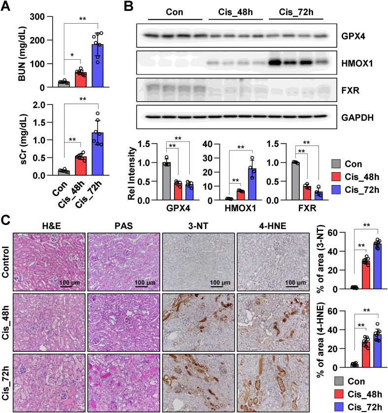
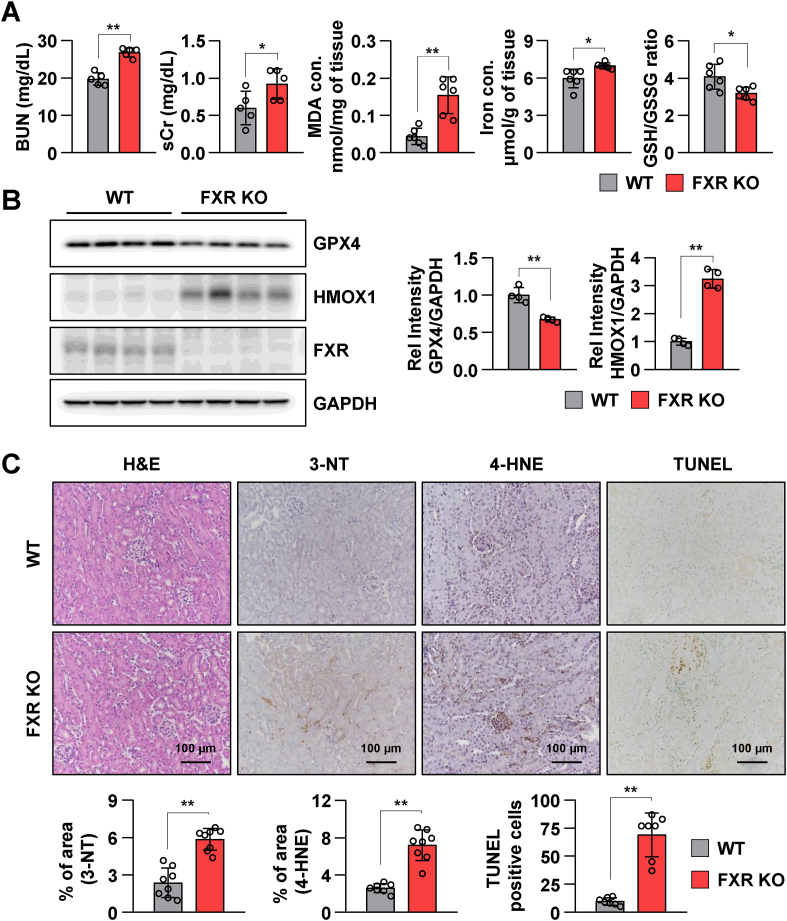
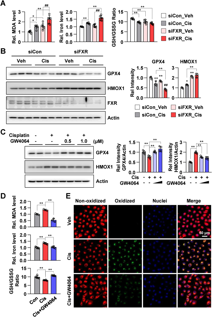
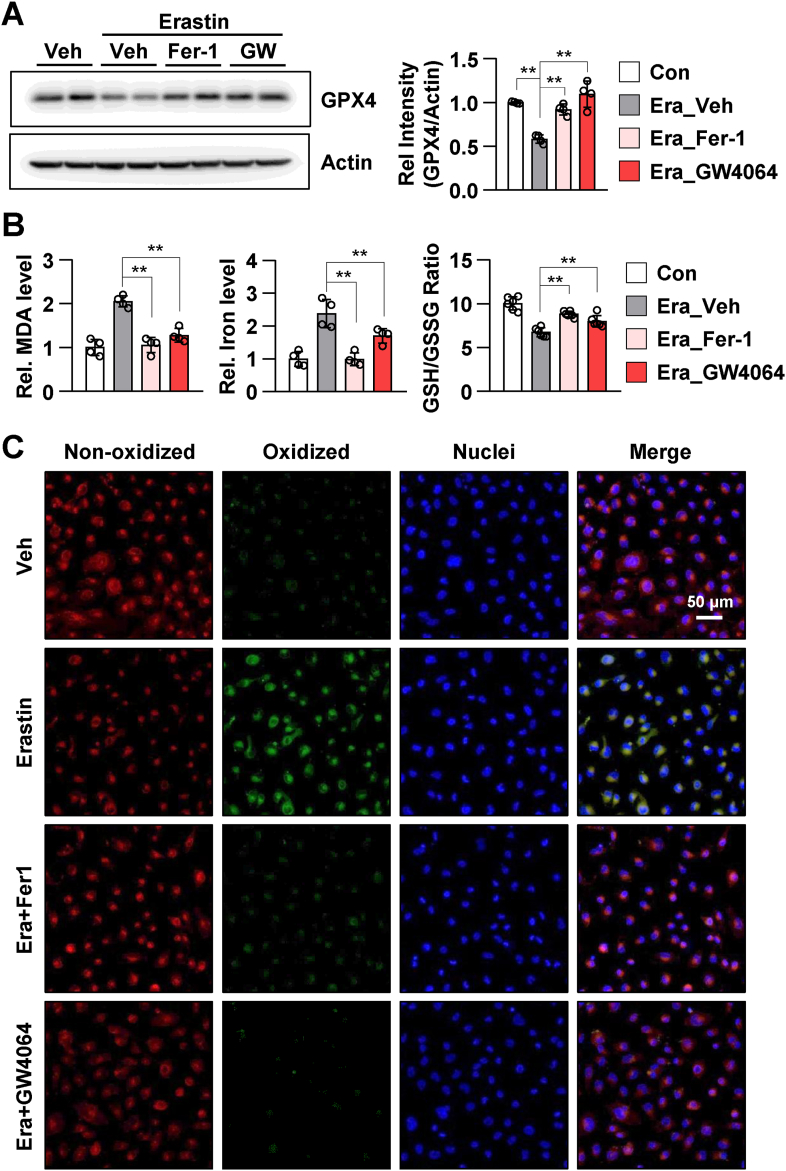
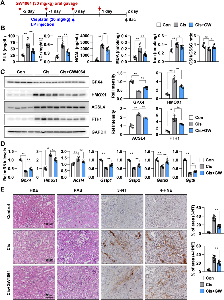
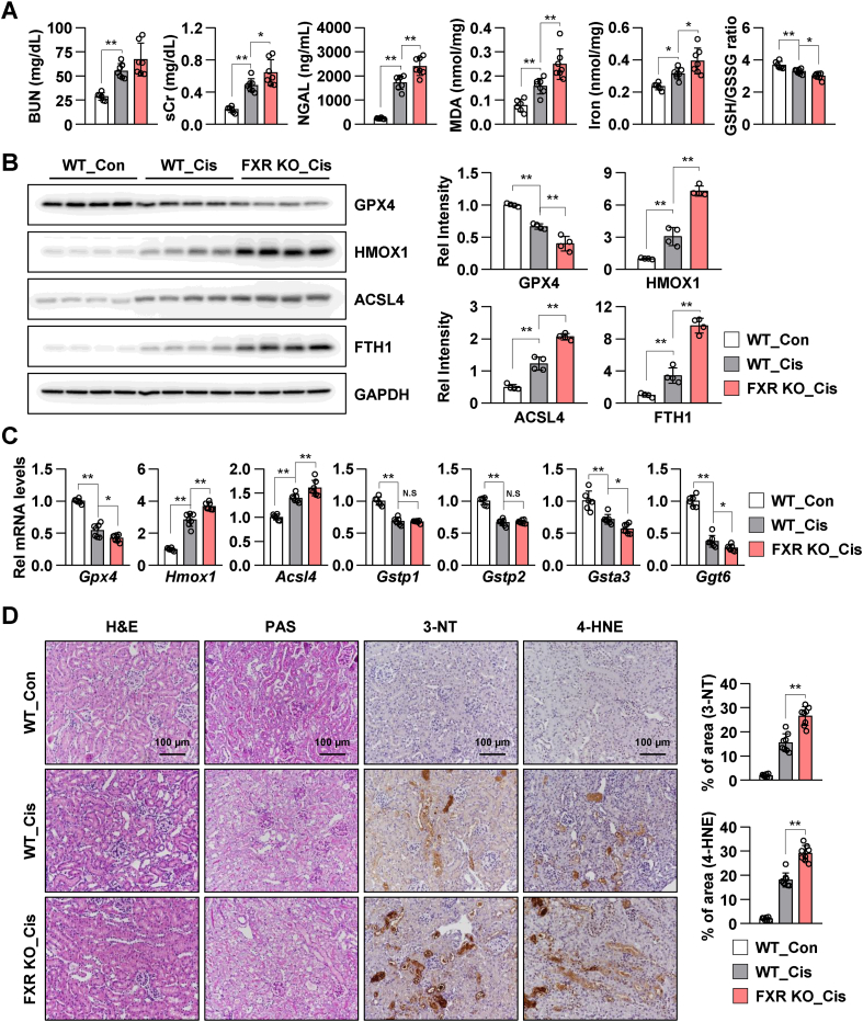
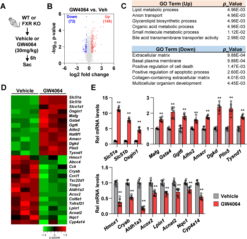
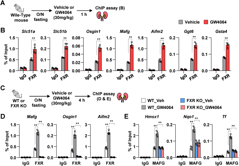
The side effects of cisplatin, a widely used chemotherapeutic agent, include nephrotoxicity. Previous studies have reported that cisplatin induces ferroptosis and lipid peroxide accumulation. Ferroptosis, a type of regulated cell death, is characterized by iron-dependent lipid peroxidation. Although previous studies have examined the regulation of ferroptosis in acute kidney injury (AKI), the regulatory mechanism of ferroptosis has not been elucidated. Here, the ability of activated farnesoid X receptor (FXR) to attenuate cisplatin-induced AKI through the regulation of ferroptosis was examined. FXR deficiency exhibited more ferroptosis responses, such as increase in lipid peroxidation, iron content and heme oxygenase 1 protein, and a decrease in glutathione/glutathione disulfide ratio and glutathione peroxidase 4 levels in HK2 cells and mice. Increased blood urea nitrogen, serum creatinine, and ferroptotic responses in the cisplatin-induced AKI mouse model were mitigated upon treatment with the FXR agonist GW4064 but were exacerbated in FXR knockout mice. RNA sequencing analysis revealed that ferroptosis-associated genes were novel targets of FXR. FXR agonist upregulated the expression of lipid and glutathione metabolism-related genes and downregulated cell death-related genes. Additionally, chromatin immunoprecipitation assays, using mice renal tissues, revealed that agonist-activated FXR could bind to its known target genes (Slc51a, Slc51b, Osgin1, and Mafg) and ferroptosis-related genes (Aifm2, Ggt6, and Gsta4). Furthermore, activated FXR-dependent MAFG, a transcriptional repressor, could bind to Hmox1, Nqo1, and Tf in the renal tissues of FXR agonist-treated mice. These findings indicate that activated FXR regulates the transcription of ferroptosis-related genes and protects against cisplatin-induced AKI.

Keywords: Acute kidney injury (AKI); Farnesoid X receptor (FXR); Ferroptosis; MAF bZIP transcription factor G (MAFG); Reactive oxidative stress (ROS).


Conflict of interest statement

The authors declare that they have no conflict of interest.

Figures

Fig. 1
Fig. 1
Cisplatin promotes renal ferroptosis and downregulates FXR expression in mice. The serum and the kidneys were collected from mice after 48 h and 72 h of cisplatin injection (20 mg/kg), (n = 6–7). (A) Serum BUN and Cr levels of each group. (B) Protein levels of GPX4, HMOX1, and FXR were detected by immunoblotting. The relative protein levels are shown. The values for the control group were set to 1. (C) Representative images of hematoxylin and eosin (H&E) and periodic acid-Schiff (PAS) staining to examine the histology and immunohistochemistry to examine the expression of 3-NT and 4-HNE. Scale bar, 100 μm. Computer-based morphometric analysis is shown (right bar graph, n = 7–8 in each group). All values are presented as the mean ± SD. Statistical significance was measured using one-way ANOVA with Bonferroni post hoc-test. *P < 0.05, **P < 0.005.
Fig. 2
Fig. 2
FXR deficiency promotes renal injury and ferroptotic responses in mice. The serum and kidney tissues were collected from wild-type (WT) and FXR knockout (FXR KO) mice (n = 6). (A) Serum BUN and Cr levels of each group. Malondialdehyde (MDA) and iron levels as well as GSH/GSSG ratio in kidney tissues. (B) Protein levels of GPX4, HMOX1, and FXR were detected by immunoblotting. The relative protein levels are shown. The values for the WT group were set to 1. (C) Representative images of hematoxylin and eosin (H&E) staining to examine the histology and immunohistochemistry to examine the expression of 3-NT and 4-HNE. Computer-based morphometric analysis is shown (bar graph, n = 8 in each group). Representative image of TUNEL staining. Quantitative analysis of positive TUNEL staining is shown (n = 7). Scale bar, 100 μm. All values are presented as the mean ± SD. Statistical significance was measured using one-way ANOVA with Bonferroni post hoc-test. *P < 0.05, **P < 0.005.
Fig. 3
Fig. 3
FXR agonist GW4064 mitigates cisplatin-induced ferroptotic responses in HK2 cells. (A) HK2 cells were transfected with siControl or siFXR as indicated; 48 h later, the cells were treated with cisplatin (10 μM) for 24 h. The levels of MDA and iron, and GSH/GSSG ratio in HK2 cells (n = 6). (B) Protein levels of GPX4, HMOX1, and FXR were detected by immunoblotting. The relative protein levels are shown. The values for vehicle-treated siControl group were set to 1 (n = 3). (C–E) After treatment with GW4064 (1 μM) for 2 h, HK2 cells were treated with cisplatin (10 μM) for another 24 h. (C) Protein levels of GPX4 and HMOX1 were detected by immunoblotting. The relative protein levels are shown. The values for the vehicle group were set to 1 (n = 4). (D) The relative levels of MDA and iron as well as GSH/GSSG ratio are shown. (E) After treatment with cisplatin and GW4064, the cells were incubated with C11-BODIPY 581/591 (2 μM) and Hoechst 33258 for counterstaining; the images were then immediately visualized by confocal microscopy. Confocal microscopy showed non-oxidized lipid (red, Texas Red) and oxidized lipid (green, FITC). scale bar, 50 μm. All values are presented as the mean ± SD. Statistical significance was measured using one- or two-way ANOVA with Bonferroni post hoc-test. *P < 0.05, **P < 0.005. (For interpretation of the references to color in this figure legend, the reader is referred to the Web version of this article.)
Fig. 4
Fig. 4
FXR agonist GW4064 mitigates erastin-induced ferroptotic responses in HK2 cells. After treatment with ferrostatin-1 (Fer-1, 5 μM) and GW4064 (1 μM) for 2 h, HK2 cells were treated with erastin (5 μM) for another 24 h. (A) Protein levels of GPX4 were detected by immunoblotting. The relative protein levels are shown. The values for the vehicle group were set to 1 (n = 4). (B) The relative levels of MDA and iron and GSH/GSSG ratio are shown. (C) After treatment with Fer-1 and GW4064, the cells were incubated with C11-BODIPY 581/591 (2 μM) and Hoechst 33258 for counterstaining; the images were then immediately visualized by confocal microscopy. Confocal microscopy showed non-oxidized lipid (red, Texas Red) and oxidized lipid (green, FITC). scale bar, 50 μm. All values are presented as the mean ± SD. Statistical significance was measured using one- or two-way ANOVA with Bonferroni post hoc-test. **P < 0.005. (For interpretation of the references to color in this figure legend, the reader is referred to the Web version of this article.)
Fig. 5
Fig. 5
FXR activation mitigates cisplatin-induced renal injury and ferroptotic response in a mouse model of cisplatin-induced AKI. WT mice were administered with GW4064 or vehicle (soybean oil) by oral gavage (single daily administration for 4 days) as shown in the experimental outline (A). The mice were injected with cisplatin (20 mg/kg) on the third day of GW4064 administration. After 48 h of cisplatin injection, the serum and the kidneys were collected. (B) BUN, sCr, and neutrophil gelatinase-associated lipocalin (NGAL) levels were measured in the serum. MDA, iron levels, and GSH/GSSG ratio were measured in the kidney tissue (n = 6–7). (C) Protein levels of GPX4, HMOX1, ACSL4, and FTH1 were detected by immunoblotting. The relative protein levels are shown. The values for the control group were set to 1 (n = 4). (D) The mRNA levels of the indicated genes were measured by qRT-PCR. The values for control group are set to 1. (E) Representative images of H&E and PAS staining to examine histology. Paraffin-embedded kidney tissue sections were stained with antibodies against 3-NT and 4-HNE (scale bar 100 μm). Computer-based morphometric analysis is shown (right bar graph, n = 8 in each group). All values are presented as the mean ± SD. Statistical significance was measured using one-way ANOVA with Bonferroni post hoc-test. *P < 0.05, **P < 0.005.
Fig. 6
Fig. 6
FXR deficiency exacerbates cisplatin-induced renal injury and ferroptotic response in a mouse model of the AKI. The WT and FXR knockout mice were intraperitoneally injected with cisplatin (20 mg/kg) or vehicle control (saline). After 48 h of cisplatin injection, the serum and the kidneys were collected. (A) BUN, sCr, and neutrophil gelatinase-associated lipocalin (NGAL) levels were measured in the serum. MDA and iron levels and GSH/GSSG ratio were measured in the kidney tissue (n = 6–7). (B) Protein levels of GPX4, HMOX1, ACSL4, and FTH1 were detected by immunoblotting. The relative protein levels are shown. The values for the WT control group were set to 1 (n = 4). (C) The mRNA levels of the indicated genes were measured by qRT-PCR. The values for the control group are set to 1. (D) Representative images of H&E and PAS staining to examine histology. Paraffin-embedded kidney tissue sections were stained with antibodies against 3-NT and 4-HNE (scale bar 100 μm). Computer-based morphometric analysis is shown (right bar graph, n = 7–8 in each group). All values are presented as the mean ± SD. Statistical significance was measured using one-way ANOVA with Bonferroni post hoc-test. *P < 0.05, **P < 0.005.
Fig. 7
Fig. 7
FXR activation upregulates the expression of lipid and glutathione metabolism-related genes. WT and FXR KO mice were fasted for 16 h and were subsequently administered GW4064 or vehicle (soybean oil) by oral gavage. After 6 h of GW4064 administration, the kidneys were collected. (A) Experimental outline. (B–D) RNA-sequencing analysis: (B) Volcano plots of RNA-seq data obtained using WT mice kidney tissues treated with vehicle or GW4064 (30 mg/kg). Blue, down-regulated genes; red, up-regulated genes. (C) Gene ontology (GO) analysis of genes either up- or down-regulated. (D) Heat maps showing changes in gene expression in WT mice treated with GW4064 compared to WT mice treated with vehicle. (E) The mRNA levels of the selected genes were measured by qRT-PCR (n = 6). The values for vehicle-treated group are set to 1. All values are presented as the mean ± SD. Statistical significance was measured using one-way ANOVA with Bonferroni post hoc-test. **P < 0.005. (For interpretation of the references to color in this figure legend, the reader is referred to the Web version of this article.)
Fig. 8
Fig. 8
FXR epigenetically activates ferroptosis-related genes. WT and FXR KO mice were fasted overnight (16 h) and were subsequently administered GW4064 or vehicle by oral gavage. After 1 h or 4 h of GW4064 administration, ChIP assay was performed using mice kidney tissues. (A and C) Experimental outline. (B) ChIP assay was performed after treatment with GW4064 for 1 h. Occupancy of FXR at the indicated genes (n = 4 mice). (D–E) ChIP assay was performed after treatment with GW4064 for 4 h. Occupancy of FXR (D) and MAFG (E) at the indicated genes (n = 4 mice). All values are presented as the mean ± SD. Statistical significance was measured using one- or two-way ANOVA with Bonferroni post hoc-test. *P < 0.05, **P < 0.005.

References

    1. Khwaja A. KDIGO clinical practice guidelines for acute kidney injury. Nephron Clin. Pract. 2012;120:c179–184. doi: 10.1159/000339789. - DOI - PubMed
    1. Lameire N.H., Bagga A., Cruz D., De Maeseneer J., Endre Z., Kellum J.A., et al. Acute kidney injury: an increasing global concern. Lancet. 2013;382:170–179. doi: 10.1016/S0140-6736(13)60647-9. - DOI - PubMed
    1. Miller R.P., Tadagavadi R.K., Ramesh G., Reeves W.B. Mechanisms of cisplatin nephrotoxicity. Toxins (Basel) 2010;2:2490–2518. doi: 10.3390/toxins2112490. - DOI - PMC - PubMed
    1. Sears S., Siskind L. Potential therapeutic targets for cisplatin-induced kidney injury: lessons from other models of AKI and fibrosis. J. Am. Soc. Nephrol. 2021 doi: 10.1681/ASN.2020101455. - DOI - PMC - PubMed
    1. Ozkok A., Edelstein C.L. Pathophysiology of cisplatin-induced acute kidney injury. BioMed Res. Int. 2014;967826 doi: 10.1155/2014/967826. 2014. - DOI - PMC - PubMed