. 2022 Jun 29;8(1):76.
doi: 10.1038/s41523-022-00443-9.

High prevalence of somatic PIK3CA and TP53 pathogenic variants in the normal mammary gland tissue of sporadic breast cancer patients revealed by duplex sequencing

Affiliations

High prevalence of somatic PIK3CA and TP53 pathogenic variants in the normal mammary gland tissue of sporadic breast cancer patients revealed by duplex sequencing

Anna Kostecka et al. NPJ Breast Cancer. .

Abstract

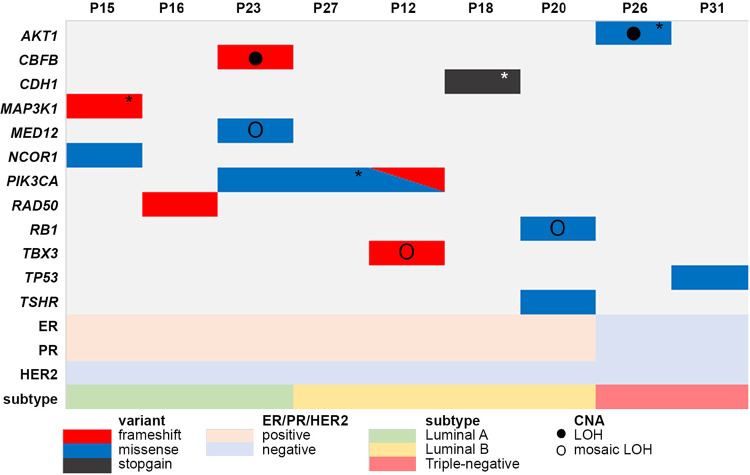
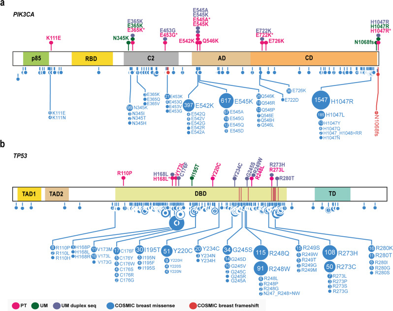
The mammary gland undergoes hormonally stimulated cycles of proliferation, lactation, and involution. We hypothesized that these factors increase the mutational burden in glandular tissue and may explain high cancer incidence rate in the general population, and recurrent disease. Hence, we investigated the DNA sequence variants in the normal mammary gland, tumor, and peripheral blood from 52 reportedly sporadic breast cancer patients. Targeted resequencing of 542 cancer-associated genes revealed subclonal somatic pathogenic variants of: PIK3CA, TP53, AKT1, MAP3K1, CDH1, RB1, NCOR1, MED12, CBFB, TBX3, and TSHR in the normal mammary gland at considerable allelic frequencies (9 × 10⁻²- 5.2 × 10⁻¹), indicating clonal expansion. Further evaluation of the frequently damaged PIK3CA and TP53 genes by ultra-sensitive duplex sequencing demonstrated a diversified picture of multiple low-level subclonal (in 10-2-10-4 alleles) hotspot pathogenic variants. Our results raise a question about the oncogenic potential in non-tumorous mammary gland tissue of breast-conserving surgery patients.


Conflict of interest statement

The authors declare no competing financial interests, but the following competing non-financial interests have been declared: J.P.D. is cofounder and shareholder in Cray Innovation AB.

Figures

Fig. 1
Fig. 1. Summary of Copy Number Alterations (CNAs) detected in the studied cohort.
Chromosomal CNAs were calculated as mean Log R Ratio (LRR) for chromosome arm and normalized to mean LRR of a sample. Results are presented as a heatmap with colors indicating gains (positive LRR values; red) and deletions (negative LRR values; blue). Hierarchical clustering was performed with Ward2 algorithm and identified six clusters. Pie charts with proportion of samples within clusters are presented in the Supplementary Fig. 3. Ctrl control cohort mammary gland, UM uninvolved mammary gland, PT tumor.
Fig. 2
Fig. 2. Somatic variants detected in the uninvolved mammary gland (UM).
Targeted sequencing revealed somatic variants of known breast cancer-associated genes (rows) present in 9–52% alleles in the UM of sporadic breast cancer patients (columns). Information on estrogen receptor (ER), progesterone receptor (PR), human epidermal growth factor receptor 2 (HER2), and biological subtype of matched primary tumor sample is included. *Variants detected in matched PT sample. CNA Copy Number Alteration status based on SNP arrays. LOH loss of heterozygosity. Description of detected variants, including genomic position and pathogenicity classification is provided in Table 2.
Fig. 3
Fig. 3. Somatic PIK3CA and TP53 variants detected in the uninvolved mammary gland (UM) and primary tumor (PT) samples.
Lollipop plots represent somatic variants of (a) PIK3CA and (b) TP53 genes detected by targeted next-generation sequencing (NGS). Upper panel represents variants detected in patient uninvolved mammary gland (UM) and tumor (PT) samples. All somatic variants detected according to the standard NGS and pathogenic/likely pathogenic variants detected by duplex sequencing in UM samples are included. Lower panel is a summary of somatic variants detected in breast tumors reported in the COSMIC database (https://cancer.sanger.ac.uk/cosmic). p85 p85-binding domain, RBD Ras-binding domain, C2 C2 domain, AD accessory domain, CD catalytic domain. TAD1, TAD2 transcription activation domain 1 and 2, DBD DNA-binding domain, DNA-binding sites are marked with red lines, TD tetramerization domain. Lollipop plots were prepared based on the images generated with the Protein paint application. *Variants detected by standard NGS in primary tumor samples and selected for duplex sequencing.
Fig. 4
Fig. 4. Oncogenic potential of the normal mammary tissue.
We used duplex sequencing to screen for ultra-low frequency variants and detected PIK3CA and TP53 hotspot alterations. The sampled normal mammary gland tissue is referred to as uninvolved glandular tissue and was not removed during surgical resection of the tumor mass. Detected variants might alter the function of the main breast cancer drivers: activate PIK3CA oncogene and impair TP53 tumor suppressor DNA-binding capacity. The presence of these changes implicates an oncogenic potential of the uninvolved mammary gland tissue and emphasizes the importance of thorough monitoring of sporadic breast cancer patients that underwent breast-conserving surgery.

References

    1. Heer E, et al. Global burden and trends in premenopausal and postmenopausal breast cancer: a population-based study. Lancet Glob. Heal. 2020;8:e1027–e1037. doi: 10.1016/S2214-109X(20)30215-1. - DOI - PubMed
    1. Coughlin SS. Epidemiology of breast cancer in women. Adv. Exp. Med. Biol. 2019;1152:9–29. doi: 10.1007/978-3-030-20301-6_2. - DOI - PubMed
    1. Kleibl Z, Kristensen VN. Women at high risk of breast cancer: molecular characteristics, clinical presentation and management. Breast. 2016;28:136–144. doi: 10.1016/j.breast.2016.05.006. - DOI - PubMed
    1. Sorlie T. Gene expression patterns of breast carcinomas distinguish tumor subclasses with clinical implications. PNAS. 2001;98:10869–10874. doi: 10.1073/pnas.191367098. - DOI - PMC - PubMed
    1. Koboldt DC, et al. Comprehensive molecular portraits of human breast tumours. Nature. 2012;490:61–70. doi: 10.1038/nature11412. - DOI - PMC - PubMed