. 2022 Jun 29;12(1):10972.
doi: 10.1038/s41598-022-13553-y.

Epidemiologically-based strategies for the detection of emerging plant pathogens

Affiliations

Epidemiologically-based strategies for the detection of emerging plant pathogens

Alexander J Mastin et al. Sci Rep. .

Abstract

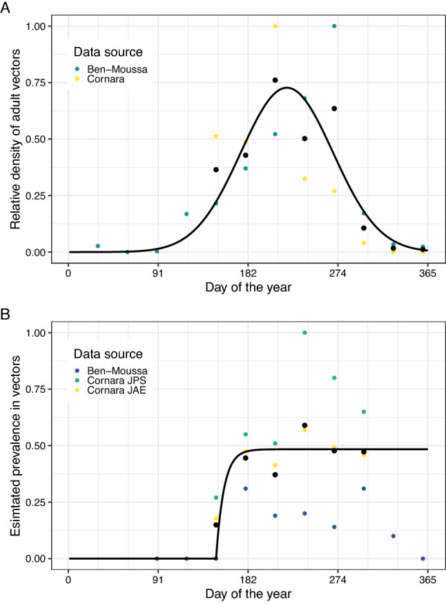
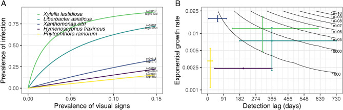
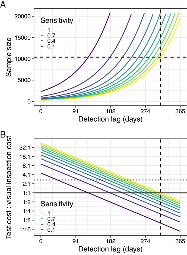
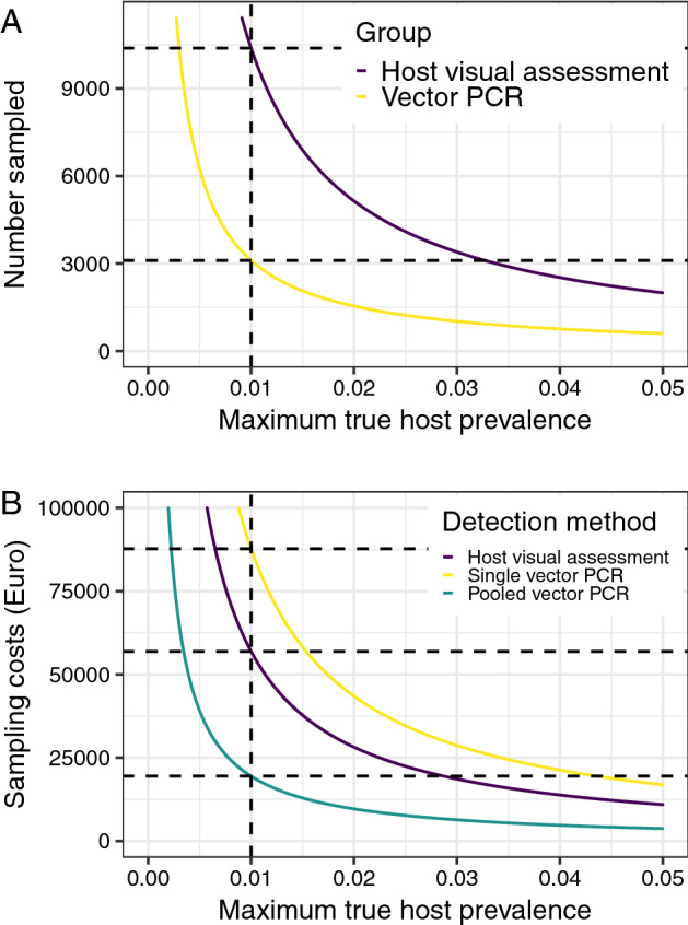
Emerging pests and pathogens of plants are a major threat to natural and managed ecosystems worldwide. Whilst it is well accepted that surveillance activities are key to both the early detection of new incursions and the ability to identify pest-free areas, the performance of these activities must be evaluated to ensure they are fit for purpose. This requires consideration of the number of potential hosts inspected or tested as well as the epidemiology of the pathogen and the detection method used. In the case of plant pathogens, one particular concern is whether the visual inspection of plant hosts for signs of disease is able to detect the presence of these pathogens at low prevalences, given that it takes time for these symptoms to develop. One such pathogen is the ST53 strain of the vector-borne bacterial pathogen Xylella fastidiosa in olive hosts, which was first identified in southern Italy in 2013. Additionally, X. fastidiosa ST53 in olive has a rapid rate of spread, which could also have important implications for surveillance. In the current study, we evaluate how well visual surveillance would be expected to perform for this pathogen and investigate whether molecular testing of either tree hosts or insect vectors offer feasible alternatives. Our results identify the main constraints to each of these strategies and can be used to inform and improve both current and future surveillance activities.


Conflict of interest statement

The authors declare no competing interests.

Figures

Figure 1
Figure 1
The true prevalence of infection can be estimated for any apparent prevalence from the rate of pathogen spread and the length of the detection lag period. (A) A detection lag period can be considered as a shift of the epidemic growth curve to the right. In this plot, time is shown on the x-axis and the proportion of infected or symptomatic hosts (the prevalence) on the y-axis. The two curves represent the true prevalence and the apparent prevalence (e.g. the proportion of hosts with symptoms, if detection is based upon visual inspection). The curves are parameterised based upon X. fastidiosa, but are intended for visualising the relative difference in true and apparent prevalences rather than the exact prevalences at different time points. The horizontal distance between the curves (i.e. in the direction of the x-axis) represents the asymptomatic period (the detection lag δ for visual inspection), and the vertical distance (in the direction of the y-axis) represents the difference between the true and apparent prevalences at any given time. (B) The ratio of the true and apparent prevalences decreases as the true prevalence increases. This plot shows the ratio of the true and apparent (detectable) prevalences (which can be interpreted as the number of asymptomatic trees per symptomatic tree) under logistic and exponential growth as time progresses. The dashed line represents the predicted ratio under continued exponential growth and the solid line represents that under logistic growth. Although during very early stage spread, the growth in both the true and apparent prevalences is broadly exponential, as the true prevalence deviates from this, the ratio of the two prevalences starts to decrease.
Figure 2
Figure 2
There is pronounced seasonal variability in the density of adult P. spumarius and the prevalence of X. fastidiosa infection amongst these. (A) Data suggest that adult P. spumarius are absent from January to March, and peak in density around August. This plot shows the modelled change in relative P. spumarius density over a year, fitted to data from two papers. The black dots show the mean density from both papers,. (B) Data suggest that the prevalence of X. fastidiosa infection in adult P. spumarius increases rapidly between June and July, to reach a steady peak for the rest of the year. This plot shows the modelled change in the prevalence of X. fastidiosa infection of P. spumarius over a year, fitted to data from three papers: “Cornara JPS”, “Cornara JAE”, and “Ben-Moussa”. The black dots show the mean prevalence estimates from all three papers.
Figure 3
Figure 3
In the early stages of the epidemic, the apparent prevalence of X. fastidiosa in vectors increases faster than that in hosts. (A) Although the total density of P. spumarius is assumed to be fixed between years, the density of infected vectors increases each year. This plot shows the modelled density of P. spumarius and the density of X. fastidiosa-infected P. spumarius over the course of 5 years. (B) The apparent prevalence of X. fastidiosa infection is higher in vectors than in hosts in the early stages of a new epidemic. This plot shows how the modelled apparent prevalence of X. fastidiosa in hosts contrasts with that in vectors, over the course of 5 years. The inset plot shows the estimates from the first 2 years in more detail.
Figure 4
Figure 4
The asymptomatic period for Xylella fastidiosa makes it very difficult to detect at an early stage when using visual inspection. (A) The highest difference between apparent and true prevalence is seen for olive quick decline syndrome, caused by X. fastidiosa. This plot shows the relationship between the apparent prevalence (on the x-axis) and the true prevalence (on the y-axis) for a number of different pathogens (associated disease): Hymenoscyphus fraxineus (ash dieback); Xanthomonas citri subsp. citri (citrus canker); Candidatus Liberbacter asiaticus (huanglongbing); X. fastidiosa ST53 (olive quick decline syndrome); Phytophthora ramorum (ramorum). (B) In order to confidently declare pest freedom, more samples are needed when the asymptomatic period and/or the spread rate are high. This plot shows the relationship between the detection lag (x-axis), the exponential growth rate (on a logarithmic scale on the y-axis), and the number of samples (also on a logarithmic scale, in the contour lines) required to be 90% confident that the true prevalence is lower than 1% given that no positive detections are made. The coloured lines indicate our best estimates of the growth rate and presymptomatic period for the pathogens considered.
Figure 5
Figure 5
The low sensitivity of current diagnostic tests when applied to presymptomatic hosts may limit their ability to detect infections at a low prevalence. (A) Diagnostic tests can result in a lower sample size than visual inspection, but if the diagnostic sensitivity is low, the test needs to be able to detect infection shortly after infection. This plot shows the impact of reducing the detection lag and the diagnostic sensitivity on the number of hosts which must be found to be negative to be 90% confident that the prevalence is lower than 1% (the sample size). As the dashed lines reflect the detection lag and required sample size under visual inspection, all solid lines below the horizontal dashed line indicate that fewer trees must be tested to declare pathogen freedom than would have to be visually inspected. (B) Lower detection sensitivities and higher costs both reduce the feasibility of a nonvisual detection method, even if the detection lag is short. This plot expands on plot A to also incorporate testing costs. We capture this by showing in solid coloured lines the relative cost of an alternative detection method at which the total costs of surveillance would be equal to those under visual inspection, for different diagnostic sensitivities. The intersections of the solid coloured lines with the solid black line (which indicates that the costs of the detection method is equal to that of visual inspection) therefore represent equal required sample sizes (and therefore match the intersections of the curves in plot A with the dashed line in that plot). The horizontal dotted line indicates the current estimated relative cost of using the host ELISA test (a cost ratio of €14.63/€5.48 = 2.67). The vertical dashed line shows the presymptomatic period for X. fastidiosa (and therefore the detection lag for visual inspection). All areas of the parameter space below the test sensitivity contour of interest indicate that the alternative detection method is cheaper to deploy than visual inspection, and all areas above indicate that visual inspection is cheaper.
Figure 6
Figure 6
In cases where X. fastidiosa is not thought to be present, fewer vectors than hosts need to be tested in order to declare pest freedom. (A) The number of hosts which need to be tested to detect at a given prevalence is over three times higher than the number of vectors. This plot shows the 90th percentile of the host prevalence in the absence of positive detections on the x-axis, and the number of individuals which would have to be sampled (and found to be negative) to achieve this on the y-axis, when hosts or vectors are sampled exclusively. The intersection of the curves and the vertical dashed line represents the sample size required to be 90% confident that the true prevalence is lower than 1% if no detections are made. (B) If vectors are pooled, the total cost of sampling hosts is around three times higher than the cost of sampling vectors. This plot shows the 90th percentile of the host prevalence in the absence of positive detections on the x-axis, and the total cost of the required sampling and testing effort to achieve this on the y-axis, when hosts or vectors are sampled exclusively. We assume that hosts are sampled with visual inspection and ELISA confirmation of suspected positives, and that vectors are tested using qPCR, either singly or pooled in batches of five.

Similar articles

Cited by

References

    1. Anderson PK, et al. Emerging infectious diseases of plants: Pathogen pollution, climate change and agrotechnology drivers. Trends Ecol. Evol. 2004;19:535–544. doi: 10.1016/j.tree.2004.07.021. - DOI - PubMed
    1. Brasier CM. The biosecurity threat to the UK and global environment from international trade in plants. Plant Pathol. 2008;57:792–808. doi: 10.1111/j.1365-3059.2008.01886.x. - DOI
    1. Waage JK, Mumford JD. Agricultural biosecurity. Philos. Trans. R. Soc. Lond. B Biol. Sci. 2008;363:863–876. doi: 10.1098/rstb.2007.2188. - DOI - PMC - PubMed
    1. IPPC. Surveillance guide—A guide to understand the principal requirements of surveillance programmes for national plant protection organizations. Second edition.http://www.fao.org/documents/card/en/c/cb7139en (2021) 10.4060/cb7139en.
    1. Parnell S, van den Bosch F, Gottwald T, Gilligan CA. Surveillance to inform control of emerging plant diseases: An epidemiological perspective. Annu. Rev. Phytopathol. 2017;55:591–610. doi: 10.1146/annurev-phyto-080516-035334. - DOI - PubMed

Publication types