Diagnostic performance of dedicated breast positron emission tomography
- PMID: 35768684
- PMCID: PMC9587931
- DOI: 10.1007/s12282-022-01381-x
Diagnostic performance of dedicated breast positron emission tomography
Abstract
Background: Dedicated breast positron emission tomography (dbPET) has been developed for detecting smaller breast cancer. We investigated the diagnostic performance of dbPET in patients with known breast cancer.
Methods: Eighty-two preoperative patients with breast cancer were included in the study (84 tumours: 11 ductal carcinomas in situ [DCIS], 73 invasive cancers). They underwent mammography (MMG), ultrasonography (US), and contrast-enhanced breast magnetic resonance imaging (MRI) before whole-body PET/MRI (WBPET/MRI) and dbPET. We evaluated the sensitivity of all modalities, and the association between the maximum standard uptake value (SUVmax) level and histopathological features.
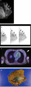
Results: The sensitivities of MMG, US, MRI, WBPET/MRI and dbPET for all tumours were 81.2% (65/80), 98.8% (83/84), 98.6% (73/74), 86.9% (73/84), and 89.2% (75/84), respectively. For 11 DCIS and 22 small invasive cancers (≤ 2 cm), the sensitivity of dbPET (84.9%) tended to be higher than that of WBPET/MRI (69.7%) (p = 0.095). Seven tumours were detected by dbPET only, but not by WBPET/MRI. Five tumours were detected by only WBPET/MRI because of the blind area of dbPET detector, requiring a wider field of view. After making the mat of dbPET detector thinner, all 22 scanned tumours were depicted. The higher SUVmax of dbPET was significantly related to the negative oestrogen receptor status, higher nuclear grade, and higher Ki67 (p < 0.001).
Conclusions: The sensitivity of dbPET for early breast cancer was higher than that of WBPET/MRI. High SUVmax was related to aggressive features of tumours. Moreover, dbPET can be used for the diagnosis and oncological evaluation of breast cancer.
Keywords: Breast cancer; Dedicated breast positron emission tomography; F-18 fluorodeoxyglucose; Magnetic resonance imaging; Standardised uptake value; Whole-body positron emission tomography.
© 2022. The Author(s).
Conflict of interest statement
The funds for this collaborative research were provided by Shimadzu Corporation. All dedicated breast positron emission tomography and WBPET were scanned free of charge at Midtown Clinic. Sadako Akashi-Tanaka received a research grant from Shimadzu Corporation.
Figures


References
-
- Ugurluer G, Yavuz S, Calikusu Z, Seyrek E, Kibar M, Serin M, et al. Correlation between 18F-FDG positron-emission tomography 18F-FDG uptake levels at diagnosis and histopathologic and immunohistochemical factors in patients with breast cancer. J Breast Health. 2016;12:112–118. doi: 10.5152/tjbh.2016.3031. - DOI - PMC - PubMed
-
- Ueda S, Tsuda H, Asakawa H, Shigekawa T, Fukatsu K, Kondo N, et al. Clinicopathological and prognostic relevance of uptake level using 18F-fluorodeoxyglucose positron emission tomography/computed tomography fusion imaging (18F-FDG PET/CT) in primary breast cancer. Jpn J Clin Oncol. 2008;38:250–258. doi: 10.1093/jjco/hyn019. - DOI - PubMed
MeSH terms
Substances
LinkOut - more resources
Full Text Sources
Medical

