Identification of cranial nerve ganglia using sectioned images and three-dimensional models of a cadaver
- PMID: 35768980
- PMCID: PMC9251397
- DOI: 10.3344/kjp.2022.35.3.250
Identification of cranial nerve ganglia using sectioned images and three-dimensional models of a cadaver
Abstract
Background: Cranial nerve ganglia, which are prone to viral infections and tumors, are located deep in the head, so their detailed anatomy is difficult to understand using conventional cadaver dissection. For locating the small ganglia in medical images, their sectional anatomy should be learned by medical students and doctors. The purpose of this study is to elucidate cranial ganglia anatomy using sectioned images and three-dimensional (3D) models of a cadaver.
Methods: One thousand two hundred and forty-six sectioned images of a male cadaver were examined to identify the cranial nerve ganglia. Using the real color sectioned images, real color volume model having a voxel size of 0.4 × 0.4 × 0.4 mm was produced.
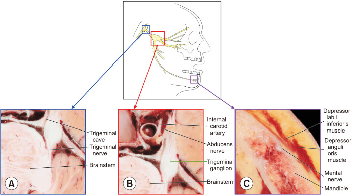
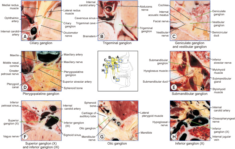
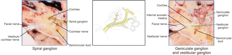
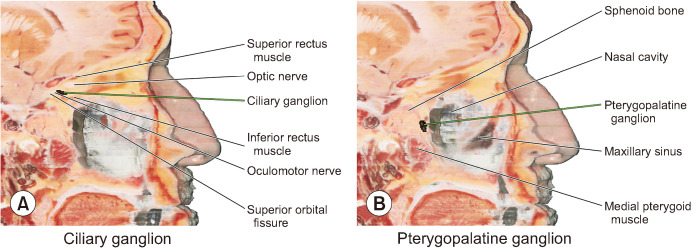
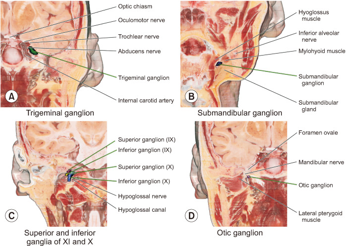
Results: The sectioned images and 3D models can be downloaded for free from a webpage, anatomy.dongguk.ac.kr/ganglia. On the images and model, all the cranial nerve ganglia and their whole course were identified. In case of the facial nerve, the geniculate, pterygopalatine, and submandibular ganglia were clearly identified. In case of the glossopharyngeal nerve, the superior, inferior, and otic ganglia were found. Thanks to the high resolution and real color of the sectioned images and volume models, detailed observation of the ganglia was possible. Since the volume models can be cut both in orthogonal planes and oblique planes, advanced sectional anatomy of the ganglia can be explained concretely.
Conclusions: The sectioned images and 3D models will be helpful resources for understanding cranial nerve ganglia anatomy, for performing related surgical procedures.
Keywords: Anatomy; Cadaver; Cranial Nerves; Cross-Sectional; Dissection; Facial Nerve; Ganglia; Glossopharyngeal Nerve; Imaging; Neuroanatomy.; Parasympathetic; Three-Dimensional.
Conflict of interest statement
No potential conflict of interest relevant to this article was reported.
Figures
Similar articles
-
Lymph Node Stations of Pancreas Which Are Identified in Real Color Sectioned Images of a Cadaver With Pancreatic Cancer.J Korean Med Sci. 2023 Nov 27;38(46):e392. doi: 10.3346/jkms.2023.38.e392. J Korean Med Sci. 2023. PMID: 38013647 Free PMC article.
-
Advanced Sectioned Images of a Cadaver Head with Voxel Size of 0.04 mm.J Korean Med Sci. 2019 Sep 2;34(34):e218. doi: 10.3346/jkms.2019.34.e218. J Korean Med Sci. 2019. PMID: 31456382 Free PMC article.
-
Whole courses of the oculomotor, trochlear, and abducens nerves, identified in sectioned images and surface models.Anat Rec (Hoboken). 2015 Feb;298(2):436-43. doi: 10.1002/ar.23048. Epub 2014 Sep 22. Anat Rec (Hoboken). 2015. PMID: 25212480
-
Visible Korean based on true color sectioned images for making realistic digital human, twenty years' record: a review.Surg Radiol Anat. 2024 Jul;46(7):935-947. doi: 10.1007/s00276-024-03381-2. Epub 2024 May 8. Surg Radiol Anat. 2024. PMID: 38717503 Review.
-
Tracking the glossopharyngeal nerve pathway through anatomical references in cross-sectional imaging techniques: a pictorial review.Insights Imaging. 2018 Aug;9(4):559-569. doi: 10.1007/s13244-018-0630-5. Epub 2018 Jun 13. Insights Imaging. 2018. PMID: 29949035 Free PMC article. Review.
References
LinkOut - more resources
Full Text Sources