. 2022 Sep 14;145(9):3274-3287.
doi: 10.1093/brain/awac164.

Monoallelic and biallelic mutations in RELN underlie a graded series of neurodevelopmental disorders

Affiliations

Monoallelic and biallelic mutations in RELN underlie a graded series of neurodevelopmental disorders

Nataliya Di Donato et al. Brain. .

Abstract

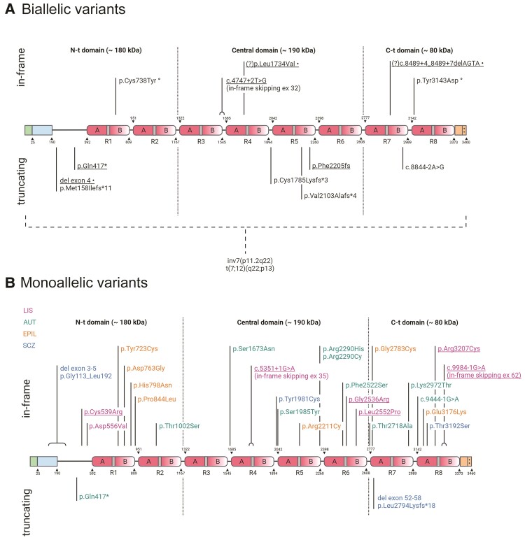
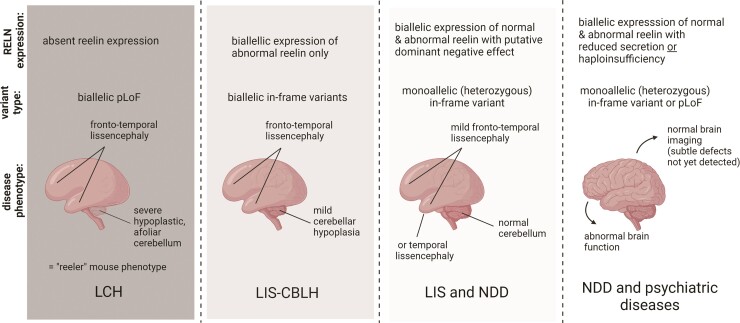
Reelin, a large extracellular protein, plays several critical roles in brain development and function. It is encoded by RELN, first identified as the gene disrupted in the reeler mouse, a classic neurological mutant exhibiting ataxia, tremors and a 'reeling' gait. In humans, biallelic variants in RELN have been associated with a recessive lissencephaly variant with cerebellar hypoplasia, which matches well with the homozygous mouse mutant that has abnormal cortical structure, small hippocampi and severe cerebellar hypoplasia. Despite the large size of the gene, only 11 individuals with RELN-related lissencephaly with cerebellar hypoplasia from six families have previously been reported. Heterozygous carriers in these families were briefly reported as unaffected, although putative loss-of-function variants are practically absent in the population (probability of loss of function intolerance = 1). Here we present data on seven individuals from four families with biallelic and 13 individuals from seven families with monoallelic (heterozygous) variants of RELN and frontotemporal or temporal-predominant lissencephaly variant. Some individuals with monoallelic variants have moderate frontotemporal lissencephaly, but with normal cerebellar structure and intellectual disability with severe behavioural dysfunction. However, one adult had abnormal MRI with normal intelligence and neurological profile. Thorough literature analysis supports a causal role for monoallelic RELN variants in four seemingly distinct phenotypes including frontotemporal lissencephaly, epilepsy, autism and probably schizophrenia. Notably, we observed a significantly higher proportion of loss-of-function variants in the biallelic compared to the monoallelic cohort, where the variant spectrum included missense and splice-site variants. We assessed the impact of two canonical splice-site variants observed as biallelic or monoallelic variants in individuals with moderately affected or normal cerebellum and demonstrated exon skipping causing in-frame loss of 46 or 52 amino acids in the central RELN domain. Previously reported functional studies demonstrated severe reduction in overall RELN secretion caused by heterozygous missense variants p.Cys539Arg and p.Arg3207Cys associated with lissencephaly suggesting a dominant-negative effect. We conclude that biallelic variants resulting in complete absence of RELN expression are associated with a consistent and severe phenotype that includes cerebellar hypoplasia. However, reduced expression of RELN remains sufficient to maintain nearly normal cerebellar structure. Monoallelic variants are associated with incomplete penetrance and variable expressivity even within the same family and may have dominant-negative effects. Reduced RELN secretion in heterozygous individuals affects only cortical structure whereas the cerebellum remains intact. Our data expand the spectrum of RELN-related neurodevelopmental disorders ranging from lethal brain malformations to adult phenotypes with normal brain imaging.

Keywords: RELN; Reelin; autism; epilepsy; lissencephaly.


Figures

Figure 1
Figure 1
RELN protein cartoon showing pathogenic and probably pathogenic variants. Protein diagrams show the domain organization of RELN with two in vivo proteolysis sites shown as dotted lines highlighting N-terminal (N-t), Central and C-terminal (C-t) domains. The known boundaries of each domain or other functional region are indicated above or below the diagram and identified by numbers corresponding to the human RELN sequence (Uniprot no. P78509). RELN repeats are numbered (R1–R8) and their composition is marked with sub-repeats A and B separated by EGF-like domains (grey). The N-terminal region contains a signal peptide (positions 0–25 in green) and F-spondin-like domain (positions 25–190 in blue) followed by a unique sequence region (positions 190–502). The C-terminal region (3373–3460 in orange) ends with a stretch of 33 amino acids rich in basic residues (++). (A) Biallelic variants in RELN. Thirteen variants are shown, including six reported in this study (underlined). Compound heterozygous variants in one individual are marked with symbols following the description of the variant; (?) indicates two VUS identified in cis in one individual, both with potential disease relevance. Two structural chromosome rearrangements disrupting RELN are indicated below the dashed bracket. (B) Monoallelic variants in RELN. Twenty-seven variants are shown, including six reported in this study (underlined). The colour legend for associated phenotypes is also shown beneath the diagram; AUT = autism; EPIL = lateral temporal epilepsy; SCZ = schizophrenia. Created with BioRender.com.
Figure 2
Figure 2
Brain MRI showing LIS-CBLH with biallelic RELN variants. Brain imaging in six individuals with biallelic RELN-related LIS-CBLH. Midline sagittal MRIs in three individuals show an abnormally thin brainstem with flat pons and very small cerebellum with an afoliar surface (arrowheads in A, C and E). Midline sagittal images in two siblings with a less severe mutation show a normal brainstem and only moderately small cerebella with some foliation evident (I, M; double arrowheads point to the small cerebella). Axial (D, F–H, J–L) and coronal (B) images shows diffuse but frontal and temporal-predominant LIS with moderately thick 5–8 mm cortex. Arrows point to representative areas of pachygyria (mild LIS) only; the cortical malformation affects all brain regions. Low resolution head CT images in another child show the small cerebellum (arrowhead in N) and mild LIS (arrows in O). The imaging pattern seen in panels AH appears similar to those shown in prior reports of individuals with biallelic mutations, while the pattern seen in panels IM are less severe. Panels A and B are from subject LP95-137a1; C and D from LP95-137a2, E and H from LR14-063, I and L from LR17-413a1, M from LR17-413a3 and N and O from LP96-078.
Figure 3
Figure 3
Brain MRI showing frontotemporal LIS with monoallelic RELN variants. Images show the same pattern of LIS in four unrelated subjects including LR02-111a2 (A–D), LR21-446 (E–H), LR15-139 (I–L) and LR18-437 (M–P). Midline sagittal images (first column) show normal brainstem and cerebellum. Axial images at the level of the lateral ventricles (second column), basal ganglia (three column) and temporal lobes (fourth column) show diffuse mild LIS (pachygyria) with only moderately thick 5–8 mm cortex and consistent gradient with the malformation most severe in the frontal and temporal lobes and becoming less severe posteriorly. The posterior parietal and occipital regions appear mildly abnormal, although this is sometimes subtle. Arrows point to representative areas of pachygyria. The hippocampi also appear normal (not shown).
Figure 4
Figure 4
Brain MRI showing temporal LIS with monoallelic RELN variants. Images show the same pattern of LIS in four subjects from two families including LR02-111a3 (A–D), LR02-111r1 (E–H), LR17-364a2 (I–L) and LR17-364f (M–P). Midline sagittal images (first column) show normal brainstem and cerebellum. Axial images at the level of the lateral ventricles (second column), basal ganglia (third column) and temporal lobes (fourth column) show moderate but definite LIS (also known as mild pachygyria) with moderately thick 5–8 mm cortex over the temporal lobes best seen in the last column (D, H, L, P). Higher images show mild pachygyria in probands from the two families (B and J) and subtle pachygyria (or normal) in their less affected adult relatives (F and N). Arrows point to representative areas of pachygyria, while arrowheads point to areas of subtle undersulcation (F and N) or to normal hippocampi (L and P).
Figure 5
Figure 5
Proposed genotype-functional correlation with RELN-related phenotypes. Our data and analysis support a strong correlation between residual RELN activity and severity of the neurodevelopmental phenotype. LCH = lissencephaly with severe cerebellar hypoplasia; NDD = neurodevelopmental disorders. Created with BioRender.com.

References

    1. D'Arcangelo G, Curran T. Reeler: New tales on an old mutant mouse. Bioessays. 1998;20:235–244. - PubMed
    1. D'Arcangelo G, Miao GG, Chen SC, Soares HD, Morgan JI, Curran T. A protein related to extracellular matrix proteins deleted in the mouse mutant reeler. Nature. 1995;374:719–723. - PubMed
    1. Falconer DS. Two new mutations, trembler and reeler, with neurological actions in the house mouse (Mus musculus l). J Genet. 1951;50:192–201. - PubMed
    1. Hong SE, Shugart YY, Huang DT, et al. Autosomal recessive lissencephaly with cerebellar hypoplasia is associated with human RELN mutations. Nat Genet. 2000;26:93–96. - PubMed
    1. Hong SE, Shugart YY, Huang DT, et al. Correction: Autosomal recessive lissencephaly with cerebellar hypoplasia is associated with human RELN mutations. Nat Genet. 2001;27:225–225. - PubMed

Publication types

Supplementary concepts