. 2022 Jun 20:2022:6394199.
doi: 10.1155/2022/6394199. eCollection 2022.

Hepatoprotective Effect of Mitochondria-Targeted Antioxidant Mito-TEMPO against Lipopolysaccharide-Induced Liver Injury in Mouse

Affiliations

Hepatoprotective Effect of Mitochondria-Targeted Antioxidant Mito-TEMPO against Lipopolysaccharide-Induced Liver Injury in Mouse

Peng-Fei Wang et al. Mediators Inflamm. .

Abstract

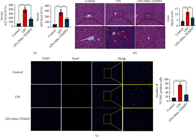
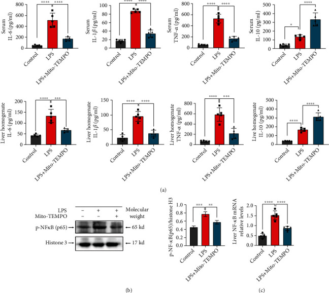
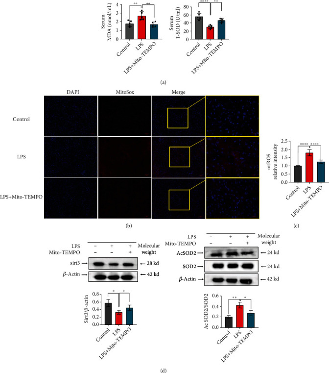
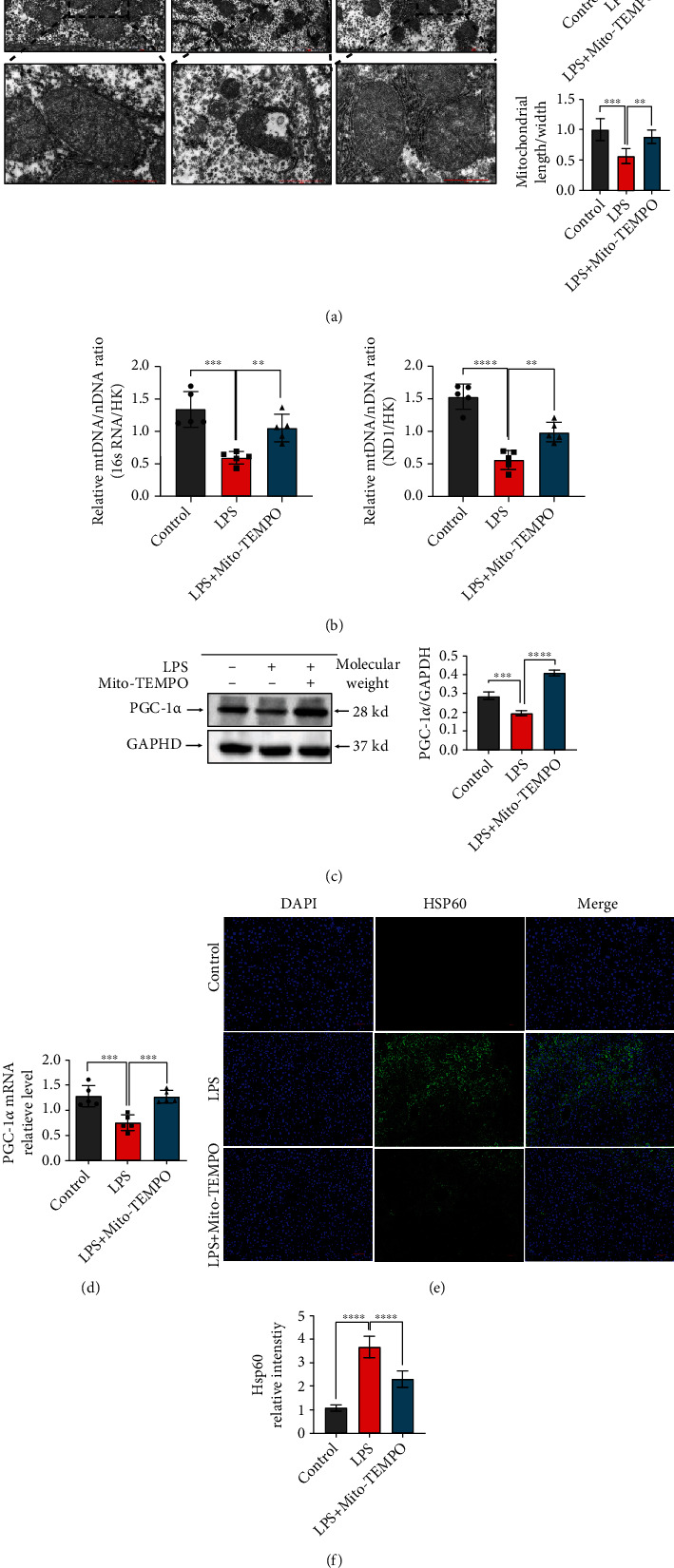
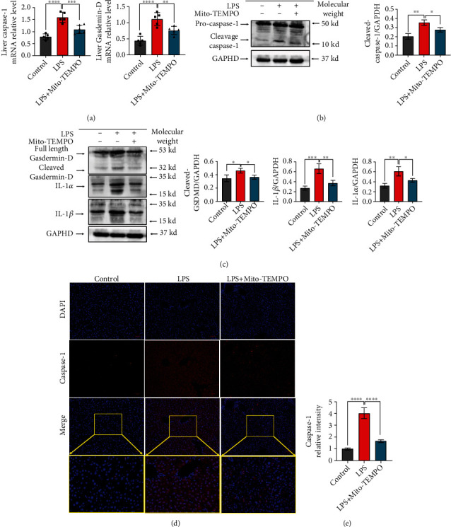
The liver is vulnerable to sepsis, and sepsis-induced liver injury is closely associated with poor survival of sepsis patients. Studies have found that the overproduction of reactive oxygen species (ROS) is the major cause of oxidative stress, which is the main pathogenic factor for the progression of septic liver injury. The mitochondria are a major source of ROS. Mito-TEMPO is a mitochondria-specific superoxide scavenger. The aim of this study was to investigate the effect of Mito-TEMPO on lipopolysaccharide- (LPS-) induced sepsis mice. We found that Mito-TEMPO pretreatment inhibited inflammation, attenuated LPS-induced liver injury, and enhanced the antioxidative capability in septic mice, as evidenced by the decreased MDA content and the increased SOD activity. In addition, Mito-TEMPO restored mitochondrial size and improved mitochondrial function. Finally, we found that the levels of pyroptosis-related proteins in the liver of LPS-treated mice were lower after pretreatment with Mito-TEMPO. The mechanisms could be related to Mito-TEMPO enhanced antioxidative capability and improved mitochondrial function, which reflects the ability to neutralize ROS.


Conflict of interest statement

The authors declare that there are no known conflicts of interest.

Figures

Figure 1
Figure 1
Mito-TEMPO mitigates liver injury in LPS-induced sepsis mice. (a) Serum ALT and AST levels of LPS-induced sepsis mice. (b) Scoring and representative pictures of hepatic H&E staining (scale bar = 100 μm, 50 μm). Liver injury scores were estimated by the following six indicators: vacuolization, nuclear condensation, nuclear fragmentation, nuclear fading, erythrocyte stasis, and inflammatory cell infiltration. Scores were assigned based on the percentage of cells showing these phenomena in five microscopic fields (×200) as follows: 0 = 0%, 1 = 0–10%, 2 = 10–50%, and 3 = 50–100%. The six subscores were summed; the higher the total score, the more severe the injury. (c) The TUNEL-positive cells in the liver of LPS-induced sepsis mice (scale bar = 50 μm, 20 μm). The proportion of positive cells was assessed in five random fields (×200). n = 5 per group. Data are presented as the mean ± SD from at least three independent experiments. P < 0.05, ∗∗P < 0.01, ∗∗∗P < 0.005, and ∗∗∗∗P < 0.0001.
Figure 2
Figure 2
Mito-TEMPO diminishes liver inflammation in LPS-induced sepsis. (a) Expression of cytokines in serum and liver tissue homogenate. (b) Nuclear proteins from mouse liver tissues were extracted, and p-NF-κB (p65) protein levels in the liver were detected by western blotting. (c) NF-κB relative mRNA levels in the liver. n = 5 per group. Data are presented as the mean ± SD from at least three independent experiments. P < 0.05, ∗∗P < 0.01, ∗∗∗P < 0.005, and ∗∗∗∗P < 0.0001.
Figure 3
Figure 3
Mito-TEMPO alleviates oxidative stress in LPS-induced sepsis. (a) MDA and SOD activity in serum from LPS-induced sepsis mice. (b, c) Measurement of mitochondrial superoxide production in the liver from LPS-induced sepsis mice using a fluorescent MitoSOX probe (scale bar = 50 μm, 20 μm). (d) The protein expression levels of Sirt3, SOD2, and Ac-SOD2 in the liver were detected by western blot. n = 5 per group. Data are presented as the mean ± SD from at least three independent experiments. P < 0.05, ∗∗P < 0.01, ∗∗∗P < 0.005, and ∗∗∗∗P < 0.0001.
Figure 4
Figure 4
Mito-TEMPO improves mitochondrial function in LPS-induced sepsis. (a) Representative transmission electron microscopy images of mitochondria in the liver of LPS-induced sepsis mice (scale bar = 2 μm, 1 μm). (b) Measurement of mtDNA copy numbers in the liver of mice. (c) The liver protein levels of PGC-1α as assessed by western blot. (d) The liver mRNA levels of PGC-1α as assessed by qPCR. (e, f) The proportion of HSP60-positive cells in the liver was determined by immunofluorescence (scale bar = 50 μm). n = 5 per group. Data are presented as the mean ± SD from at least three independent experiments. P < 0.05, ∗∗P < 0.01, ∗∗∗P < 0.005, and ∗∗∗∗P < 0.0001.
Figure 5
Figure 5
Mito-TEMPO alleviates caspase-1-dependent pyroptosis in the liver upon LPS-induced sepsis. (a) The mRNA levels of caspase-1 and Gasdermin-D. (b, c) The levels of pyroptosis-related proteins as detected by western blot. (d, e) Immunofluorescence assays of caspase-1 (scale bar = 50 μm, 20 μm). n = 5 per group. Data are presented as the mean ± SD from at least three independent experiments. P < 0.05, ∗∗P < 0.01, ∗∗∗P < 0.005, and ∗∗∗∗P < 0.0001.

References

    1. Singer M., Deutschman C. S., Seymour C. W., et al. The third international consensus definitions for sepsis and septic shock (Sepsis-3) JAMA . 2016;315(8):801–810. doi: 10.1001/jama.2016.0287. - DOI - PMC - PubMed
    1. Islam M. S., Yu H., Miao L., Liu Z., He Y., Sun H. Hepatoprotective effect of the ethanol extract of Illicium henryi against acute liver injury in mice induced by lipopolysaccharide. Antioxidants (Basel) . 2019;8(10):p. 446. doi: 10.3390/antiox8100446. - DOI - PMC - PubMed
    1. Yan J., Li S., Li S. The role of the liver in sepsis. International Reviews of Immunology . 2014;33(6):498–510. doi: 10.3109/08830185.2014.889129. - DOI - PMC - PubMed
    1. Sun J., Zhang J., Wang X., et al. Gut-liver crosstalk in sepsis-induced liver injury. Critical Care . 2020;24(1):p. 614. doi: 10.1186/s13054-020-03327-1. - DOI - PMC - PubMed
    1. Bai L., Kong M., Duan Z., Liu S., Zheng S., Chen Y. M2-like macrophages exert hepatoprotection in acute-on-chronic liver failure through inhibiting necroptosis-S100A9-necroinflammation axis. Cell Death & Disease . 2021;12(1):p. 93. doi: 10.1038/s41419-020-03378-w. - DOI - PMC - PubMed