

Patellar medial closing-wedge osteotomy in patello-femoral instability: Indications and outcomes
- PMID: 35769286
- PMCID: PMC9234004
- DOI: 10.1016/j.jor.2022.06.005
Patellar medial closing-wedge osteotomy in patello-femoral instability: Indications and outcomes
Abstract
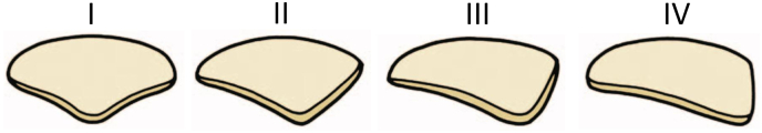
Background: Patellofemoral (PF) instability is a syndrome involving several factors and conditions. Trochlear dysplasia (TD) has been shown to be the feature most frequently diagnosed in patients with OPI (Objective Patellar Instability). Even the patella might have an altered shape. Articulating during growth with an abnormal trochlea might lead the patellar posterior surface to modify accordingly, becoming congruent with the trochlear shape. A mismatch between the two articulating surfaces has a role in patellofemoral dislocations.Rarely and in case of a highly dysplastic patella, reshaping only the trochlear groove might lead subsequently to an incongruence between the two bones.When isolated sulcus-deepening trochleoplasty does not restore a congruent patellofemoral tracking tested perioperatively, further surgical procedures might be required.
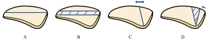
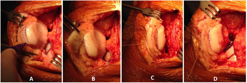
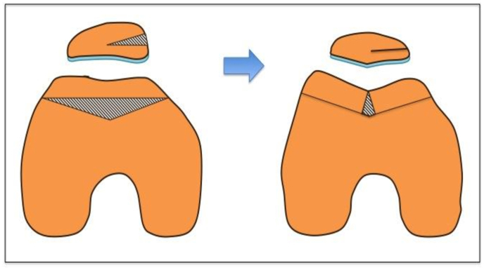
Methods: A medial closing wedge patellar osteotomy (MCWPO) added to sulcus deepening trochleoplasty should be performed to create a congruent patellofemoral joint.Indications for this procedure are Wiberg type III or IV patella with an intraoperative abnormal patellar tracking after the sulcus deepening trochleoplasty procedure has been concluded.
Results: From January 2012 to August 2020, 21 (6 right and 15 left knees) sulcus deepening trochleoplasties with a concomitant medial closing wedge patellar osteotomy have been performed. The mean follow up was 35,25 months (ranging from 10 to 60 months). The average Kujala, IKDC and Lysholm score were respectively 70,14 ± 15,51; 55,75 ± 7,12 and 77,12 ± 14,80. No further patellar dislocation has been assessed in those patients during follow up.
Conclusion: This technique has shown good and promising clinical outcomes and should be considered when treating patients with a still unsatistactory patellofemoral tracking after the sulcus deepening trochleoplasty has been performed due to a patella-trochlea shape mismatch.
Keywords: Patella; Patellar closing wedge; Patellar osteotomy; Patollofemoral instability; Trochlea dysplasia.
© 2022 Professor P K Surendran Memorial Education Foundation. Published by Elsevier B.V. All rights reserved.
Conflict of interest statement
Edoardo Giovannetti de Sanctis has no conflict of interest; David H. Dejour receive Royalties from ARTHREX.
Figures
References
-
- Dejour H., Walch G., Nove-Josserand L., Guier C. Factors of patellar instability: an anatomic radiographic study. Knee Surg Sports Traumatol Arthrosc. 1994;2(1):19–26. - PubMed
-
- Roentgenographic G.W. And anatomic studies on the patellofemoral joint with special reference to chondromalacia patellae. Acta Orthop Scand. 1941;12:319–410.
-
- Baumgartl F. Anatomical and clinical importance of the femoropatellar joint. Zentralbl Chir. . 1966;91(14):506–516. - PubMed
-
- Barnett A.J., Gardner R.O., Lankester B.J., Wakeley C.J., Eldridge J.D. Magnetic resonance imaging of the patella: a comparison of the morphology of the patella in normal and dysplastic knees. J Bone Joint Surg Br. 2007;89(6):761–765. Jun. - PubMed
LinkOut - more resources
Full Text Sources
