

Meta-Heuristic Algorithm-Tuned Neural Network for Breast Cancer Diagnosis Using Ultrasound Images
- PMID: 35769710
- PMCID: PMC9234296
- DOI: 10.3389/fonc.2022.834028
Meta-Heuristic Algorithm-Tuned Neural Network for Breast Cancer Diagnosis Using Ultrasound Images
Abstract
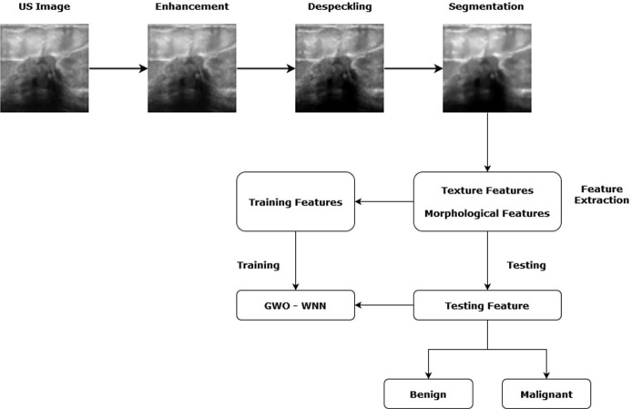
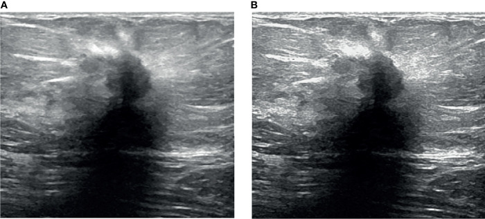
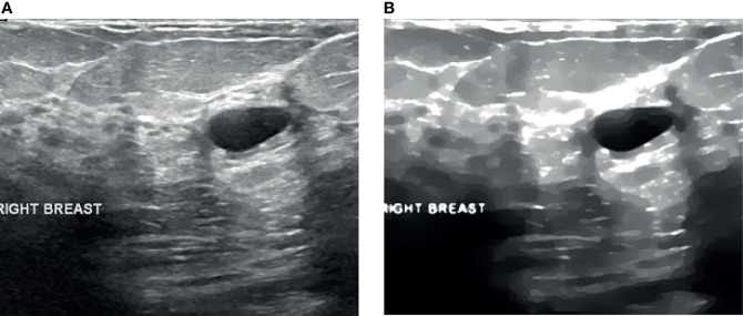
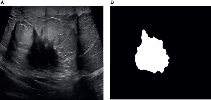
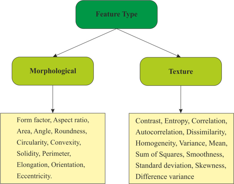
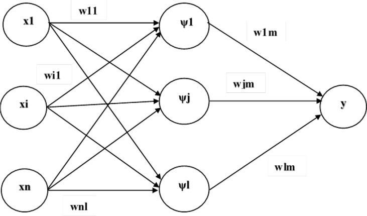
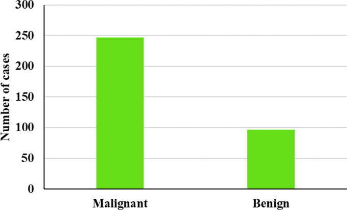
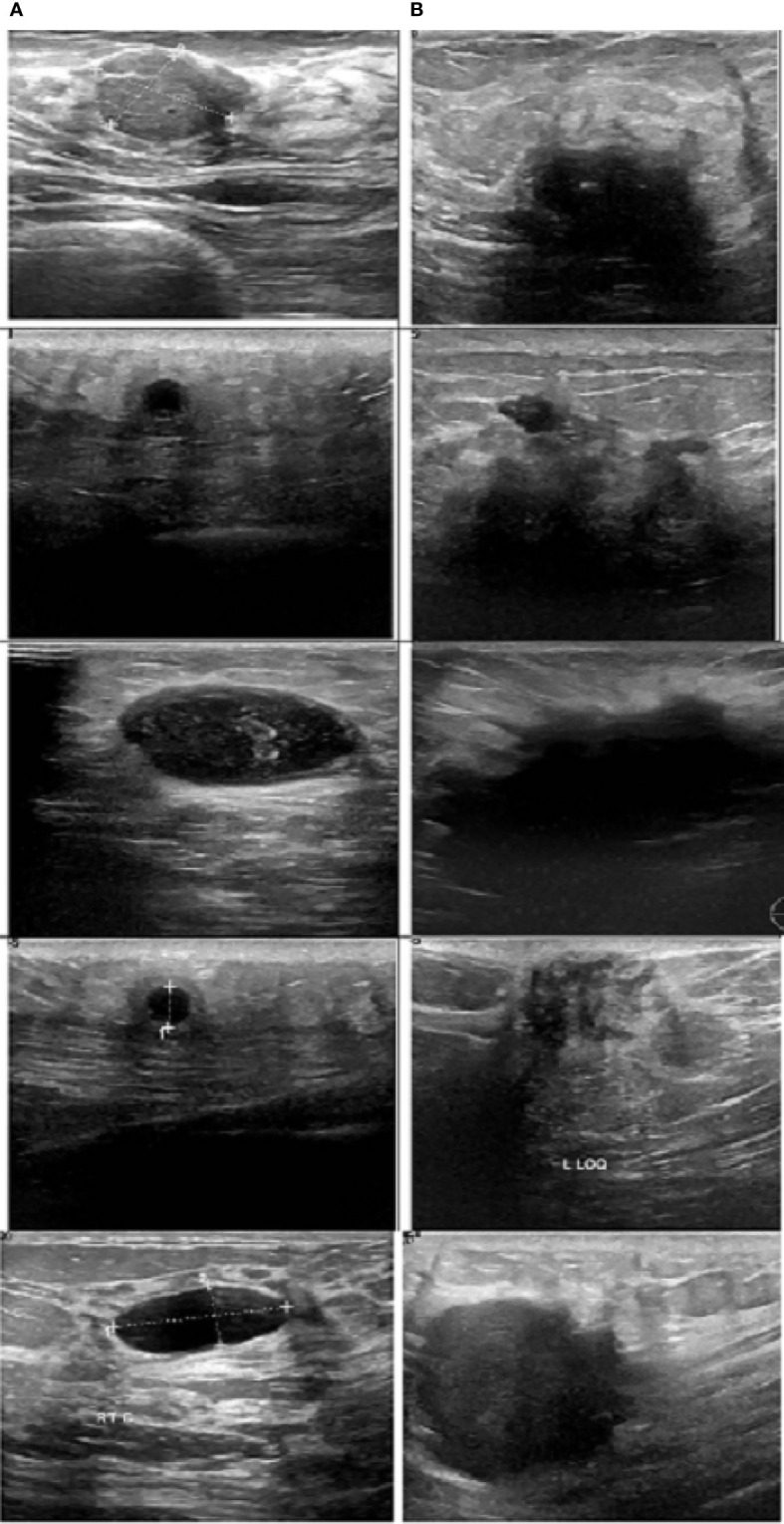
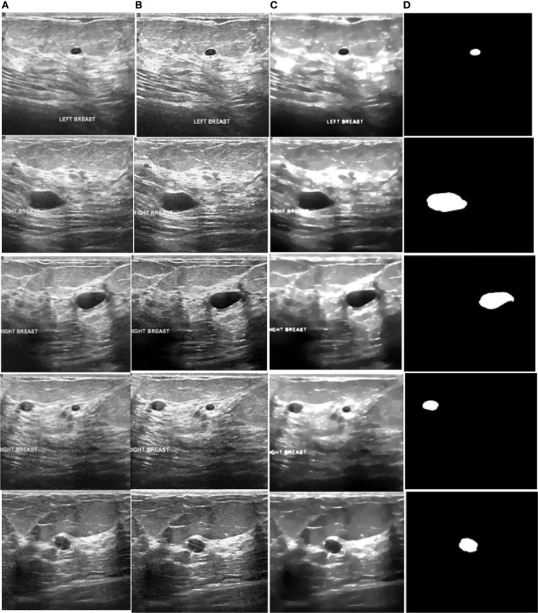
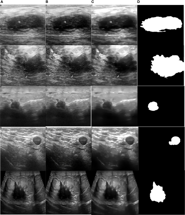
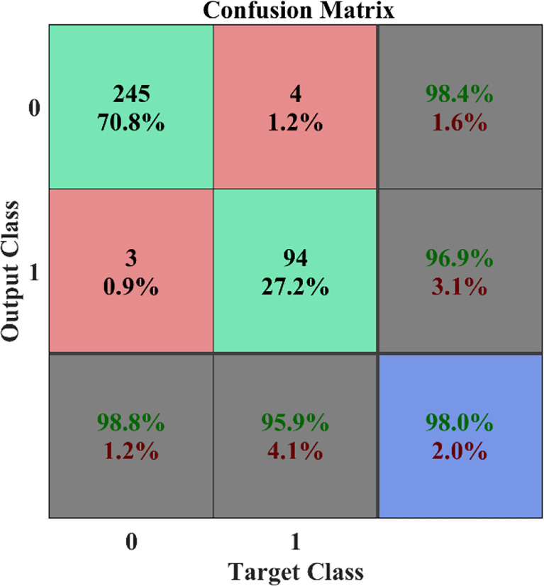
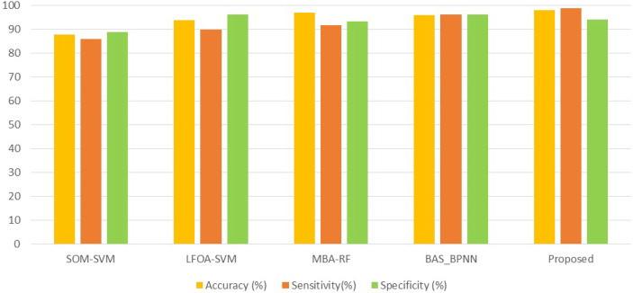
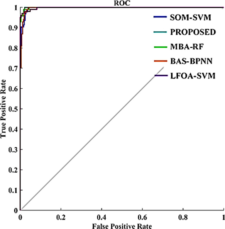
Breast cancer is the most menacing cancer among all types of cancer in women around the globe. Early diagnosis is the only way to increase the treatment options which then decreases the death rate and increases the chance of survival in patients. However, it is a challenging task to differentiate abnormal breast tissues from normal tissues because of their structure and unclear boundaries. Therefore, early and accurate diagnosis and classification of breast lesions into malignant or benign lesions is an active domain of research. Over the decade, numerous artificial neural network (ANN)-based techniques were adopted in order to diagnose and classify breast cancer due to the unique characteristics of learning key features from complex data via a training process. However, these schemes have limitations like slow convergence and longer training time. To address the above mentioned issues, this paper employs a meta-heuristic algorithm for tuning the parameters of the neural network. The main novelty of this work is the computer-aided diagnosis scheme for detecting abnormalities in breast ultrasound images by integrating a wavelet neural network (WNN) and the grey wolf optimization (GWO) algorithm. Here, breast ultrasound (US) images are preprocessed with a sigmoid filter followed by interference-based despeckling and then by anisotropic diffusion. The automatic segmentation algorithm is adopted to extract the region of interest, and subsequently morphological and texture features are computed. Finally, the GWO-tuned WNN is exploited to accomplish the classification task. The classification performance of the proposed scheme is validated on 346 ultrasound images. Efficiency of the proposed methodology is evaluated by computing the confusion matrix and receiver operating characteristic (ROC) curve. Numerical analysis revealed that the proposed work can yield higher classification accuracy when compared to the prevailing methods and thereby proves its potential in effective breast tumor detection and classification. The proposed GWO-WNN method (98%) gives better accuracy than other methods like SOM-SVM (87.5), LOFA-SVM (93.62%), MBA-RF (96.85%), and BAS-BPNN (96.3%).
Keywords: breast cancer detection; computer-aided diagnosis; supervised learning; texture features; ultrasound imaging; wavelet neural network.
Copyright © 2022 A, M, Bourouis, Band, Mosavi, Agrawal and Hamdi.
Conflict of interest statement
The authors declare that the research was conducted in the absence of any commercial or financial relationships that could be construed as a potential conflict of interest.
Figures
References
-
- Cengiz. Cancer Facts & Figures Vol. Vol. 5. . Atlanta, GA: American Cancer Society; (2019).
-
- Zarychta P, Badura P, Pietka E. Comparative Analysis of Selected Classifiers in Posterior Cruciate Ligaments Computer Aided Diagnosis. Bull Pol Acad Sci: Tech Sci (2017) 65:63–70. doi: 10.1515/bpasts-2017-0008 - DOI
-
- Kurek J, Świderski B, Osowski S, Kruk M, Barhoumi W. Deep Learning Versus Classical Neural Approach to Mammogram Recognition. Bull Pol Acad Sci Tech Sci (2018) 66:831–40. doi: 10.24425/bpas.2018.125930 - DOI
-
- Zhao Q, Qiu Y, Zhou G, Cichocki A. Comparative Study on the Classification Methods for Breast Cancer Diagnosis. Bull Pol Acad Sci: Tech Sci (2018) 66:841–8.
-
- Iwendi C, Maddikunta PKR, Gadekallu TR, Lakshmanna K, Bashir AK, Piran MJ. A Metaheuristic Optimization Approach for Energy Efficiency in the Iot Networks. Softw: Pract Exper (2021) 51:2558–71. doi: 10.1002/spe.2797 - DOI
LinkOut - more resources
Full Text Sources