. 2022 Jun 13:12:924725.
doi: 10.3389/fcimb.2022.924725. eCollection 2022.

Oral SARS-CoV-2 Inoculation Causes Nasal Viral Infection Leading to Olfactory Bulb Infection: An Experimental Study

Affiliations

Oral SARS-CoV-2 Inoculation Causes Nasal Viral Infection Leading to Olfactory Bulb Infection: An Experimental Study

Rumi Ueha et al. Front Cell Infect Microbiol. .

Abstract

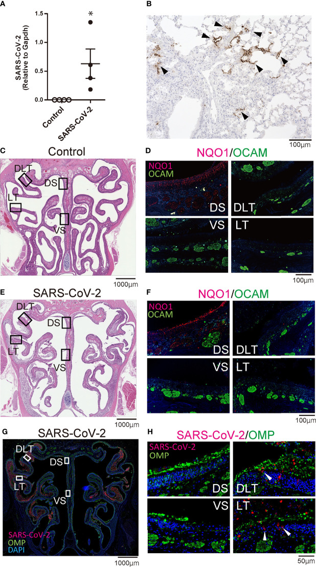
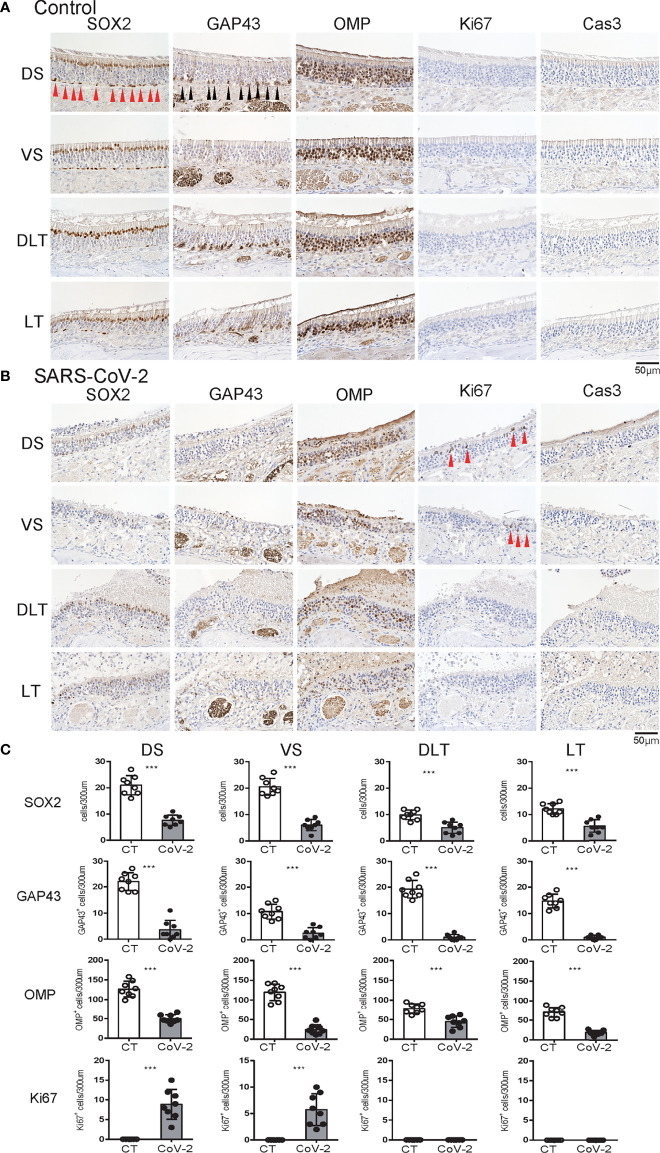
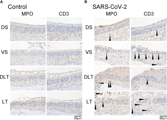
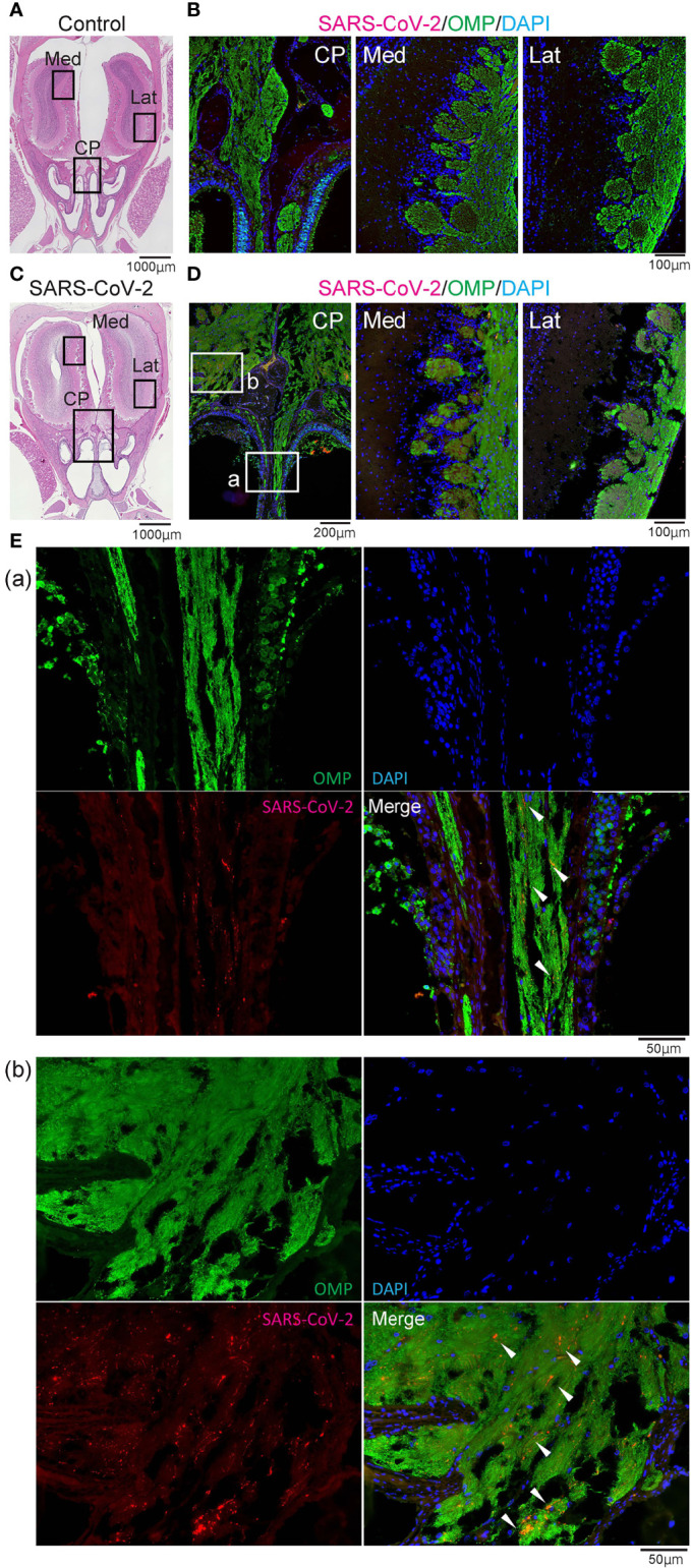
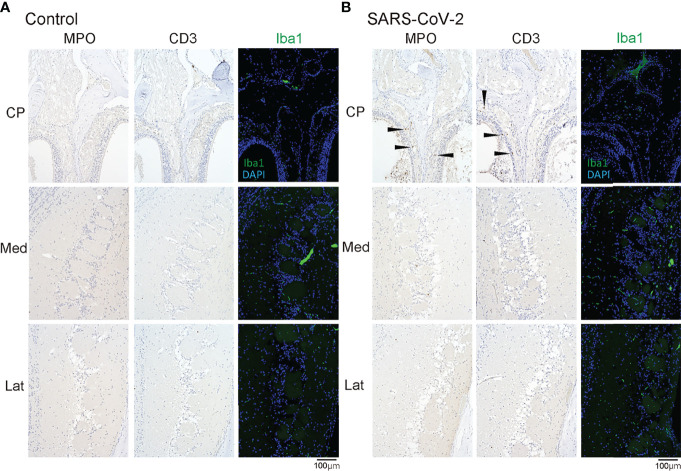
Severe acute respiratory syndrome coronavirus 2 (SARS-CoV-2) infections can cause long-lasting anosmia, but the impact of SARS-CoV-2 infection, which can spread to the nasal cavity via the oral route, on the olfactory receptor neuron (ORN) lineage and olfactory bulb (OB) remains undetermined. Using Syrian hamsters, we explored whether oral SARS-CoV-2 inoculation can lead to nasal viral infection, examined how SARS-CoV-2 affects the ORN lineage by site, and investigated whether SARS-CoV-2 infection can spread to the OB and induce inflammation. On post-inoculation day 7, SARS-CoV-2 presence was confirmed in the lateral area (OCAM-positive) but not the nasal septum of NQO1-positive and OCAM-positive areas. The virus was observed partially infiltrating the olfactory epithelium, and ORN progenitor cells, immature ORNs, and mature ORNs were fewer than in controls. The virus was found in the olfactory nerve bundles to the OB, suggesting the nasal cavity as a route for SARS-CoV-2 brain infection. We demonstrated that transoral SARS-CoV-2 infection can spread from the nasal cavity to the central nervous system and the possibility of central olfactory dysfunction due to SARS-CoV-2 infection. The virus was localized at the infection site and could damage all ORN-lineage cells.

Keywords: COVID-19; SARS-CoV-2; olfactory bulb; olfactory dysfunction; olfactory epithelium; oral inoculation; zone.


Conflict of interest statement

The authors declare that the research was conducted in the absence of any commercial or financial relationships that could be construed as a potential conflict of interest.

Figures

Figure 1
Figure 1
Infectious findings in the lungs and nasal cavity. (A) Severe acute respiratory syndrome coronavirus 2 (SARS-CoV-2) gene detection with RT- quantitative polymerase chain reaction (*P < 0.05). (B) Immunohistochemistry staining of SARS-CoV-2 in the lung (shown in brown, arrow heads). (C, D) Representative images of the olfactory epithelium from control hamsters (C: hematoxylin and eosin stain, D: immunohistological staining of NQO1 and OCAM). The boxes in (C) indicate the regions of the olfactory epithelium shown in D; the dorsal nasal septum (DS) area, ventral nasal septum (VS) area, dorsal lateral turbinate (DLT) area, and lateral turbinate (LT) area. Only the DS area appears positive for NQO1. (E, F) Representative images of the olfactory epithelium from SARS-CoV-2 hamsters. (G) Immunohistochemistry staining of the olfactory epithelium in SARS-CoV-2 hamsters. The virus is mainly present in the lateral region of the nasal cavity. (H) Double stained images of SARS-CoV-2 and OMP in each region of the olfactory epithelium. No double positive cells for SARS-CoV-2 and OMP are present in the olfactory epithelium. The arrow heads show SARS-CoV-2 positive cells.
Figure 2
Figure 2
Effects of SARS-CoV-2 infection on the olfactory receptor neuron lineage. (A, B) Representative images of immunohistological staining in control hamsters (A) and severe acute respiratory syndrome coronavirus 2 (SARS-CoV-2) hamsters (B). Sex-determining region Y-box 2 (SOX2)+ progenitor cells, growth-associated protein 4 (GAP43)+ immature olfactory receptor neurons (ORNs), olfactory marker protein (OMP)+ ORNs, Ki67+ proliferating cells, and cleaved caspase-3 (Cas3)+ apoptotic cells are shown in brown. Each cell, except for the numerous OMP+ cells, is indicated by arrows. Tissue sections were counterstained with the nuclear dye hematoxylin (blue). (C) Numbers of SOX2+ ORN progenitors and Ki67+ actively proliferating cells per 300 μm of the basal layer and OMP+ mature ORNs, GAP43+ immature ORNs, and Cas3+ apoptotic cells per 300 μm of olfactory epithelium in each area are counted in control or SARS-CoV-2 hamsters. ***P < 0.001.
Figure 3
Figure 3
Inflammatory cell infiltration in the olfactory mucosa. (A, B) Representative images of immunohistological staining of MPO and CD3 in control hamsters (A) and severe acute respiratory syndrome coronavirus 2 (SARS-CoV-2) hamsters (B). (B) In the SARS-CoV-2 group, some MPO+ and CD3+ cells are present in the olfactory epithelium.
Figure 4
Figure 4
Representative images of SARS-CoV-2 and OMP staining in the olfactory nerve bundles and olfactory bulb. Representative coronal section images of the olfactory bulb area. (A, B) Images of control hamsters (A: hematoxylin and eosin stain, B: immunohistological staining of severe acute respiratory syndrome coronavirus 2 (SARS-CoV-2), olfactory marker protein (OMP), and DAPI). The boxes in (A) indicate the areas shown enlarged in B; the cribriform plate area (CP), medial area (Med), and lateral area (Lat). (C–E) Images of SARS-CoV-2 hamsters (C: hematoxylin and eosin stain, D, E: immunohistological staining of SARS-CoV-2, OMP, and DAPI). The boxes in (C) indicate the areas shown enlarged in (D), and the boxes in (D) are shown enlarged in (E). SARS-CoV-2 is present in the olfactory nerve of the nasal mucosa and the olfactory nerve fiber layer around the olfactory bulb.
Figure 5
Figure 5
MPO/CD3/Iba1 positive cells in the olfactory bulb area. Representative immunohistological images of MPO and CD3 in the olfactory bulb area (A: control hamster, B: SARS-CoV-2 hamster). Some MPO+ cells and CD3+ cells are observed in the olfactory mucosa around the cribriform plate (arrow heads). CP, cribriform plate area; Iba1, ionized calcium-binding adaptor molecule 1; Lat, lateral area; Med, medial area; SARS-CoV-2, severe acute respiratory syndrome coronavirus 2.

Similar articles

Cited by

References

    1. Boscolo-Rizzo P., Menegaldo A., Fabbris C., Spinato G., Borsetto D., Vaira L. A., et al. . (2021). Six-Month Psychophysical Evaluation of Olfactory Dysfunction in Patients With COVID-19. Chem. Senses 46, 1–9. doi: 10.1093/chemse/bjab006 - DOI - PMC - PubMed
    1. Brann D. H., Tsukahara T., Weinreb C., Lipovsek M., Van den Berge K., Gong B., et al. . (2020). Non-Neuronal Expression of SARS-CoV-2 Entry Genes in the Olfactory System Suggests Mechanisms Underlying COVID-19-Associated Anosmia. Sci. Adv. 6 (31), eabc5801. doi: 10.1126/sciadv.abc5801 - DOI - PMC - PubMed
    1. Bryche B., St Albin A., Murri S., Lacote S., Pulido C., Ar Gouilh M., et al. . (2020). Massive Transient Damage of the Olfactory Epithelium Associated With Infection of Sustentacular Cells by SARS-CoV-2 in Golden Syrian Hamsters. Brain Behav. Immun. 89, 579–586. doi: 10.1016/j.bbi.2020.06.032 - DOI - PMC - PubMed
    1. Butowt R., von Bartheld C. S. (2021). Anosmia in COVID-19: Underlying Mechanisms and Assessment of an Olfactory Route to Brain Infection. Neuroscientist: Rev. J. Bringing Neurobiol. Neurol. Psychiatry 27 (6), 582–603. doi: 10.1177/1073858420956905 - DOI - PMC - PubMed
    1. Cantuti-Castelvetri L., Ojha R., Pedro L. D., Djannatian M., Franz J., Kuivanen S., et al. . (2020). Neuropilin-1 Facilitates SARS-CoV-2 Cell Entry and Infectivity. Science 370 (6518), 856–860. doi: 10.1126/science.abd2985 - DOI - PMC - PubMed

Publication types