Characterization of the B-Cell Epitopes of Echinococcus granulosus Histones H4 and H2A Recognized by Sera From Patients With Liver Cysts
- PMID: 35770070
- PMCID: PMC9234146
- DOI: 10.3389/fcimb.2022.901994
Characterization of the B-Cell Epitopes of Echinococcus granulosus Histones H4 and H2A Recognized by Sera From Patients With Liver Cysts
Abstract
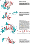
Cystic echinococcosis (CE) is a zoonotic disease worldwide distributed, caused by the cestode Echinococcus granulosus sensu lato (E. granulosus), with an incidence rate of 50/100,000 person/year and a high prevalence in humans of 5-10%. Serology has variable sensitivity and specificity and low predictive values. Antigens used are from the hydatid fluid and recombinant antigens have not demonstrated superiority over hydatid fluid. A cell line called EGPE was obtained from E. granulosus sensu lato G1 strain from bovine liver. Serum from CE patients recognizes protein extracts from EGPE cells with higher sensitivity than protein extracts from hydatid fluid. In the present study, EGPE cell protein extracts and supernatants from cell colonies were eluted from a protein G affinity column performed with sera from 11 CE patients. LC-MS/MS proteomic analysis of the eluted proteins identified four E. granulosus histones: one histone H4 in the cell extract and supernatant, one histone H2A only in the cell extract, and two histones H2A only in the supernatant. This differential distribution of histones could reflect different parasite viability stages regarding their role in gene transcription and silencing and could interact with host cells. Bioinformatics tools characterized the linear and conformational epitopes involved in antibody recognition. The three-dimensional structure of each histone was obtained by molecular modeling and validated by molecular dynamics simulation and PCR confirmed the presence of the epitopes in the parasite genome. The three histones H2A were very different and had a less conserved sequence than the histone H4. Comparison of the histones of E. granulosus with those of other organisms showed exclusive regions for E. granulosus. Since histones play a role in the host-parasite relationship they could be good candidates to improve the predictive value of serology in CE.
Keywords: Echinococcus granulosus; Histones; cell extract; epitopes; extracellular.
Copyright © 2022 Maglioco, Agüero, Valacco, Valdez, Paulino and Fuchs.
Conflict of interest statement
The authors declare that the research was conducted in the absence of any commercial or financial relationships that could be construed as a potential conflict of interest.
Figures


References
-
- Avila H. G., Maglioco A., Getiser M. L., Ferreyra M. P., Ferrari F., Klinger E., et al. (2021). First Report of Cystic Echinococcosis Caused by Echinococcus Granulosus Sensu Strict/G1 in Felix Catus From the Patagonian Region of Argentina. Parasitol. Res. 120, 747–750. doi: 10.1007/s00436-021-07048-4 - DOI - PubMed
-
- Baranova S. V., Dmitrenok P. S., Zubkova A. D., Ivanisenko N. V., Odintsova E. S., Buneva V. N., et al. (2018). Antibodies Against H3 and H4 Histones From the Sera of HIV-Infected Patients Catalyze Site-Specific Degradation of These Histones. J. Mol. Recognit. 31, e2703. doi: 10.1002/jmr.2703 - DOI - PubMed
Publication types
MeSH terms
Substances
Supplementary concepts
LinkOut - more resources
Full Text Sources

