Review
. 2022 Jun 13:13:909518.
doi: 10.3389/fphys.2022.909518. eCollection 2022.

Extracellular Vesicles in Pathogenesis and Treatment of Metabolic Associated Fatty Liver Disease

Affiliations
Review

Extracellular Vesicles in Pathogenesis and Treatment of Metabolic Associated Fatty Liver Disease

Ji Sun et al. Front Physiol. .

Abstract

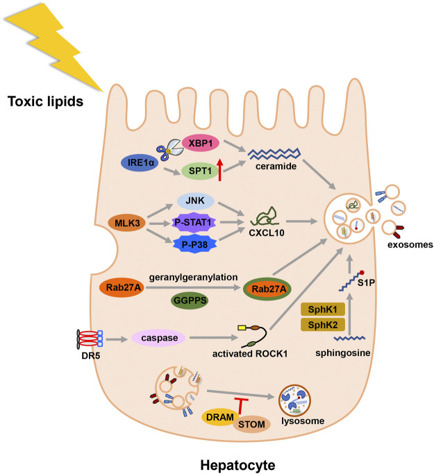
Metabolic associated fatty liver disease (MAFLD) is the most common chronic liver disease worldwide due to the sedentary and overeating lifestyle. Yet, the pathophysiology of MAFLD is still unclear and no drug has been approved for MAFLD treatment. Extracellular vesicles (EVs) are heterogenous membrane-bound particles released from almost all types of cells. These nano-sized particles mediate intercellular communication through their bioactive cargos including nucleic acids, proteins, and lipids. The EVs modulate metabolic homeostasis via communication between adipose tissue and liver. The dysregulation of lipid metabolism leads to inflammation in liver and the number and compounds of EVs are changed during MAFLD. The injured hepatocytes secrete EVs to induce the migration of bone marrow-derived monocytes and the activation of macrophages in liver. The EVs secreted by different cells regulate the alteration of hepatic stellate cell (HSC) phenotypes and HSC activation gives rise to liver fibrosis. Based on the participation of EVs in MAFLD progression, we discuss the prospects of EVs as a therapeutic target and their application in drug delivery.

Keywords: extracellular vesicles; inflammation; lipids; metabolic associated fatty liver disease; pathogenesis; treatment.


Conflict of interest statement

The authors declare that the research was conducted in the absence of any commercial or financial relationships that could be construed as a potential conflict of interest.

Figures

FIGURE 1
FIGURE 1
Biogenesis and release of various EVs. Generally, EVs are divided into exosomes, microvesicles and apoptotic bodies. Exosomal formation begins with endocytic process. After the endosomal membrane invagination, intraluminal vesicles (ILVs) are formed. The endosomes containing ILVs are referred as multivesicular bodies (MVBs). Subsequently, MVBs have two fates. They can be degraded in lysosomes. Alternatively, MVBs merge with plasma membrane and release ILVs as exosomes. Microvesicles are formed through direct budding process. Apoptotic bodies are formed during apoptosis.
FIGURE 2
FIGURE 2
Illustration for the biogenesis fo exosomes in MAFLD. Toxic lipid accumulation is the major hit of MAFLD, which causes more secretion of exosomes by multiple mechanisms. Abbreviation: death receptor 5 (DR5), Rho-associated kinase 1 (ROCK1), geranylgeranyl diphosphate synthase (Ggpps), mixed lineage kinase 3 (MLK3), (C-X-C motif) ligand 10 (CXCL10), inositol requiring enzyme 1α (IRE1α), X-box binding protein-1 (XBP1), serine palmitoyltransferase1 (SPT1), sphingosine-1-phosphate (S1P), sphingosine kinase 1 (SphK1), and sphingosine kinase 2 (SphK2), damage-regulated autophagy modulator (DRAM), stomatin (STOM).
FIGURE 3
FIGURE 3
Illustration for how EVs participate in the development of MAFLD. Under the condition of metabolic disturbance, cells secrete EVs to transfer information, which contain protein, lipid, miRNA, lncRNA or DNA. Adipocytes and macrophages in adipose tissue transfer EVs to liver and regulate the lipid metabolism and fibrotic phenotype in liver. Inside liver, EVs link up the communication of various hepatic cells and promote MAFLD progression. EVs secreted outside the liver can be consumed by adipocytes and monocytes, then regulating energy metabolism and inflammatory condition. Abbreviation: aldo-keto-reductase 1B7 (Akr1b7), cluster of differentiation 36 (CD36), interleukin- 6 (IL-6), monocyte chemoattractant protein-1 (MCP-1), migration inhibitory factor (MIF), connective tissue growth factor (CCN2), mitochondrial DNA (mtDNA), sphingosine-1-phosphate (S1P), tumor necrosis factor-related apoptosis-inducing ligand (TRAIL), integrin β1(ITGβ1), C-X-C motif ligand 10 (CXCL10).

References

    1. Afrisham R., Sadegh-Nejadi S., Meshkani R., Emamgholipour S., Bagherieh M., Paknejad M. (2021). Anti-inflammatory Effects of Plasma Circulating Exosomes Obtained from Normal-Weight and Obese Subjects on Hepatocytes. Emiddt 21, 478–484. 10.2174/1871530320666200505121426 - DOI - PubMed
    1. Andreu Z., Yáñez-Mó M. a. (2014). Tetraspanins in Extracellular Vesicle Formation and Function. Front. Immunol. 5, 442. 10.3389/fimmu.2014.00442 - DOI - PMC - PubMed
    1. Arraud N., Linares R., Tan S., Gounou C., Pasquet J.-M., Mornet S., et al. (2014). Extracellular Vesicles from Blood Plasma: Determination of Their Morphology, Size, Phenotype and Concentration. J. Thromb. Haemost. 12, 614–627. 10.1111/jth.12554 - DOI - PubMed
    1. Azzu V., Vacca M., Virtue S., Allison M., Vidal-Puig A. (2020). Adipose Tissue-Liver Cross Talk in the Control of Whole-Body Metabolism: Implications in Nonalcoholic Fatty Liver Disease. Gastroenterology 158, 1899–1912. 10.1053/j.gastro.2019.12.054 - DOI - PubMed
    1. Baietti M. F., Zhang Z., Mortier E., Melchior A., Degeest G., Geeraerts A., et al. (2012). Syndecan-syntenin-ALIX Regulates the Biogenesis of Exosomes. Nat. Cell. Biol. 14, 677–685. 10.1038/ncb2502 - DOI - PubMed

LinkOut - more resources