. 2022 Jun 23:16:2057-2067.
doi: 10.2147/OPTH.S370800. eCollection 2022.

Analysis of Lacrimal Duct Morphology from Cone-Beam Computed Tomography Dacryocystography in a Japanese Population

Affiliations

Analysis of Lacrimal Duct Morphology from Cone-Beam Computed Tomography Dacryocystography in a Japanese Population

Jutaro Nakamura et al. Clin Ophthalmol. .

Abstract

Purpose: The dacryoendoscope is a practical instrument for the examination and the treatment of lacrimal duct obstruction. Nevertheless, as it is a rigid fiberscope, manipulation of the endoscope is somewhat affected by the patient's lacrimal duct alignment and the skeletal structure of the face. The morphology and inclination of the lacrimal duct vary among individuals and ethnic groups. We aimed to evaluate the alignment of the lacrimal duct from the perspective of endoscopic maneuverability in a Japanese population.

Methods: This retrospective study analyzed the cone-beam computed tomography dacryocystography (CBCT-DCG) images of 102 patients diagnosed with unilateral primary acquired nasolacrimal duct obstruction (PANDO) at Ehime University Hospital from December 2015 to May 2021. The following parameters of the lacrimal duct on the contralateral side of unilateral PANDO were investigated: (1) angle formed by the superior orbital rim-internal common punctum-nasolacrimal duct opening, (2) angle formed by the lacrimal sac and the nasolacrimal duct, (3) length of the lacrimal sac, and (4) length of the nasolacrimal duct.

Results: Measurements of the above parameters were (1) 10.2° ± 7.8° (range, -11° to +27°), (2) -6.3° ± 14.1° (range, -43° to +40°), (3) 8.9 ± 2.3 mm (range, 4.3-17.1), and (4) 13.2 ± 2.7 mm (range, 5.7-20.7), respectively. The Shapiro-Wilk test demonstrated that the values of all parameters, except (3), followed a normal distribution (p = 0.55, 0.30, 0.0002, and 0.39, respectively). No significant difference was found between the female and male groups (p > 0.05).

Conclusion: This study reported anthropometric analysis data of the morphology of the lacrimal ducts using CBCT-DCG in a Japanese population. In our cohort, the line from the superior orbital rim through the internal common punctum to the nasolacrimal duct opening inclined anteriorly in 92% of the patients.

Keywords: cone-beam computed tomography; dacryocystography; dacryoendoscope; endoscopic-assisted nasolacrimal duct intubation; primary acquired nasolacrimal duct obstruction.


Conflict of interest statement

The authors declare that they have no competing interests. The authors alone are responsible for the content and writing of the paper.

Figures

Figure 1
Figure 1
SOR–ICP line as an anatomical limitation. The line formed by the SOR–ICP is the anatomical limit where the tip of a straight probe can reach most anteriorly after entering the LS through the ICP. The red arrow indicates when the LS and NLD inclined anteriorly to the line formed by the SOR–ICP. Manipulating straight probes poses a risk for forming an iatrogenic false passage posterior to the original lacrimal duct.
Figure 2
Figure 2
SOR interference during manipulation of the dacryoendoscope. SOR interferes the manipulation of the endoscope. It is difficult to tilt the probe horizontally than the SOR. However, the endoscope can stand more vertically than the SOR.
Figure 3
Figure 3
CBCT-DCG sagittal image sectioning the lacrimal duct. The figure shows a dacryocystographic image of the nasolacrimal duct on the right side from a patient diagnosed with left-sided unilateral primary acquired nasolacrimal duct obstruction. The original image was converted to monochrome to facilitate observation of the contrast media.
Figure 4
Figure 4
Parameters measured in CBCT-DCG images. (A) Angle formed by SOR–ICP–NLD opening. In this case, the line connecting the ICP–NLD opening was inclined anteriorly at 4 relative to the line connecting the SOR–ICP. (B) Angle formed by LS and NLD. In this case, the long axis of the NLD was inclined posteriorly at 10° relative to the LS. (C) Length of LS and NLD, with 6.15 mm and 14.30 mm, respectively.
Figure 5
Figure 5
Example of a large SOR–ICP–NLD opening angle. On the left side is the original image. On the right side, a relatively large angle of 24° is seen, which is due to the elevation of the SOR and the anterior inclination of the NLD.
Figure 6
Figure 6
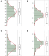
Test results for the normal distribution of the measured parameters. (A) SOR–ICP–NLD opening angle: The average angle was 10.7° ± 7.4° (range, −6° to +27°) and followed a normal distribution (p = 0.55). (B) LS–NLD angle: The mean angle was −6.84° ± 13.7° (range, −43° to +34°) and followed a normal distribution (p = 0.28). (C) LS length: The mean length was 8.9 ± 2.2 mm (range, 5.4–17.1) and did not follow a normal distribution (p = 0.078). (D) NLD length: The mean length was 13.3 ± 2.7 mm (range, 5.7–20.7) and followed a normal distribution (p = 0.17). The p-value was determined by the Shapiro–Wilk test. A p-value of >0.05 indicated a normal distribution.
Figure 7
Figure 7
Case in which the NLD is inclined anteriorly to the LS (anterior bending type). On the left is the original image. On the right side, the long axis of the NLD was anteriorly inclined by +14° relative to that of the LS.
Figure 8
Figure 8
Case in which the NLD is inclined posteriorly to the LS (posterior bending type). On the left is the original image. On the right side, the long axis of the NLD was posteriorly inclined by −21° relative to the LS.
Figure 9
Figure 9
Dacryoendoscope with an anteriorly bent-tip probe and a curve-tipped probe. Left: CK10® (FiberTech Co., Ltd., Tokyo, Japan); external diameter, 0.9mm; field of view, 60°; image quality, 10,000 pixels. Middle: CH15C® (FiberTech Co., Ltd., Tokyo, Japan); external diameter, 0.82 mm; field of view, 70°; image quality, 15,000 pixels. Right: LAC-06-FY® (Machida Endoscope Co., Ltd., Chiba, Japan); external diameter, 0.9 mm; field of view, 65°; image quality, 10,000 pixels.

References

    1. Mandeville JT, Woog JJ. Obstruction of the lacrimal drainage system. Curr Opin Ophthalmol. 2002;13(5):303–309. doi:10.1097/00055735-200210000-00003 - DOI - PubMed
    1. Montecalvo RM, Zegel HG, Barnett FJ, et al. Evaluation of the lacrimal apparatus with digital subtraction macrodacryocystography. Radiographics. 1990;10(3):483–490. doi:10.1148/radiographics.10.3.2188309 - DOI - PubMed
    1. Singh S, Ali MJ, Paulsen F. Dacryocystography: from theory to current practice. Ann Anat. 2019;224:33–40. doi:10.1016/j.aanat.2019.03.009 - DOI - PubMed
    1. Cakli H, Cingi C, Ay Y, Oghan F, Ozer T, Kaya E. Use of cone beam computed tomography in otolaryngologic treatments. Eur Arch Otorhinolaryngol. 2012;269(3):711–720. doi:10.1007/s00405-011-1781-x - DOI - PubMed
    1. Mozzo P, Procacci C, Tacconi A, Martini PT, Andreis IA. A new volumetric CT machine for dental imaging based on the cone-beam technique: preliminary results. Eur Radiol. 1998;8(9):1558–1564. doi:10.1007/s003300050586 - DOI - PubMed