. 2022 Jun 13:16:817948.
doi: 10.3389/fnbot.2022.817948. eCollection 2022.

Brain-Inspired Spiking Neural Network Controller for a Neurorobotic Whisker System

Affiliations

Brain-Inspired Spiking Neural Network Controller for a Neurorobotic Whisker System

Alberto Antonietti et al. Front Neurorobot. .

Abstract

It is common for animals to use self-generated movements to actively sense the surrounding environment. For instance, rodents rhythmically move their whiskers to explore the space close to their body. The mouse whisker system has become a standard model for studying active sensing and sensorimotor integration through feedback loops. In this work, we developed a bioinspired spiking neural network model of the sensorimotor peripheral whisker system, modeling trigeminal ganglion, trigeminal nuclei, facial nuclei, and central pattern generator neuronal populations. This network was embedded in a virtual mouse robot, exploiting the Human Brain Project's Neurorobotics Platform, a simulation platform offering a virtual environment to develop and test robots driven by brain-inspired controllers. Eventually, the peripheral whisker system was adequately connected to an adaptive cerebellar network controller. The whole system was able to drive active whisking with learning capability, matching neural correlates of behavior experimentally recorded in mice.

Keywords: active whisking; central pattern generator (CPG); facial nuclei; neurorobotic architecture; point neuron model; trigeminal ganglion; trigeminal nuclei; vibrissae.


Conflict of interest statement

The authors declare that the research was conducted in the absence of any commercial or financial relationships that could be construed as a potential conflict of interest.

Figures

Figure 1
Figure 1
The rodent whisker system. (A) Virtual robotic mouse implemented in the NRP, with two whiskers per side. L0 and R0 are the lower left and right whiskers, L1 and R1 are the upper whiskers. (B) Block diagram of the rodent whisker system, including sensory and motor pathways, and its integration with higher-order areas (thalamus and cortex). (C) SNN implementation of the mouse peripheral whisker system; numbers in each block represent the size of the neural populations included in that brain region. Arrows represent excitatory connections, circles inhibitory connections.
Figure 2
Figure 2
SNN implementation of the cerebellum. Whisking sensory signals are conveyed to the cerebellar MFs from TG pressure and TN phase neurons, while the reward signal during correct GO trials reaches the IO neurons; the cerebellum controls the output motor response (head movement) according to DCN activity (i.e., generation of a response, head raise, when the firing rate exceeds a set threshold). Arrows and circles represent excitatory and inhibitory connections, respectively.
Figure 3
Figure 3
(A) The experimental protocol: during GO trials, a sensory cue (a small bar, depicted as a black dot) is placed in the left whisker field of the mouse. Correct responses lead to a reward (water drop). During NOGO trials, the sensory cue is placed in the right whisker field, and a response does not result in any reward. (B) Spiking activity of motor neurons for protraction and retraction during one trial. Twenty protractors neurons for each whisker and 20 retractor neurons for each side (L and R) fire under the control of the CPG neuron at 4 Hz. The resulting displacement of each whisker is depicted in the upper part of the panel. (C) Angular displacement of the four whiskers during one GO and one NOGO trial. During the GO trial, in the first second, left whiskers hit the sensory cue bar, placed in the left whisker field. On the other hand, during the NOGO trial, the right whiskers hit the sensory cue bar placed in the right whisker field.
Figure 4
Figure 4
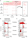
(A) Spiking activity of the Trigeminal Ganglion (TG) neurons during GO and NOGO trials. Each row represents the activity of one neuron, and different shades of red are used to plot the activity of the four groups of TG neurons. The inset shows a magnified portion of the full scatterplot, focusing on a single protraction-retraction movement of the whiskers. (B) Firing rates of TG populations were measured experimentally during a single protraction-retraction movement, as reported in Ahissar and Knutsen (2016). Vertical dashed lines represent the four main events: the start of the protraction, contact of the whisker against an object, the start of the retraction, detach of the whisker from the object. (C) Firing rates were recorded from the simulation of the SNN model of TG populations. The length of each bin is 10 ms. Colors are the same as (A,B).
Figure 5
Figure 5
(A) Learning curves recorded in the experiment performed by Rahmati et al. (2014). The upper row shows the Hit rate (i.e., the percentage of correct responses in GO trials) along with sessions, where each session is composed of 10 trials. The lower row shows the False alarms (i.e., the percentage of incorrect responses in NOGO trials) along with sessions. The blue and red curves show the mean values for control animals and knock-out (L7-PP2B) mice. Shaded areas show the standard deviation. (B) Learning curves recorded from the in-silico experiments (10 control and 10 knock-out models). Colors are the same as in (A). (C) Spiking activity of the DCN neurons during GO (green) and NOGO (red) trials, in the first session (left column) and after 10 sessions of training (right column). The first row reports the activity of one Control simulation, while the second row reports one knock-out simulation. Each dot is a spike of one of the 36 DCN in the cerebellar network. The order of GO and NOGO trials is randomized for each session and simulation, but all sessions have 5 GO and 5 NOGO trials.

References

    1. Adibi M. (2019). Whisker-mediated touch system in rodents: from neuron to behavior. Front. Syst. Neurosci. 13:40. 10.3389/fnsys.2019.00040 - DOI - PMC - PubMed
    1. Aguero C., Koenig N., Chen I., Boyer H., Peters S., Hsu J., et al. . (2015). Inside the virtual robotics challenge: simulating real-time robotic disaster response. IEEE Trans. Autom. Sci. Eng. 12, 494–506. 10.1109/TASE.2014.2368997 - DOI
    1. Ahissar E., Knutsen P. M. (2016). Vibrissal Location Coding. Paris: Atlantis Press. 725–735. 10.2991/978-94-6239-133-8_53 - DOI
    1. Antonietti A., Casellato C., Garrido J. A., Luque N. R., Naveros F., Ros E., et al. . (2016). Spiking neural network with distributed plasticity reproduces cerebellar learning in eye blink conditioning paradigms. IEEE Trans. Biomed. Eng. 63, 210–219. 10.1109/TBME.2015.2485301 - DOI - PubMed
    1. Antonietti A., Martina D., Casellato C., D'Angelo E., Pedrocchi A. (2019). Control of a humanoid NAO robot by an adaptive bioinspired cerebellar module in 3D motion tasks. Comput. Intell. Neurosci. 2019:4862157. 10.1155/2019/4862157 - DOI - PMC - PubMed

LinkOut - more resources