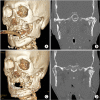
Coronoidectomy for reduction of superolateral dislocation of mandible condyle
- PMID: 35770361
- PMCID: PMC9247446
- DOI: 10.5125/jkaoms.2022.48.3.182
Coronoidectomy for reduction of superolateral dislocation of mandible condyle
Abstract
Superolateral dislocation of the condyle is a rare mandibular fracture. The treatment goal is to return the dislocated condyle to its original position to recover normal function. This study reports on superolateral dislocation of the condyle with mandibular body fracture. The mandibular body was completely separated, and the medial pole of the condyle head was fractured. The condyle segment was unstable and easily dislocated after reduction. The temporalis muscle on the condyle segment might have affected the dislocation of the condyle. A coronoidectomy was performed to disrupt the function of the temporalis muscle on the condyle segment in order to successfully reduce the dislocated condyle. Coronoidectomy is a simple procedure with minimal complications. We successfully performed a coronoidectomy to reduce the superolateral displaced condyle to its original position to achieve normal function. Coronoidectomy can be effectively used for reduction of superolaterally displaced condyles combined with severe maxilla-mandibular fractures.
Keywords: Condyle fracture; Coronoidectomy; Temporalis muscle.
Conflict of interest statement
No potential conflict of interest relevant to this article was reported.
Figures


References
-
- Rahman T, Hashmi GS, Ansari MK. Traumatic superolateral dislocation of the mandibular condyle: case report and review. Br J Oral Maxillofac Surg. 2016;54:457–9. doi: 10.1016/j.bjoms.2015.08.002. https://doi.org/10.1016/j.bjoms.2015.08.002. - DOI - PubMed
-
- Patil SG, Patil BS, Joshi U, Rudagi BM, Aftab A. Superolateral dislocation of bilateral intact mandibular condyles: a rare case series. J Maxillofac Oral Surg. 2017;16:212–8. doi: 10.1007/s12663-016-0928-0. https://doi.org/10.1007/s12663-016-0928-0. - DOI - PMC - PubMed
-
- Kim BC, Lee YC, Cha HS, Lee SH. Characteristics of temporomandibular joint structures after mandibular condyle fractures revealed by magnetic resonance imaging. Maxillofac Plast Reconstr Surg. 2016;38:24. doi: 10.1186/s40902-016-0066-0. https://doi.org/10.1186/s40902-016-0066-0. - DOI - PMC - PubMed
-
- Allen FJ, Young AH. Lateral displacement of the intact mandibular condyle. A report of five cases. Br J Oral Surg. 1969;7:24–30. doi: 10.1016/S0007-117X(69)80057-0. https://doi.org/10.1016/s0007-117x(69)80057-0. - DOI - PubMed
-
- Özalp B, Elbey H, Durgun M, Selçuk CT. An alternative surgical procedure for anterosuperior dislocation of intact mandibular condyle. J Craniofac Surg. 2014;25:e382–4. doi: 10.1097/SCS.0000000000000965. https://doi.org/10.1097/SCS.0000000000000965. - DOI - PubMed

