

Level dependent alterations in human facet cartilage mechanics and bone morphometry with spine degeneration
- PMID: 35770853
- PMCID: PMC9800647
- DOI: 10.1002/jor.25407
Level dependent alterations in human facet cartilage mechanics and bone morphometry with spine degeneration
Abstract
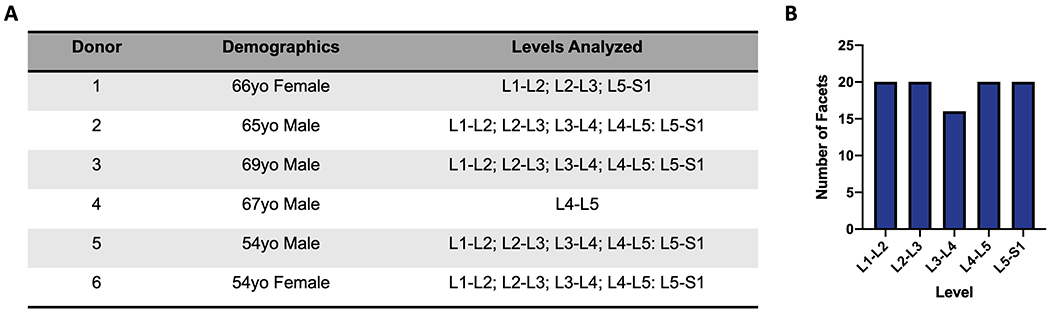
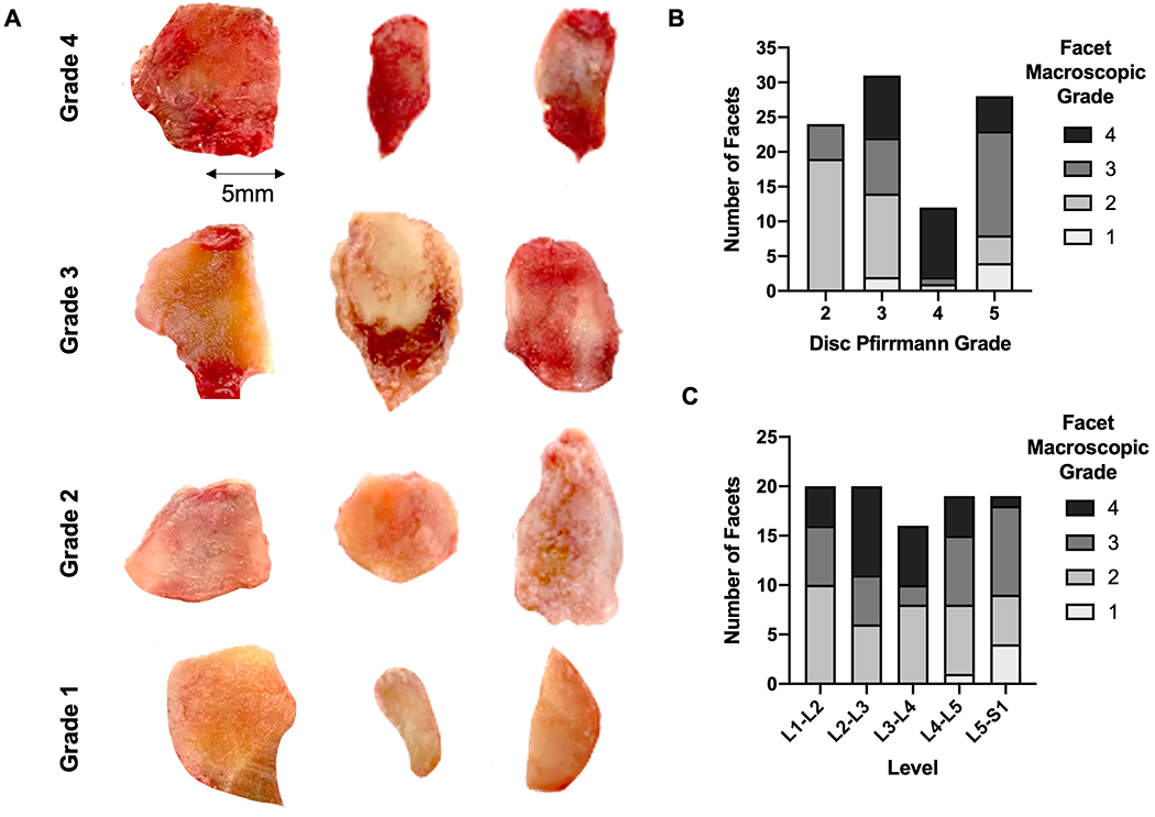
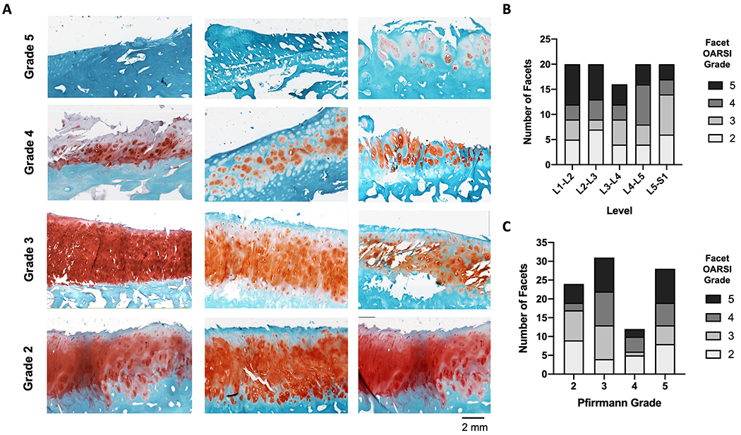
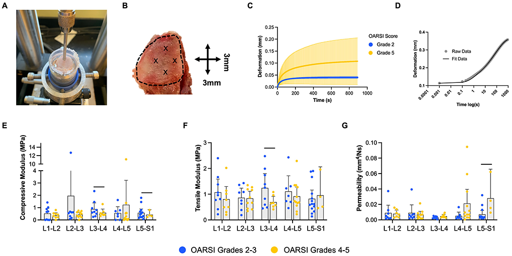
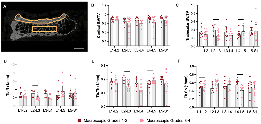
The zygapophyseal joints of the spine, also known as the facet joints, are paired diarthrodial joints posterior to the intervertebral disc and neural elements. The pathophysiology of facet osteoarthritis (OA), as well as crosstalk between the disc and facets, remains largely understudied compared to disc degeneration. The purpose of this study was to characterize alterations to human facet cartilage and subchondral bone across a spectrum of degeneration and to investigate correlations between disc and facet degeneration. Human lumbar facet articular surfaces from six independent donors were subject to creep indentation mechanical testing to quantify cartilage mechanical properties, followed by microcomputed tomography (µCT) analyses for subchondral bone morphometry. The degenerative state of each articular surface was assessed via macroscopic scoring and via Osteoarthritis Research Society International histopathology scoring. Our data suggest reduced facet cartilage compressive and tensile moduli and increased permeability with increasing degenerative grade, particularly at the lower levels of the spine. µCT analyses revealed spinal level-dependent alterations to the subchondral bone, with an increase in trabecular bone at the L4-L5 level, but a decrease at the upper levels of the lumbar spine with increasing degenerative grade. Cortical bone volume fraction was generally decreased with increasing degenerative grade across spinal levels. Correlation analysis revealed several associations between quantitative measures of disc degeneration and facet OA. This study showed that alterations in the mechanical properties of facet cartilage and in the structural properties of facet subchondral bone correlated with aspects of disc degeneration and were highly dependent on spinal level.
Keywords: biomechanics; indentation; intervertebral disc degeneration; osteoarthritis; zygapophyseal joint.
© 2022 Orthopaedic Research Society. Published by Wiley Periodicals LLC.
Conflict of interest statement
Conflict of Interest
Robert L. Mauck is a Co-Editor in Chief of JOR Spine.
Figures
Similar articles
-
The effect of disc degeneration and facet joint osteoarthritis on the segmental flexibility of the lumbar spine.Spine (Phila Pa 1976). 2000 Dec 1;25(23):3036-44. doi: 10.1097/00007632-200012010-00011. Spine (Phila Pa 1976). 2000. PMID: 11145815
-
Micro-computed tomography, scanning electron microscopy and energy X-ray spectroscopy studies of facet joint degeneration: A comparison to clinical imaging.Micron. 2017 Sep;100:50-59. doi: 10.1016/j.micron.2017.04.011. Epub 2017 May 1. Micron. 2017. PMID: 28500930
-
The relationship between structural changes in paraspinal muscles and intervertebral disc and facet joint degeneration in the lumbar spine of rats.J Orthop Surg Res. 2024 Jan 12;19(1):58. doi: 10.1186/s13018-024-04548-8. J Orthop Surg Res. 2024. PMID: 38217024 Free PMC article.
-
Spinal facet joint biomechanics and mechanotransduction in normal, injury and degenerative conditions.J Biomech Eng. 2011 Jul;133(7):071010. doi: 10.1115/1.4004493. J Biomech Eng. 2011. PMID: 21823749 Free PMC article. Review.
-
Intervertebral disc degeneration and osteoarthritis: a common molecular disease spectrum.Nat Rev Rheumatol. 2023 Mar;19(3):136-152. doi: 10.1038/s41584-022-00888-z. Epub 2023 Jan 26. Nat Rev Rheumatol. 2023. PMID: 36702892 Review.
Cited by
-
Mechanical crosstalk between the intervertebral disc, facet joints, and vertebral endplate following acute disc injury in a rabbit model.JOR Spine. 2023 Oct 24;6(4):e1287. doi: 10.1002/jsp2.1287. eCollection 2023 Dec. JOR Spine. 2023. PMID: 38156057 Free PMC article.
-
Automated magnetic resonance imaging-based grading of the lumbar intervertebral disc and facet joints.JOR Spine. 2024 Jul 15;7(3):e1353. doi: 10.1002/jsp2.1353. eCollection 2024 Sep. JOR Spine. 2024. PMID: 39011368 Free PMC article.
-
MRI-based degeneration grades for lumbar facet joints do not correlate with cartilage mechanics.JOR Spine. 2023 Jan 11;6(2):e1246. doi: 10.1002/jsp2.1246. eCollection 2023 Jun. JOR Spine. 2023. PMID: 37361329 Free PMC article.
-
Restoration of physiologic loading after engineered disc implantation mitigates immobilization-induced facet joint and paraspinal muscle degeneration.Acta Biomater. 2025 Jan 15;192:128-139. doi: 10.1016/j.actbio.2024.12.014. Epub 2024 Dec 9. Acta Biomater. 2025. PMID: 39653318
References
-
- Casiano VE, Dydyk AM, Varacallo M. 2021. Back Pain. In: StatPearls. Treasure Island (FL): StatPearls Publishing; [cited 2021 Dec 10] Available from: http://www.ncbi.nlm.nih.gov/books/NBK538173/.
-
- Humzah MD, Soames RW. 1988. Human intervertebral disc: structure and function. The Anatomical record 220(4):337–56. - PubMed
-
- Varlotta GP, Lefkowitz TR, Schweitzer M, et al. 2011. The lumbar facet joint: a review of current knowledge: part 1: anatomy, biomechanics, and grading. Skeletal radiology 40(1):13–23. - PubMed
Publication types
MeSH terms
Grants and funding
LinkOut - more resources
Full Text Sources
Medical
