. 2022:2497:129-140.
doi: 10.1007/978-1-0716-2309-1_9.

Simultaneous Acquisition of Mitochondrial Calcium Retention Capacity and Swelling to Measure Permeability Transition Sensitivity

Affiliations

Simultaneous Acquisition of Mitochondrial Calcium Retention Capacity and Swelling to Measure Permeability Transition Sensitivity

Arielys M Mendoza et al. Methods Mol Biol. 2022.

Abstract

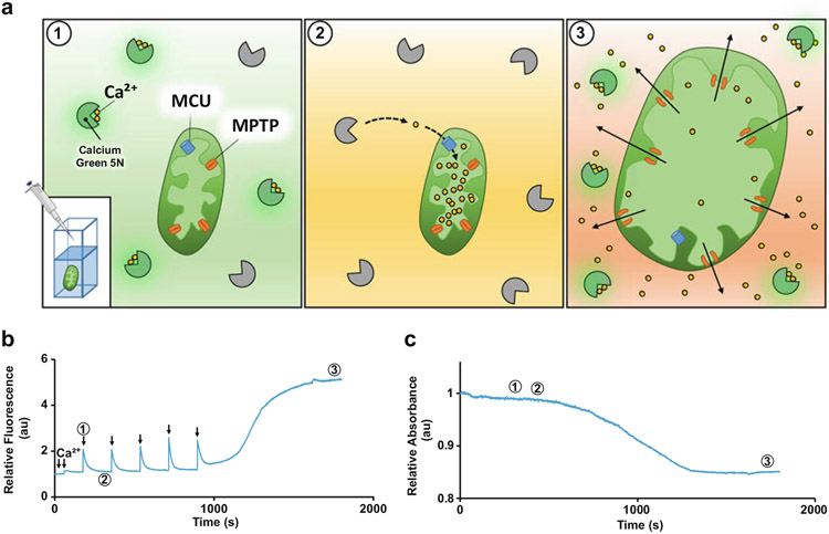
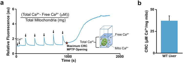
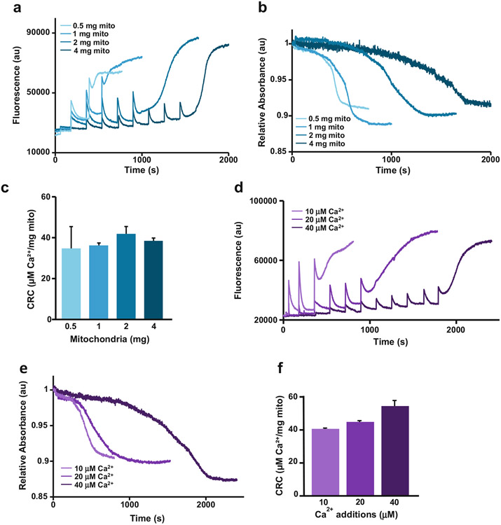
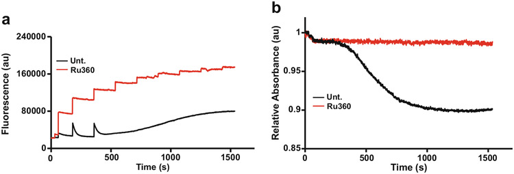
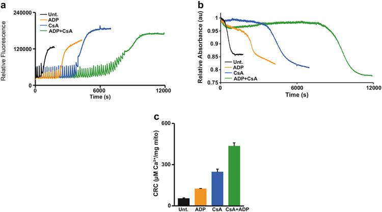
The loss of mitochondrial cristae integrity and mitochondrial swelling are hallmarks of multiple forms of necrotic cell death. One of the most well-studied and relevant inducers of mitochondrial swelling is matrix calcium (Ca2+). Respiring mitochondria will intake available Ca2+ into their matrix until a threshold is reached which triggers the opening of the mitochondrial permeability transition pore (MPTP). Upon opening of the pore, mitochondrial membrane potential dissipates and the mitochondria begin to swell, rendering them dysfunctional. The total amount of Ca2+ taken up by a mitochondrion prior to the engagement of the MPTP is referred to as mitochondrial Ca2+ retention capacity (CRC). The CRC/swelling assay is a useful tool for observing the dose-dependent event of mitochondrial dysfunction in real-time. In this technique, isolated mitochondria are treated with specific boluses of Ca2+ until they reach CRC and undergo swelling. A fluorometer is utilized to detect an increase in transmitted light passing through the sample as the mitochondria lose cristae density, and simultaneously measures calcium uptake by way of a Ca2+-specific membrane impermeable fluorescent dye. Here we provide a detailed protocol describing the mitochondrial CRC/swelling assay and we discuss how varying amounts of mitochondria and Ca2+ added to the system affect the dose-dependency of the assay. We also report how to validate the assay by using MPTP and calcium uptake inhibitors and troubleshooting common mistakes that occur with this approach.

Keywords: CRC; Calcium Green 5 N; Calcium retention capacity; Cell death; Fluorometry; Mitochondria; Mitochondrial dysfunction; Mitochondrial permeability transition pore (MPTP); Mitochondrial swelling.


Figures

Fig. 1
Fig. 1
Schematic representation of mitochondrial CRC/swelling assay with representative traces. (a) Schematic representing the events within the cuvette during the mitochondrial CRC/swelling assay. Numbers coordinate to events displayed on the traces. (b) and (c) 2 mg of mitochondria continuously dosed with 20 μM Ca2+ (arrows) over specified increments of time until CRC and swelling events occur. Excitement of calcium green 5 N (CG5N) is shown by sharp increase in fluorescence at the time of calcium treatment (event 1). Mitochondrial uptake is represented by a subsequent decrease in fluorescence, demonstrating that Ca2+ becomes unbound from the dye and enters the mitochondrial matrix (event 2). CRC is achieved once there is a release of Ca2+ from the mitochondria demonstrated by the steady increase and eventual plateau in fluorescence (event 3). Mitochondrial swelling is indicated by the sharp decrease in absorbance, which represents the opening of the MPTP
Fig. 2
Fig. 2
Quantification of the CRC Assay. Mitochondrial CRC is the maximum amount of Ca2+ taken up by the mitochondria prior to MPTP opening. (a) Representative trace of the CRC assay and equation for quantifying CRC. The red star indicates the point of CRC. (b) The CRC assay was repeated using three individual liver mitochondria samples, and the amount of μg Ca2+/mg mitochondria (mito) was averaged then graphed (n = 3) error bars (S.E.M)
Fig. 3
Fig. 3
Dose-dependency of mitochondria and Ca2+ concentrations for the CRC/Mitochondrial swelling assay. (a) and (b) Varying amounts of mitochondria (0.5, 1, 2, 4 mg) subjected to the CRC/swelling assay using the same additions of 20 μM Ca2+. Representative traces demonstrate that increasing the concentration of mitochondria in the cuvette correlates with an increase in total Ca2+ required to induce MPTP opening. (c) Quantification of figure a showing that the CRC remains constant although increasing amounts of mitochondria were used (n = 3). (d) and (e) 2 mg of mitochondria subjected to the CRC/swelling assay using increasing concentrations of Ca2+ additions (10, 20, 40 μM). Representative traces show that MPTP opening is achieved sooner with higher additions of Ca2+, demonstrating the dose-dependent nature of CRC assay. (f) Quantification of figure d showing that the CRC remains constant although increasing amounts of Ca2+ were used (n = 3) error bars (S.E.M)
Fig. 4
Fig. 4
MCU Inhibitor RU360 prevents uptake of Ca2+. (a) and (b) 2 mg of mitochondria were incubated with or without mitochondrial calcium uniporter (MCU) inhibitor 10 μM Ru360 and subjected to the CRC/swelling assay. Untreated (Unt.) mitochondria are able to take up Ca2+ until the MPTP engages, while Ru360 treated mitochondria are unable to intake Ca2+ and never undergo MPTP opening
Fig. 5
Fig. 5
Desensitization of the MPTP with CsA and ADP. (a) and (b) Representative CRC and swelling traces of 2 mg mitochondria stimulated with 40 μM Ca2+ treated with cyclosporin A (2 μM CsA) and/or adenosine phosphate (300 μM ADP). Mitochondria incubated with CsA and/or ADP have greater CRC than untreated (Unt.) mitochondria. (c) Quantification of figure a. (n = 3) error bars (S.E.M)

References

    1. Bernardi P, Lisa FD (2015) The mitochondrial permeability transition pore: molecular nature and role as a target in cardioprotection. J Mol Cell Cardiol 78:100–106 - PMC - PubMed
    1. Gunter TE, Buntinas L, Sparagna G et al. (2000) Mitochondrial calcium transport: mechanisms and functions. Cell Calcium 28(5–6):285–296 - PubMed
    1. Briston T, Roberts M, Lewis S et al. (2017) Mitochondrial permeability transition pore: sensitivity to opening and mechanistic dependence on substrate availability. Sci Rep 7:10492. - PMC - PubMed
    1. Karch J, Kwong JQ, Burr AR et al. (2013) Bax and Bak function as the outer membrane component of the mitochondrial transition pore. PNAS 111(29):10396–10397
    1. Kroemer G, Reed J (2000) Mitochondrial control of cell death. Nat Med 6:513–519 - PubMed

Publication types

MeSH terms

Substances

LinkOut - more resources