. 2022 Jun 30;17(6):e0269404.
doi: 10.1371/journal.pone.0269404. eCollection 2022.

An Rtn4/Nogo-A-interacting micropeptide modulates synaptic plasticity with age

Affiliations

An Rtn4/Nogo-A-interacting micropeptide modulates synaptic plasticity with age

S Kragness et al. PLoS One. .

Abstract

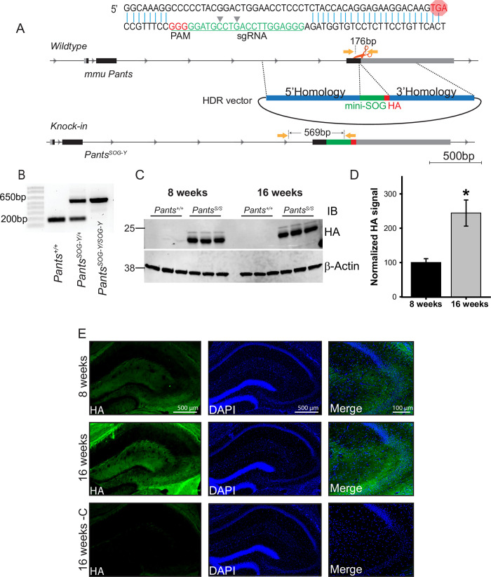
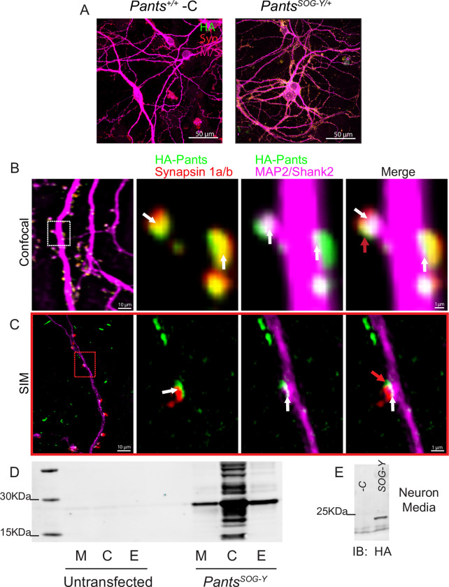
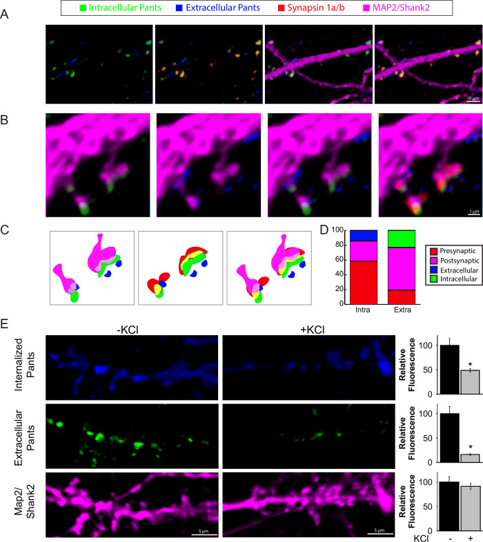
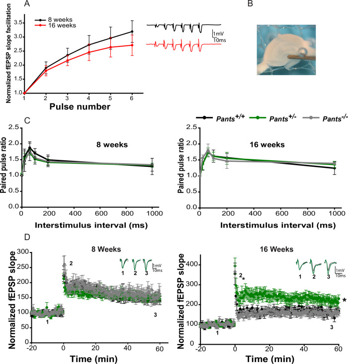
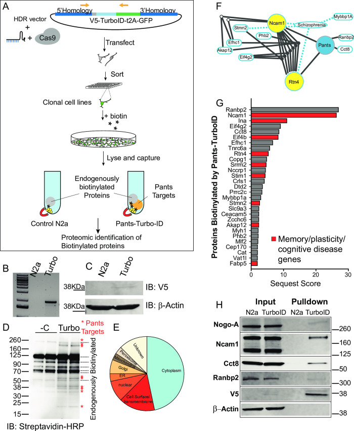
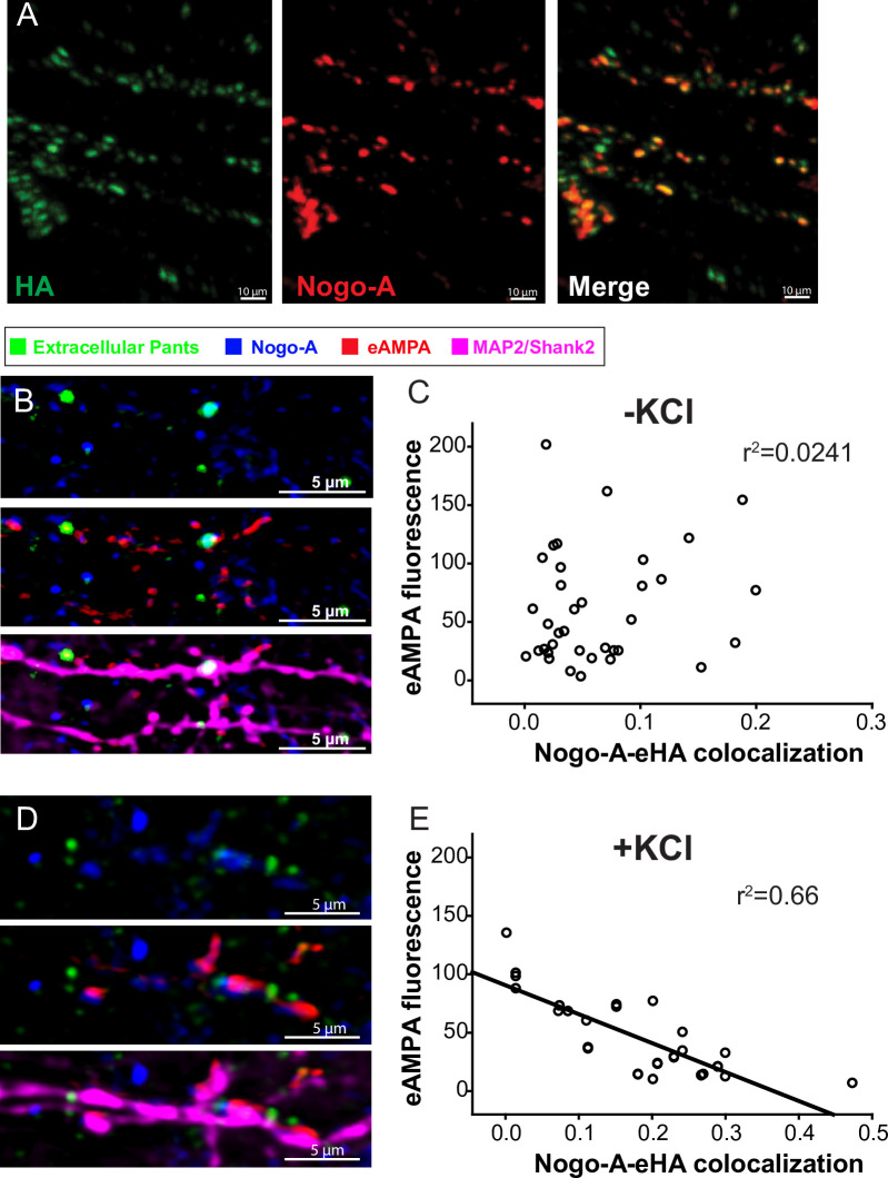
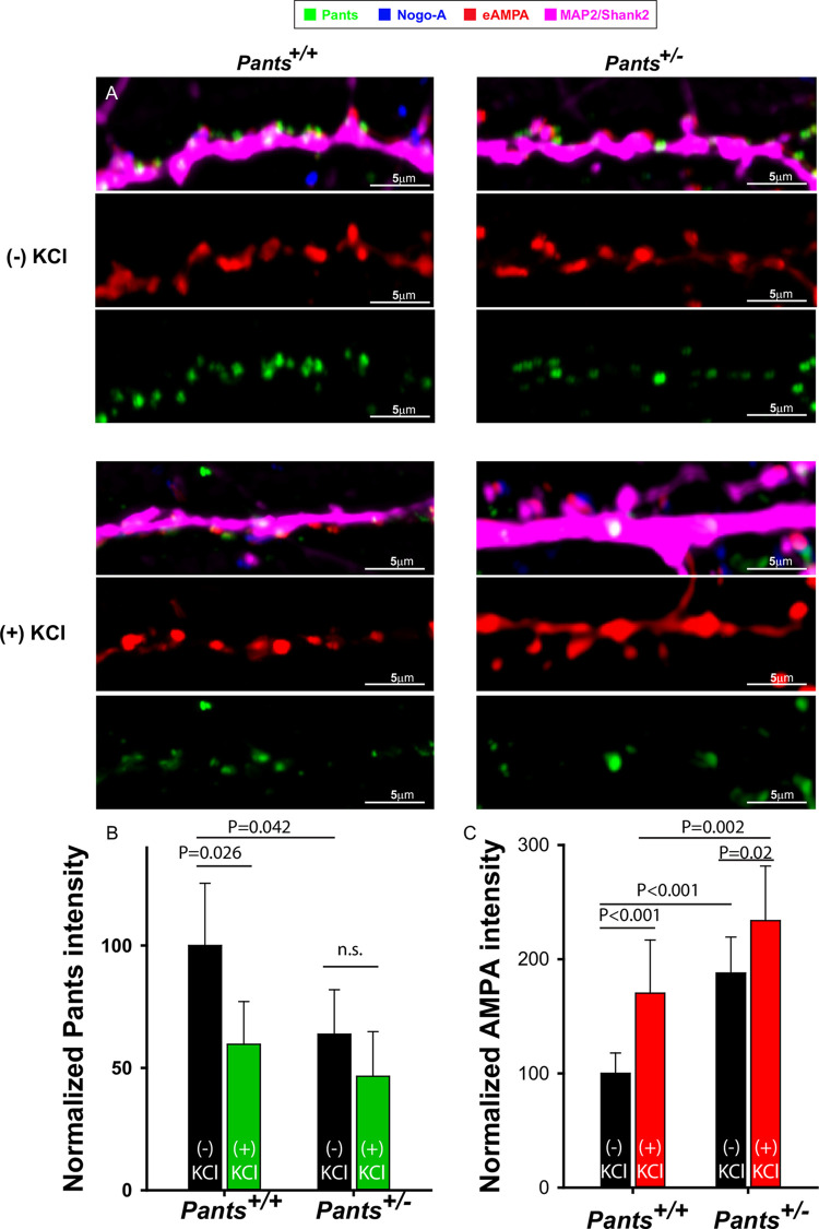
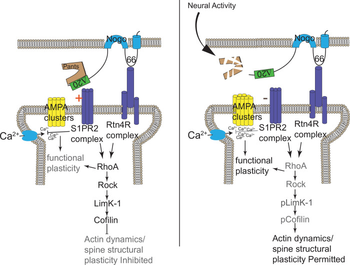
Micropeptides, encoded from small open reading frames of 300 nucleotides or less, are hidden throughout mammalian genomes, though few functional studies of micropeptides in the brain are published. Here, we describe a micropeptide known as the Plasticity-Associated Neural Transcript Short (Pants), located in the 22q11.2 region of the human genome, the microdeletion of which conveys a high risk for schizophrenia. Our data show that Pants is upregulated in early adulthood in the mossy fiber circuit of the hippocampus, where it exerts a powerful negative effect on long-term potentiation (LTP). Further, we find that Pants is secreted from neurons, where it associates with synapses but is rapidly degraded with stimulation. Pants dynamically interacts with Rtn4/Nogo-A, a well-studied regulator of adult plasticity. Pants interaction with Nogo-A augments its influence over postsynaptic AMPA receptor clustering, thus gating plasticity at adult synapses. This work shows that neural micropeptides can act as architectural modules that increase the functional diversity of the known proteome.


Conflict of interest statement

The authors have declared that no competing interests exist.

Figures

Fig 1
Fig 1. Generation and characterization of a transgenic mouse line to study endogenous Pants.
A) CRISPR/Cas9 strategy to target the miniSOG and HA tags to the C-terminus of the endogenous Pants locus. B) Genotyping verification of insert in mouse tail biopsies. C) Quantitative Western blot of Pants immunoprecipitated from hippocampus of PantsSOG-Y and WT control mice at 8 and 16 weeks immunoblotted with HA antibody. D) Quantification of HA bands from (c), normalized to β-actin controls (n = 3, P = 0.004). E) Immunohistochemistry with HA antibody shows Pants localization in the hippocampus at 8 and 16 weeks.
Fig 2
Fig 2. Synaptic localization and release of Pants.
A) Immunocytochemistry with HA antibody shows Pants localization throughout cultured hippocampal neurons from PantsSOG-Y/+ mice, but not wildtype controls. B) Immunocytochemistry of 18–21 div primary neurons from PantsSOG-Y hippocampus stained for HA, a Pre-synaptic marker (Synapsin 1a/b) and post-synaptic markers (MAP2 and Shank2) imaged with confocal microscopy. White arrows indicate areas of colocalization, and red arrows indicate suspected extra-synaptic pants that fails to co-localize with any markers. C) Structured Illumination Microscope imaging of the same staining from (A) to obtain better resolution. D) HA Western blot of HA IP from Nero2a cellular lysates (C), extracellular matrix (E) fractions, and proteins precipitated from cell media (M) after transfection with a construct for over-expressing the PantsSOG-Y fusion protein compared to untransfected controls. E) HA Western blot of HA IP of media collected from primary PantsSOG-Y hippocampal neurons or Pants+/+ negative control neurons.
Fig 3
Fig 3. Extrasynaptic Pants is downregulated in response to neuronal activity.
A) Contrasting extracellular/intracellular immunocytochemistry staining for HA in cultured hippocampal neurons. B-C) Co-localization of intra- and extra-cellular Pants with synaptic markers D) Quantification of Pants co-localization with pre- and post-synaptic markers correlation coefficients. E) Differentiated neuronal extracellular and internalized HA-Pants staining with and without 10 seconds 60mM KCl stimulation prior to fixation. Graphs at the right show quantification of HA fluorescence in dendritic ROIs, normalized to MAP2/Shank2 fluorescence. n = 3 P<0.001(extracellular) and P = 0.005 (internalized)P values generated by Mann-Whitney Rank Sum test.
Fig 4
Fig 4. Pants exerts an age-dependent, overdominant, negative effect on hippocampal Mossy Fiber LTP.
A) Frequency Facilitation at mossy fibers from acute hippocampal slices at 8 and 16 weeks of age (n = 5). B) electrode placement for mossy fiber LTP measurement. C) Paired-pulse facilitation at MF synapses at 8 and 16 weeks of age (n = 5). D) 100 Hz LTP recorded at MF synapses from 8 week old (left), and 16 week old (right) Pants+/+(black), Pants+/- (dark green), and Pants-/- (light green) acute hippocampal slices. 8 week (n = 6 mice, 18 slices for Pants+/+, n = 10 mice, 30 slices for Pants+/-, and n = 5 mice, 15 slices for Pants-/-. All P > 0.503). 16 week (n = 4 mice, 18 slices for Pants+/+, n = 5 mice, 18 slices for Pants+/-, n = 5 mice, 19 slices for Pants-/-. P<0.001 for Pants+/+ vs. Pants+/-. P = 0.157 for Pants+/+ vs. Pants-/-. P<0.001 for Pants+/- vs. Pants-/-, Mann-Whitney).
Fig 5
Fig 5. Endogenous proximity labeling proteomics reveals the Pants interactome.
A) experimental design: A homomology-driven repair construct targeting the V5 epitope tag fused to the TurboID proximity labeling biotinylation enzyme followed by the self-cleaving t2A peptide and GFP to the 3’ end of the endogenous Pants locus was co-transfected with a vector carrying the sgRNA and Cas9 into Neuro2A cells. Single cells expressing GFP were then sorted into the wells of 96-well plates, generating clonal knock-in cell lines. B) Knock-in was verified by genotyping PCR of cell lines using a forward primer in the Pants gene and a reverse in GFP. C) Expression of the Pants-V5-TurboID fusion protein in cell lines was verified by Western blotting of cell lysates with an antibody to the V5 epitope. D) Treatment of Pants-Turbo cells with 50 μM biotin for 18 hours resulted in specific biotinylation of proteins not seen in N2A controls, as verified by Streptavidin-HRP immunoblotting. E) Pie chart showing localization pattern of Pants targets identified by proximity-labeling proteomics. F) IPA analysis of functional linkage between unique proteomic hits, showing SZ connections and inter-gene connections. G) Proteins uniquely biotinylated in Pants-TurboID cell line, ordered by Sequest Score. Those implicated in plasticity, memory or cognitive disease are highlighted in red. H) Validation of proteomics results by biotinylation followed by streptavidin pulldown and Western blot with selected antibodies specific to Pants targets identified by Mass spectrometry.
Fig 6
Fig 6. Pants interacts with Rtn4/Nogo in vitro and in vivo.
A) Immunocytochemical co-localization of Pants and Nogo-A in cultured PantsSOG-Y hippocampal neurons B) AMPA receptor clustering at synapses where Pants colocalizes with Nogo-A versus Pants-free synapses in the absence of stimulation. C) Correlation between Pants-Nogo-A co-localization and GluR1 AMPA receptor fluorescence in the membrane of spine ROIs in the absence of stimulation. D) AMPA receptor clustering at synapses where Pants colocalizes with Nogo-A versus Pants-free synapses immediately after KCl stimulation. E) Correlation between Pants-Nogo-A co-localization and GluR1 AMPA receptor fluorescence in the membrane of spine ROIs immediately after KCl stimulation. n = 3 samples, 40 ROIs each.
Fig 7
Fig 7. AMPA receptor clustering at spines increases in Pants +/- neurons.
A) Representative dendrites from Pants+/+ (left) or Pants+/- (right) hippocampal neurons stained with Nogo-A, an antibody to the extracellular domain of the GluR1 AMPA receptor, and a Nogo-A antibody. Staining was performed in the absence (top) or presence (bottom) of KCl stimulation. B) Quantification of Pants fluorescence intensity in neuronal processes C) Quantification of AMPA clustering at Nogo-A-associated spines in Pants+/+ and Pants-/- neurons in the presence or absence of KCl stimulation. N = 3 samples/ 30 ROIs, Mann-Whitney.
Fig 8
Fig 8. Model for the mechanism of Pants control of plasticity.
Pants stabilizes the interaction of Nogo-A with receptors, inhibiting clustering of postsynaptic AMPA receptors at synaptic sites. Upon stimulation, Pants is degraded at synapses, reducing Nogo-A signaling and allowing AMPA receptor clustering at individual synapses.

References

    1. Schneider M, Debbane M, Bassett AS, Chow EW, Fung WL, van den Bree M, et al.. Psychiatric disorders from childhood to adulthood in 22q11.2 deletion syndrome: results from the International Consortium on Brain and Behavior in 22q11.2 Deletion Syndrome. Am J Psychiatry. 2014;171(6):627–39. doi: 10.1176/appi.ajp.2013.13070864 - DOI - PMC - PubMed
    1. Chun S, Du F, Westmoreland JJ, Han SB, Wang YD, Eddins D, et al.. Thalamic miR-338-3p mediates auditory thalamocortical disruption and its late onset in models of 22q11.2 microdeletion. Nat Med. 2017;23(1):39–48. doi: 10.1038/nm.4240 - DOI - PMC - PubMed
    1. Chun S, Westmoreland JJ, Bayazitov IT, Eddins D, Pani AK, Smeyne RJ, et al.. Specific disruption of thalamic inputs to the auditory cortex in schizophrenia models. Science. 2014;344(6188):1178–82. doi: 10.1126/science.1253895 - DOI - PMC - PubMed
    1. Devaraju P, Yu J, Eddins D, Mellado-Lagarde MM, Earls LR, Westmoreland JJ, et al.. Haploinsufficiency of the 22q11.2 microdeletion gene Mrpl40 disrupts short-term synaptic plasticity and working memory through dysregulation of mitochondrial calcium. Mol Psychiatry. 2016. doi: 10.1038/mp.2016.75 - DOI - PMC - PubMed
    1. Earls LR, Bayazitov IT, Fricke RG, Berry RB, Illingworth E, Mittleman G, et al.. Dysregulation of presynaptic calcium and synaptic plasticity in a mouse model of 22q11 deletion syndrome. J Neurosci. 2010;30(47):15843–55. doi: 10.1523/JNEUROSCI.1425-10.2010 - DOI - PMC - PubMed

Publication types