. 2023 Jan 1;72(1):71-84.
doi: 10.2337/db21-1086.

Characterization and Clinical Association of Autoantibodies Against Perilipin 1 in Patients With Acquired Generalized Lipodystrophy

Affiliations

Characterization and Clinical Association of Autoantibodies Against Perilipin 1 in Patients With Acquired Generalized Lipodystrophy

Fernando Corvillo et al. Diabetes. .

Abstract

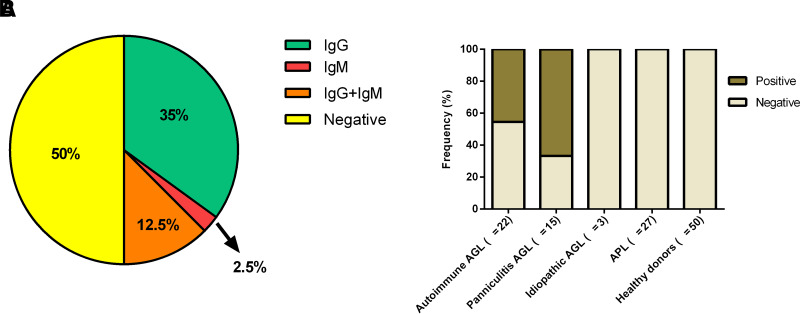
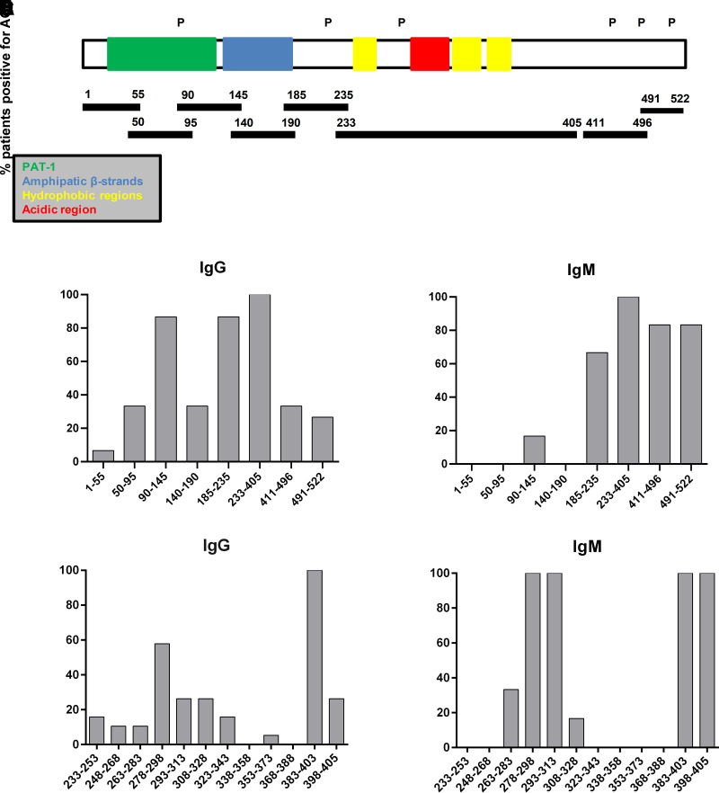
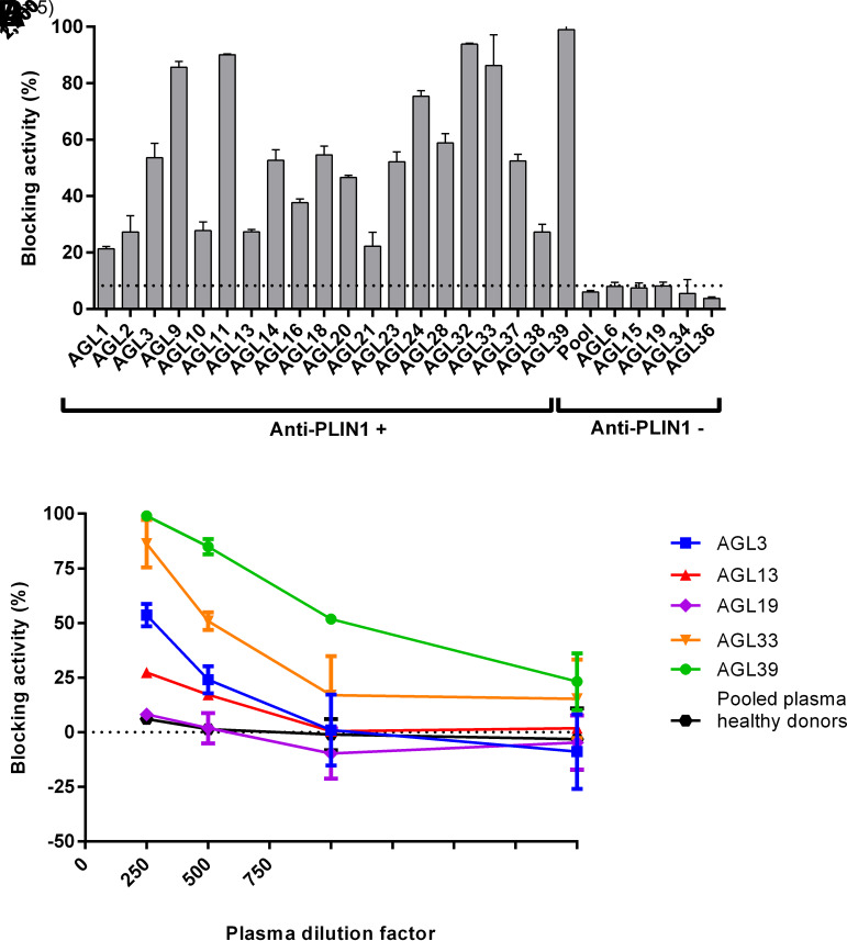
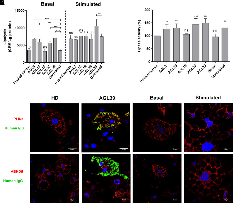
Acquired generalized lipodystrophy (AGL) is a rare condition characterized by massive loss of adipose tissue through the body, causing severe metabolic complications. Autoimmune destruction of adipocytes is strongly suspected based on the frequent association of AGL with autoimmune disorders. In 2018, autoantibodies against perilipin 1 (PLIN1) were identified in three patients with autoimmune-associated AGL. However, the pathogenic mechanism and clinical impact of anti-PLIN1 remain unsolved. The prevalence of anti-PLIN1 autoantibodies in an AGL cohort of 40 patients was 50% (20 of 40). Among positive patients, 10 had the autoimmune variety and 10 had panniculitis-associated AGL. The IgG isotype was predominant, although some IgM antibodies were detected. Epitope-mapping studies did not identify a single, major epitope. Instead, autoantibodies typically bound to several different peptides, among which the central (233-405) domain was detected in all antibody-positive patients, for both IgG and IgM autoantibodies. In-depth epitope mapping indicated that anti-PLIN1 autoantibodies predominantly recognize the αβ-hydrolase domain containing 5 (ABHD5) binding site (383-405). Autoantibodies dose-dependently blocked the binding of PLIN1 to ABHD5 and caused a dislocation of ABHD5 toward the cytosol, leading to an increase in lipolysis and lipase activities. Finally, anti-PLIN1 titers significantly correlated with the amount of fat loss, metabolic control impairment, and severity of liver injury. Our data strongly support that anti-PLIN1 autoantibodies are a diagnostic biomarker and a cause of lipodystrophy in patients with AGL.


Figures

Figure 1
Figure 1
Frequency of anti-PLIN1 autoantibodies. A: Distribution of patients AGL positive for isolated IgG or IgM autoantibodies, copositive for IgG and IgM, and negative for anti-PLIN1 antibodies. B: Frequency of anti-PLIN1 in patients with different AGL types (autoimmune, panniculitis, and idiopathic) or APL and healthy donors.
Figure 2
Figure 2
Epitope-mapping studies. A: Schematic representation of PLIN1 domain distribution and location of PLIN1 fragments used for epitope mapping. The ∼100-amino-acid hydrophobic PAT domain is located at the extreme N terminus, followed by amphipathic β-strands. The C terminus domain is composed of three hydrophobic sequences and one acidic region comprised of amino acids 291–318. The positions of the phosphorylation sites are indicated by a “P.” The reactivity of IgG (B) or IgM (C) autoantibodies from 20 patients with AGL was tested with recombinant PLIN1 fragments, covering almost all domains of PLIN1. IgG (D) and IgM (E) anti-PLIN1 reactivity against 12 overlapping peptides spanning amino acids 233–405. Results are expressed as the percentage of total positive patients for each PLIN1 peptide.
Figure 3
Figure 3
Blocking activity of anti-PLIN1 autoantibodies. A: Detection of blocking activity of anti-PLIN1 autoantibodies using serum samples at 1/250 dilution incubated with a fixed amount (100 ng) of recombinant PLIN1. The unspecific blocking activity (8.31%) was established using the mean value obtained for 50 healthy donors. B: Dose-dependent inhibition of the interaction of PLIN1 and ABHD5 in the presence of sera samples from anti-PLIN1–positive patients (AGL3, AGL13, AGL33, and AGL39). Serum from AGL19 and pooled serum from five healthy donors were negative for anti-PLIN1 autoantibodies. Serially diluted serum samples were mixed with a fixed amount (100 ng) of recombinant PLIN1. For both images, results were expressed as mean ± SD from two independent experiments.
Figure 4
Figure 4
Functional effects of anti-PLIN1 autoantibodies on lipolysis and lipase activity. Preadipocytes (3T3-L1) were incubated for 180 min with human serum at 1/10 dilution from four patients with AGL (AGL3, AGL13, AGL33, and AGL39) with anti-PLIN1 autoantibodies, one patient without antibodies (AGL19), and a pool of healthy donors. A: Results of the radiometric assessment of basal and stimulated lipolysis. Data represent mean ± SD for triplicates of two independent experiments for each sample. Statistical significance was assessed with one-way ANOVA assay comparing the mean of each column with the mean from untreated cells. B: Results of lipase activity (nanomoles of glycerol), represented as the percentage over maximum activity from cells treated with healthy donor serum. Data represent mean ± SD for triplicates of two independent experiments for each sample. C: Confocal microscopic analysis of mouse preadipocytes revealed colocalization of PLIN1 and IgG from patient AGL39 on the lipid droplet surface (merged image, in yellow). However, no colocalization was observed when using serum from healthy donors (HD). ABHD5 was localized mainly on lipid droplets under treatment with serum from HD or untreated (basal conditions). Under stimulated conditions or in the presence of serum from AGL39, ABHD5 staining was localized on cytosol. DNA was stained with DAPI (blue); IgG binding was detected using FITC-conjugated rabbit anti-human IgG (green); and PLIN1 or ABHD5 was detected with biotin-labeled rabbit IgG followed by Texas Red–labeled streptavidin (red). Scale bars, 20 μm. *P < 0.05; **P < 0.01; ***P < 0.001; ns, not significant.

Comment in

References

    1. Brown RJ, Araujo-Vilar D, Cheung PT, et al. . The diagnosis and management of lipodystrophy syndromes: a multi-society practice guideline. J Clin Endocrinol Metab 2016;101:4500–4511 - PMC - PubMed
    1. Haque WA, Shimomura I, Matsuzawa Y, Garg A. Serum adiponectin and leptin levels in patients with lipodystrophies. J Clin Endocrinol Metab 2002;87:2395. - PubMed
    1. Misra A, Garg A. Clinical features and metabolic derangements in acquired generalized lipodystrophy: case reports and review of the literature. Medicine (Baltimore) 2003;82:129–146 - PubMed
    1. Araújo-Vilar D, Santini F. Diagnosis and treatment of lipodystrophy: a step-by-step approach. J Endocrinol Invest 2019;42:61–73 - PMC - PubMed
    1. Corvillo F, Aparicio V, López-Lera A, et al. . Autoantibodies against perilipin 1 as a cause of acquired generalized lipodystrophy. Front Immunol 2018;9:2142. - PMC - PubMed

Publication types

MeSH terms