. 2022 Jul 7;185(14):2422-2433.e13.
doi: 10.1016/j.cell.2022.06.005. Epub 2022 Jun 9.

Antibody escape of SARS-CoV-2 Omicron BA.4 and BA.5 from vaccine and BA.1 serum

Collaborators, Affiliations

Antibody escape of SARS-CoV-2 Omicron BA.4 and BA.5 from vaccine and BA.1 serum

Aekkachai Tuekprakhon et al. Cell. .

Abstract

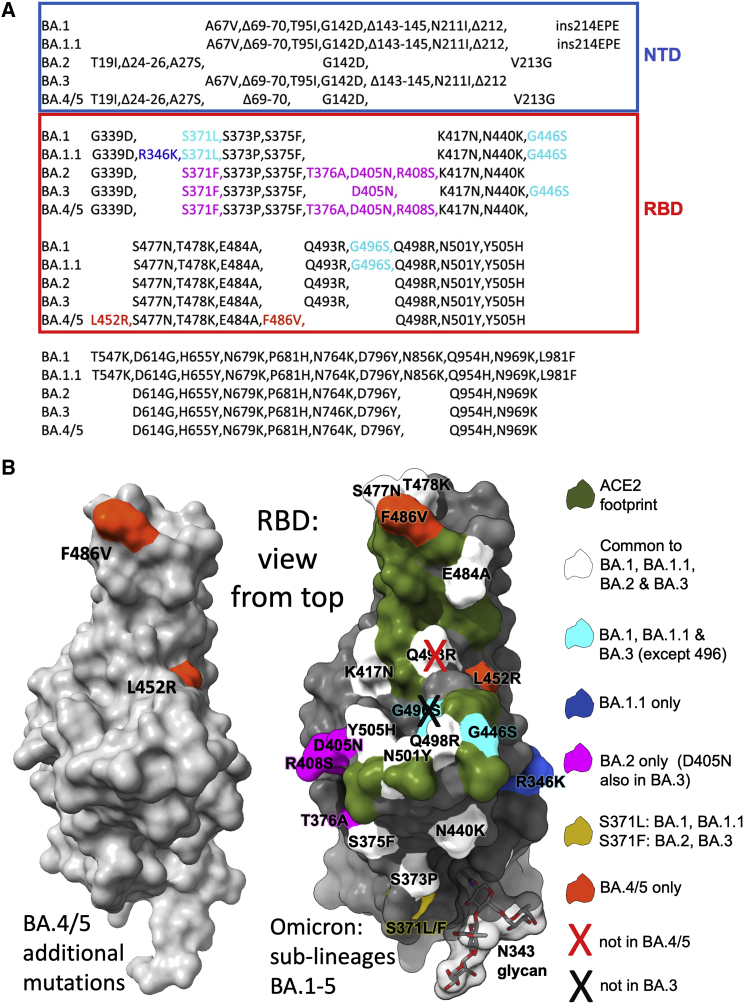
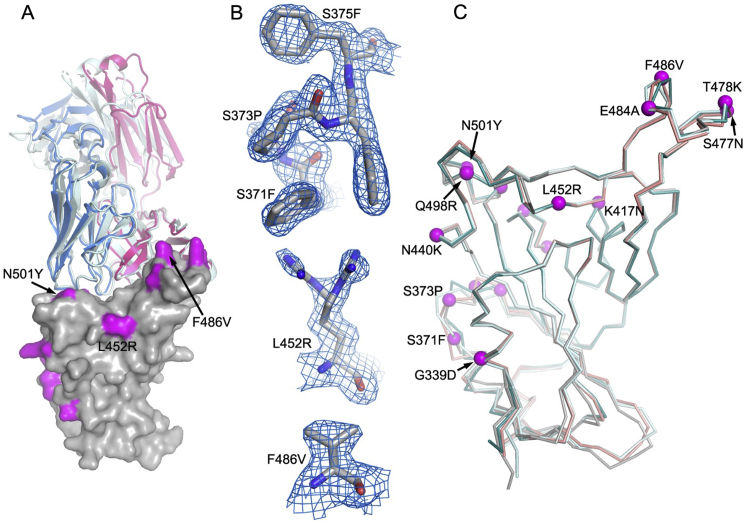
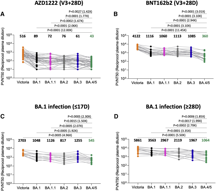
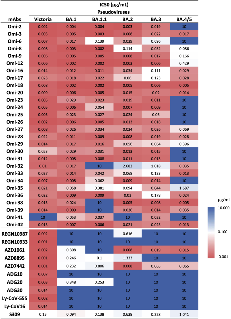
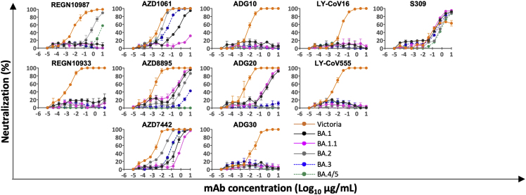
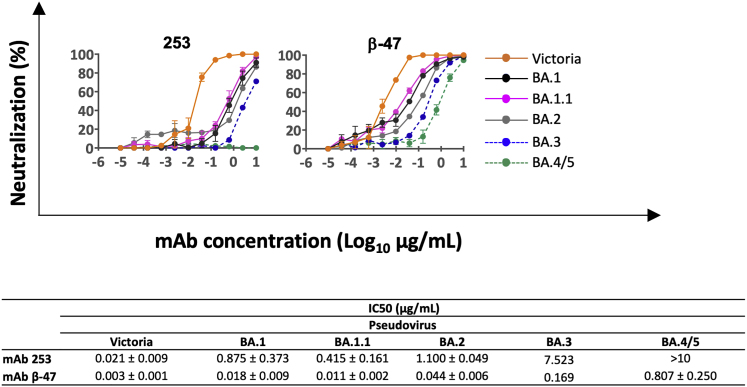
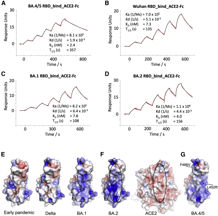
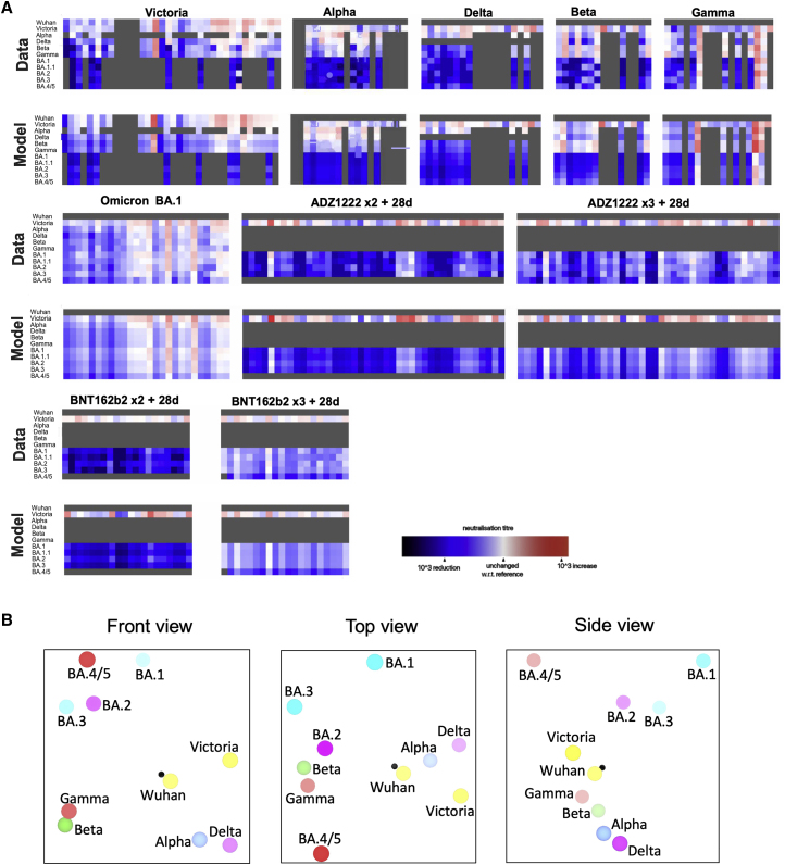
The Omicron lineage of SARS-CoV-2, which was first described in November 2021, spread rapidly to become globally dominant and has split into a number of sublineages. BA.1 dominated the initial wave but has been replaced by BA.2 in many countries. Recent sequencing from South Africa's Gauteng region uncovered two new sublineages, BA.4 and BA.5, which are taking over locally, driving a new wave. BA.4 and BA.5 contain identical spike sequences, and although closely related to BA.2, they contain further mutations in the receptor-binding domain of their spikes. Here, we study the neutralization of BA.4/5 using a range of vaccine and naturally immune serum and panels of monoclonal antibodies. BA.4/5 shows reduced neutralization by the serum from individuals vaccinated with triple doses of AstraZeneca or Pfizer vaccine compared with BA.1 and BA.2. Furthermore, using the serum from BA.1 vaccine breakthrough infections, there are, likewise, significant reductions in the neutralization of BA.4/5, raising the possibility of repeat Omicron infections.

Keywords: BA.4; BA.5; COVID-19; Omicron; SARS-CoV-2; VoC; antibody escape; variant.


Conflict of interest statement

Declaration of interests G.R.S. sits on the GSK Vaccines Scientific Advisory Board and is a founding member of RQ Biotechnology. Oxford University holds intellectual property related to the Oxford-AstraZeneca vaccine and SARS-CoV-2 mAb discovered in G.R.S.’s laboratory. A.J.P. is Chair of UK Dept. health and Social Care’s (DHSC) Joint Committee on Vaccination & Immunisation (JCVI) but does not participate in the JCVI COVID-19 committee and is a member of the WHO’s SAGE. The views expressed in this article do not necessarily represent the views of DHSC, JCVI, or WHO. The University of Oxford has entered into a partnership with AstraZeneca on coronavirus vaccine development. T.L. is named as an inventor on a patent application covering this SARS-CoV-2 vaccine and was a consultant to Vaccitech for an unrelated project while the study was conducted. S.J.D. is a scientific advisor to the Scottish Parliament on COVID-19.

Figures

None
Graphical abstract
Figure 1
Figure 1
The Omicron sublineage compared with BA.4/5 (A) Comparison of S protein mutations of Omicron BA.1, BA.1.1, BA.2, BA.3, and BA.4/5 with NTD and RBD boundaries indicated. (B) Position of RBD mutations (gray surface with the ACE2 footprint in dark green). Mutations common to all Omicron lineages are shown in white (Q493R, which is reverted in BA.4/5, is shown with a cross), those common to BA.1 and BA.1.1 in cyan, those unique to BA.1.1 in blue, and those unique to BA.2 in magenta. Residue 371 (yellow) is mutated in all Omicron viruses but differs between BA.1 and BA.2. The N343 glycan is shown as sticks with a transparent surface.
Figure S1
Figure S1
Overall structure of BA.4 RBD/Beta-27 complex, related to Table S1 and STAR Methods (A) Comparison of BA.4 RBD/Beta-27 (the bound nanobody C1 is omitted for clarity) with Beta RBD/Beta-27 (PDB: 7PS1) by overlapping the RBDs. The RBD is shown as a gray surface with mutation sites highlighted in magenta. The heavy chain and light chain are drawn as red and blue ribbons, respectively, for the BA.4 RBD/Beta-27 complex; Beta-27 in the Beta RBD complex is colored in pale cyan. The overall binding modes of the Fab in the two complexes are very similar, although there are some differences in the side-chain orientations at the interface, such as R403, N417, and Q493 of the RBD. The light-chain CDR3 becomes flexible in the BA.4 complex. (B) Electron density maps. Residues 371–375 that carry the S371L/F, S373P, and S375F mutations are flexible in the BA.1 and BA.2 RBD/Fab complexes (PDB: 7ZF3 and 7ZF8) but are well ordered in this high BA.4/5 resolution structure (top panel). L452R has double conformation (middle panel), and F486V has a well-defined density (bottom panel). (C) Comparison of the RBD of BA.4 (gray) with those of BA.1 (teal), BA.2 (cyan), and Beta (salmon). Mutation sites in BA.4 are shown as magenta spheres.
Figure 2
Figure 2
Pseudoviral neutralization assays of BA.4/5 by vaccine and BA.1 immune serum (A and B) IC50 values for the indicated viruses using serum obtained from vaccinees 28 days following their third dose of vaccine (A) AstraZeneca AZD1222 (n = 41) or (B) 4 weeks after the third dose of Pfizer BNT162b2 (n = 19). (C and D) Serum from volunteers suffering breakthrough BA.1 infection taken (C) early, i.e., ≤17 days from symptom onset (median 12 days) n = 12 and (D) late, i.e., ≥28 days from symptom onset (median 45 days) n = 14. Comparison is made with neutralization titers to Victoria an early pandemic strain, BA.1, BA.1.1, BA.2, and BA.3. Geometric mean titers are shown above each column. The Wilcoxon matched-pairs signed-rank test was used for the analysis, and two-tailed p values were calculated.
Figure 3
Figure 3
IC50 values for Omicron and commercial mAbs See also Figures S2, S3, S4, and S5.
Figure S2
Figure S2
Pseudoviral neutralization assays against Omicron monoclonal antibodies, related to Figure 3 where IC50 titers are shown Neutralization curves for a panel of 28 monoclonal antibodies made from samples taken from vaccinees infected with BA.1. Titration curves for BA.4/5 are compared with Victoria, BA.1, BA.1.1, BA.2, and BA.3, and mAbs we propose to be affected by the L452R and F486V mutations are indicated as are those belonging to the IGVH3-53/66 gene families.
Figure S3
Figure S3
Surface plasmon resonance (SPR) analysis of the interaction between BA.2 or BA.4/5 RBD and selected mAbs, related to Figure 3 (A–F) Sensorgrams (red: original binding curve; black: fitted curve) showing the interactions between BA.2 or BA.4/5 RBD and selected mAbs, with kinetics data shown. (G–K) Binding of BA.4/5 RBD is severely reduced compared with that of BA.2, so the binding could not be accurately determined, as shown by a single injection of 200 nM RBD over sample flow cells containing the mAb indicated.
Figure 4
Figure 4
Surface plasmon resonance (SPR) analysis of the interaction between BA.2 or BA.4/5 RBD and selected mAbs (A) Binding of BA.4/5 RBD is severely reduced compared with that of BA.2, so the binding could not be accurately determined, as shown by a single injection of 200 nM RBD over sample flow cells containing IgG Omi-31. (B, C, and E–I) Sensorgrams (red: original binding curve; black: fitted curve) showing the interactions between BA.2 or BA.4/5 RBD and selected mAbs, with kinetics data shown. (D) Determination of the affinity of BA.4/5 RBD to Omi-12 using a 1:1 binding equilibrium analysis. See also Figures 3 and S3.
Figure 5
Figure 5
Interactions between mAb and BA.4/5 mutation sites Overall structure (left panel) and interactions (≤4 Å) with BA.4/5 mutation sites (right panel) for (A) BA.1-RBD/Omi-31 (PDB: 7ZFB), (B) BA.1-RBD/Omi-32 (PDB: 7ZFE), (C) BA.1-RBD/Omi-25 (PDB: 7ZFD), (D) Wuhan-RBD/AZD8895 (PDB: 7L7D), (E) BA.1-RBD/Omi-3 (PDB: 7ZF3), and (F) BA.1-RBD/Omi-42 (PDB: 7ZR7) complexes. In the left panels, RBD is shown as surface representation, with BA.4/5 mutation sites highlighted in magenta and the additional two mutation sites of BA.4/5 at 452 and 486 in cyan and Fab LC as blue and HC as red ribbons. In the right panel, side chains of RBD, Fab HC, and LC are drawn as gray, red, and blue sticks, respectively. In (B), the L452R mutation (cyan sticks) is modeled to show that a salt bridge to D99 of CDR-H3 may be formed (yellow broken sticks). (F) shows that the Fab of Omi-42 does not contact either of the two BA.4/5 mutation sites. See also Figure S1.
Figure S4
Figure S4
Pseudoviral neutralization assays against commercial monoclonal antibodies, related to Figure 3 where IC50 titers are shown Pseudoviral neutralization assays with mAbs developed for human use.
Figure S5
Figure S5
Neutralization curves for IGVH1-58 mAb, related to Figure 3 Pseudoviral neutralization curves for early pandemic mAb 253 (Dejnirattisai et al., 2021a) and Beta-47 (Liu et al., 2021b) against Victoria and the panel of Omicron lineage constructs.
Figure 6
Figure 6
ACE2 RBD affinity (A–D) SPR sensorgrams showing ACE2 binding of BA.4/5 RBD (A) in comparison with binding to ancestral (Wuhan) (B), BA.1 (C), and BA.2 RBD (D). The data for Wuhan, BA.1, and BA.2 have been reported previously in Nutalai et al. (2022). (E–G) Electrostatic surfaces, (E) from left to right, early pandemic, Delta, and BA.1 RBD. (F) Open book view of BA.2 RBD and ACE2 of the BA.2 RBD/ACE2 complex (PDB: 7ZF7) and (G) BA.4/5 RBD (PDB: 7ZXU). The lozenges on ACE2 and RBD show the interaction areas.
Figure 7
Figure 7
Antigenic mapping (A) Neutralization data and model (log titer values) used to calculate antigenic maps in (B). Columns represent sera collected from inoculated volunteers or infected patients. Rows are challenge strains: Victoria, Alpha, Delta, Beta, Gamma, BA.1, BA1.1, BA.2, BA.3, and BA.4/5 in order. Values are colored according to their deviation from the reference value. The reference value is calculated on a serum-type basis as the average of neutralization titers from the row that gives this the highest value. (B) Orthogonal views of the antigenic map showing BA.4/5 in the context of the positions of previous VoC and BA.1, BA.1.1, BA.1, and BA.2, calculated from pseudovirus neutralization data. Distance between two positions is proportional to the reduction in neutralization titer when one of the corresponding strains is challenged with a serum derived by infection by the other. No scale is provided since the figures are projections of a three-dimensional distribution; however, the variation can be calibrated by comparison with (i) BA.1 to BA.2, which is 2.93× reduced, and (ii) BA.2 to BA.4/5, which is 3.03× reduced. The third dimension may be inferred by fading of the colors with greater distance from the viewer.

References

    1. Aricescu A.R., Lu W., Jones E.Y. A time- and cost-efficient system for high-level protein production in mammalian cells. Acta Crystallogr. D Biol. Crystallogr. 2006 Oct;62:1243–1250. doi: 10.1107/S0907444906029799. - DOI - PubMed
    1. Barnes C.O., Jette C.A., Abernathy M.E., Dam K.A., Esswein S.R., Gristick H.B., Malyutin A.G., Sharaf N.G., Huey-Tubman K.E., Lee Y.E., et al. SARS-CoV-2 neutralizing antibody structures inform therapeutic strategies. Nature. 2020;588:682–687. - PMC - PubMed
    1. Cele S., Jackson L., Khoury D.S., Khan K., Moyo-Gwete T., Tegally H., San J.E., Cromer D., Scheepers C., Amoako D.G., et al. Omicron extensively but incompletely escapes Pfizer BNT162b2 neutralization. Nature. 2022;602:654–656. - PMC - PubMed
    1. Cele S., Karim F., Lustig G., San J.E., Hermanus T., Tegally H., Snyman J., Moyo-Gwete T., Wilkinson E., Bernstein M., et al. SARS-CoV-2 prolonged infection during advanced HIV disease evolves extensive immune escape. Cell Host Microbe. 2022;30:154–162. e5. - PMC - PubMed
    1. Cerutti G., Guo Y., Zhou T., Gorman J., Lee M., Rapp M., Reddem E.R., Yu J., Bahna F., Bimela J., et al. Potent SARS-CoV-2 neutralizing antibodies directed against spike N-terminal domain target a single Supersite. Cell Host Microbe. 2021;29:819–833. e7. - PMC - PubMed

Publication types