. 2022 Aug;298(8):102206.
doi: 10.1016/j.jbc.2022.102206. Epub 2022 Jun 27.

Incorporation of fucose into glycans independent of the GDP-fucose transporter SLC35C1 preferentially utilizes salvaged over de novo GDP-fucose

Affiliations

Incorporation of fucose into glycans independent of the GDP-fucose transporter SLC35C1 preferentially utilizes salvaged over de novo GDP-fucose

Edyta Skurska et al. J Biol Chem. 2022 Aug.

Abstract

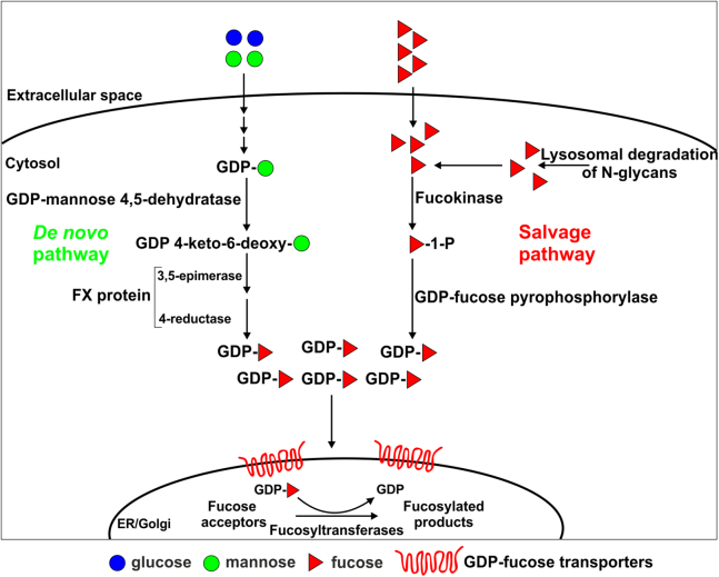
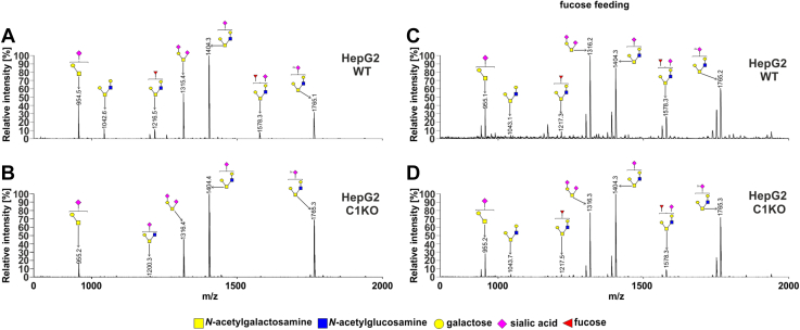
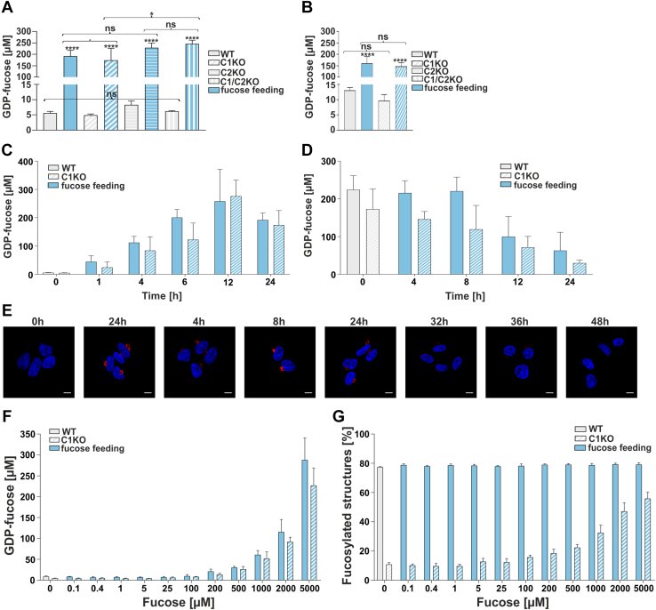
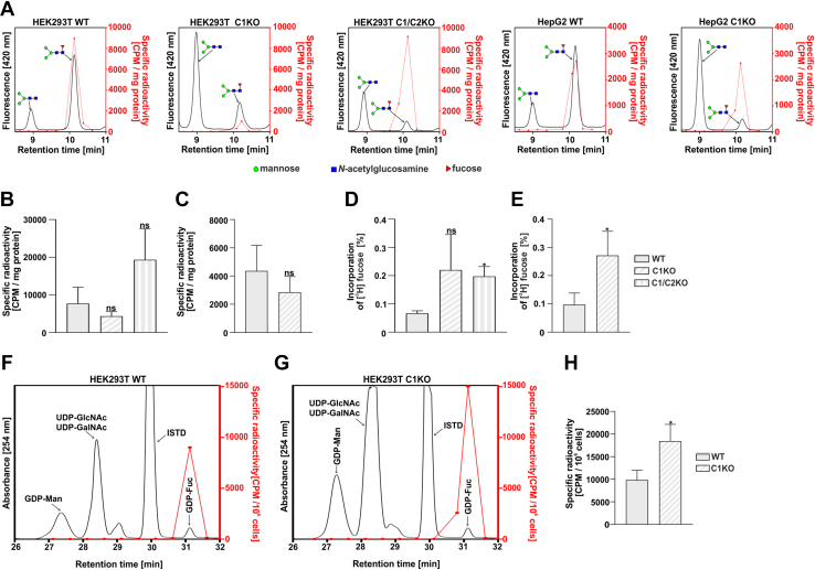
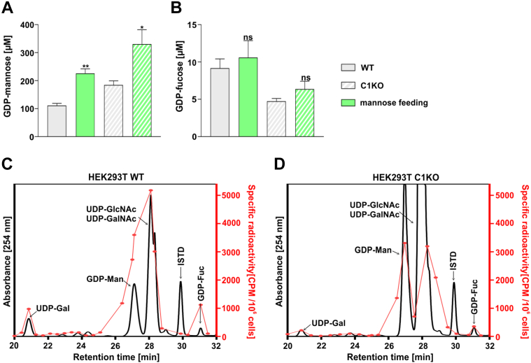
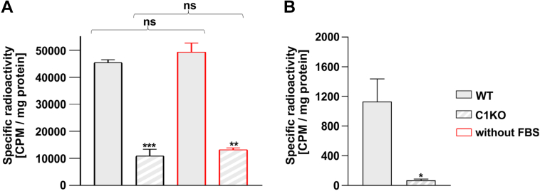
Mutations in the SLC35C1 gene encoding the Golgi GDP-fucose transporter are known to cause leukocyte adhesion deficiency II. However, improvement of fucosylation in leukocyte adhesion deficiency II patients treated with exogenous fucose suggests the existence of an SLC35C1-independent route of GDP-fucose transport, which remains a mystery. To investigate this phenomenon, we developed and characterized a human cell-based model deficient in SLC35C1 activity. The resulting cells were cultured in the presence/absence of exogenous fucose and mannose, followed by examination of fucosylation potential and nucleotide sugar levels. We found that cells displayed low but detectable levels of fucosylation in the absence of SLC35C1. Strikingly, we show that defects in fucosylation were almost completely reversed upon treatment with millimolar concentrations of fucose. Furthermore, we show that even if fucose was supplemented at nanomolar concentrations, it was still incorporated into glycans by these knockout cells. We also found that the SLC35C1-independent transport preferentially utilized GDP-fucose from the salvage pathway over the de novo biogenesis pathway as a source of this substrate. Taken together, our results imply that the Golgi systems of GDP-fucose transport discriminate between substrate pools obtained from different metabolic pathways, which suggests a functional connection between nucleotide sugar transporters and nucleotide sugar synthases.

Keywords: GDP-fucose synthesis; Golgi; LADII; N-linked glycosylation; SLC35C1; SLC35C2; cell metabolism; fucose supplementation; glycoprotein biosynthesis; membrane protein.


Conflict of interest statement

Conflict of interest The authors declare that they have no conflicts of interest with the contents of this article.

Figures

Figure 1
Figure 1
Simplified representation of GDP-fucose metabolism in mammalian cells. GDP-fucose is synthesized in two independent pathways, that is, de novo (from GDP-mannose) and salvage (from fucose).
Figure 2
Figure 2
Optimization of the conditions of fucose supplementation. AAL dot blot analysis of the wildtype and SLC35C1 knockout HEK293T (A) and HepG2 (B) cells after 5 days of supplementation with (+) or without (−) different fucose concentrations. C, AAL staining (red) of HEK293T SLC35C1 knockout supplemented with 5 mM fucose during 48 h. 0 h—control cells (no fucose supplementation, image reused in 6E panel). Cell nuclei were counterstained with DAPI. The scale bar represents 10 μm. DF, schematic representation of our HPLC-based method for quantification of-α1,6-fucosylation of N-glycans. G, quantitative analysis of α-1,6-fucosylation of N-glycans in HEK293T wildtype and SLC35C1 knockout cells supplemented with different fucose concentrations for indicated periods. AAL, Aleuria aurantia lectin; DAPI, 4',6-diamidino-2-phenylindole; HEK293T, human embryonic kidney 293T cell line.
Figure 3
Figure 3
Quantification of the percentage of the core-fucosylated N-glycan structures. HPLC quantification of N-glycans derived from endogenous HEK293T (A) and HepG2 (B) glycoproteins. N-glycans decorating SEAP glycoprotein overexpressed by HEK293T (C) and HepG2 (D) cell lines. ∗p < 0.05; ∗∗p < 0.01; ∗∗∗p < 0.001; and ∗∗∗∗p < 0.0001 as determined using one-way ANOVA with the Tukey post hoc test. Data are presented as mean ± SD. Each sample was run in three technical replicates. HEK293T, human embryonic kidney 293T cell line; ns, not significant, SEAP, secreted alkaline phosphatase.
Figure 4
Figure 4
O-glycosylation fingerprinting of HEK293T cells. MALDI-TOF mass spectra of permethylated mucin-type Bn-O-glycans secreted to the culture medium were permethylated and analyzed in a positive-ion mode. Structural assignments based on biosynthetic knowledge were prepared using the GlycoWorkBench tool (2.1; EuroCarbDB). HEK293T, human embryonic kidney 293T cell line.
Figure 5
Figure 5
O-glycosylation fingerprinting of HepG2 cells. MALDI-TOF mass spectra of permethylated mucin-type Bn-O-glycans secreted to the culture medium were permethylated and analyzed in a positive-ion mode. Structural assignments based on biosynthetic knowledge were prepared using the GlycoWorkBench tool (2.1; EuroCarbDB).
Figure 6
Figure 6
Quantification of intracellular GDP-fucose concentration. Intracellular GDP-fucose concentration in HEK293T (A) and HepG2 (B) cell lines. ∗p < 0.05; ∗∗p < 0.01; ∗∗∗p < 0.001; and ∗∗∗∗p < 0.0001 as determined using one-way ANOVA with the Tukey post hoc test. Data are represented as mean ± SD. Each sample was run in three biological replicates. C, time-course analysis (0–24 h) of GDP-fucose synthesis in wildtype and SLC35C1 knockout HEK293T cells fed with 5 mM fucose. Each sample was run in three biological replicates. D, analysis of GDP-fucose degradation in wildtype and SLC35C1 knockout HEK293T cells. The cells were first cultured for 24 h in the presence of 5 mM fucose and then for another 24 h in a fucose-free medium. Each sample was run in three biological replicates. E, disappearance of the fucosylation phenotype visualized by AAL staining (red). 0 h—control cells (no fucose supplementation, image reused in 2C panel). Cell nuclei were counterstained with DAPI. The scale bar represents 10 μm. F, dependence of the intracellular GDP-fucose concentration on the concentration of the fucose supplemented to the culture medium (HEK293T). G, dependence of the percentage of the core fucosylated N-glycans on the concentration of the fucose supplemented to the culture medium (HEK293T). Each sample was run in three biological replicates. AAL, Aleuria aurantia lectin; DAPI, 4',6-diamidino-2-phenylindole; HEK293T, human embryonic kidney 293T cell line; ns, not significant.
Figure 7
Figure 7
Metabolic labeling of cellular N-glycans with radioactive fucose.A, exemplary representative HPLC chromatograms of digested N-glycans isolated from indicated cell lines. The fluorescence signal from the 2-AB-labeled N-glycans (black solid lines) was overlaid with the radioactivity data (red dots and solid lines). Total radioactivity of the fucosylated N-glycans normalized for the amount of the starting material (total protein) from HEK293T (B) and HepG2 (C) cell lines. Percentage of the radioactive fucose incorporated into the fucosylated N-glycans isolated from HEK293T (D) and HepG2 (E) cell lines. To calculate the values, the sum of radioactivity measured in the HPLC fractions corresponding to the fucosylated species was divided by the total radioactivity of the fucose added to the culture medium. Comparison of the relative incorporation of the radioactive fucose derived from the salvage pathway in the wildtype (F) and SLC35C1 knockout (G) HEK293T cells. H, total radioactivity of the GDP-fucose normalized for total number of HEK293T cells. ∗p < 0.05 as determined using two-tailed unpaired t test with Welch’s correction. Data are presented as mean ± SD. Each sample was run in three biological replicates. 2-AB, 2-aminobenzamide; ISTD, internal standard (GDP-glucose); ns, not significant.
Figure 8
Figure 8
Quantification of intracellular GDP-mannose and GDP-fucose concentration in cells fed with mannose. Intracellular concentrations of GDP-mannose (A) and GDP-fucose (B) in the wildtype and SLC35C1 knockout HEK293T cells cultured for 48 h in the absence and presence of 5 mM mannose. ∗p < 0.05; ∗∗p < 0.01 as determined using two-tailed unpaired t test with Welch’s correction. Data are presented as mean ± SD. Each sample was run in three biological replicates. Comparison of relative incorporation of the radioactive fucose derived from the de novo pathway in wildtype (C) and SLC35C1 knockout (D) HEK293T cells. HEK293T, human embryonic kidney 293T cell line; ISTD, internal standard (GDP-glucose); ns, not significant.
Figure 9
Figure 9
Relative contribution of the de novo and salvage pathways in glycan fucosylation.A, incorporation of the radioactive fucose derived from the GDP-fucose produced in the salvage pathway into N-glycans measured for the wildtype (full bars) and SLC35C1 knockout (dashed bars) HEK293T cells cultured in the presence (black contours) and absence (red contours) of fetal bovine serum (FBS). B, incorporation of the radioactive fucose derived from the GDP-fucose produced in the de novo pathway into N-glycans measured for the wildtype (full bar) and SLC35C1 knockout (dashed bar) HEK293T cells. Radioactivity was normalized for the amount of the starting material (total protein). ∗p < 0.05; ∗∗∗p < 0.001 as determined using two-tailed unpaired t test with Welch’s correction. Data are presented as mean ± SD. Each sample was run in three biological replicates. HEK293T, human embryonic kidney 293T cell line; ns, not significant.

References

    1. Varki A., Cummings R.D., Esko J.D., Stanley P., Hart G.W., Aebi M., et al., editors. Essentials of Glycobiology, Cold Spring Harbor Laboratory Press. Cold Spring Harbor; New York: 2015-2017. - PubMed
    1. Yurchenco P.D., Atkinson P.H. Equilibration of fucosyl glycoprotein pools in HeLa cells. Biochemistry. 1977;16:944–953. - PubMed
    1. Yurchenco P.D., Atkinson P.H. Fucosyl-glycoprotein and precursor polls in HeLa cells. Biochemistry. 1975;14:3107–3114. - PubMed
    1. Moriwaki K., E. Miyoshi E. Fucosylation and gastrointestinal cancer. World J. Hepatol. 2010;2:151–161. - PMC - PubMed
    1. Wiese T.J., Dunlap J.A., Yorek M.A. L-fucose is accumulated via a specific transport system in eukaryotic cells. J. Biol. Chem. 1994;269:22705–22711. - PubMed

Publication types

MeSH terms

Supplementary concepts

LinkOut - more resources