

Crystal structure of Trypanosoma cruzi heme peroxidase and characterization of its substrate specificity and compound I intermediate
- PMID: 35772495
- PMCID: PMC9358470
- DOI: 10.1016/j.jbc.2022.102204
Crystal structure of Trypanosoma cruzi heme peroxidase and characterization of its substrate specificity and compound I intermediate
Abstract
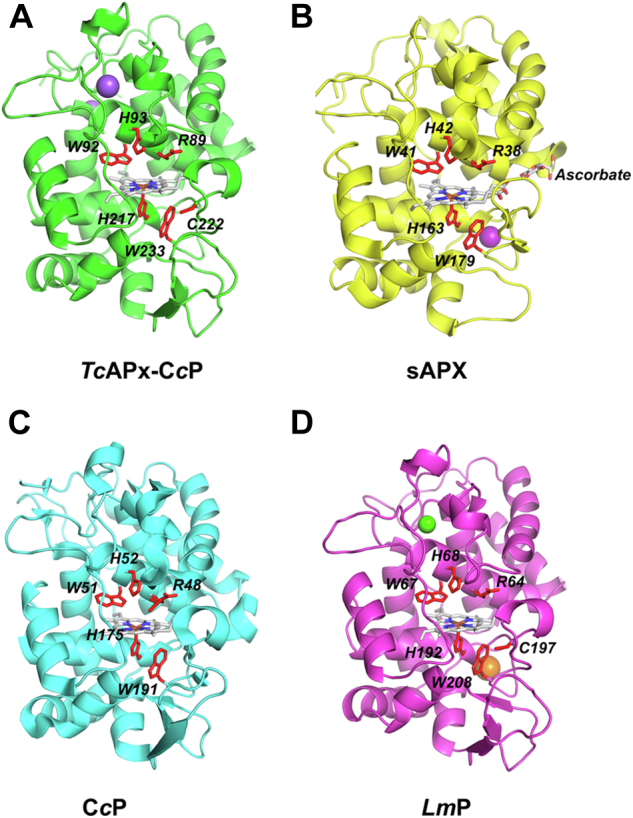
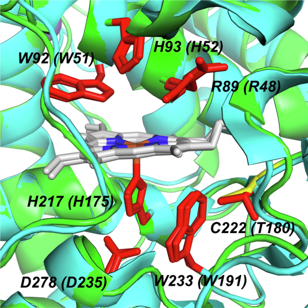
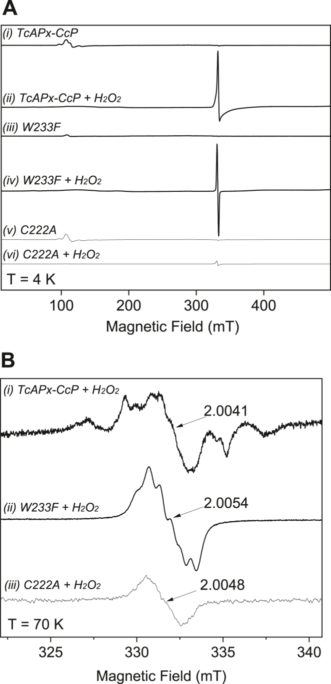
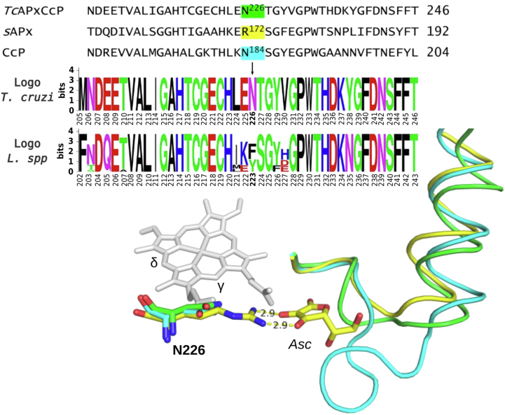
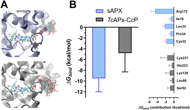
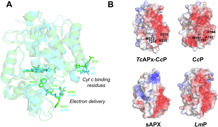
The protozoan parasite Trypanosoma cruzi is the causative agent of American trypanosomiasis, otherwise known as Chagas disease. To survive in the host, the T. cruzi parasite needs antioxidant defense systems. One of these is a hybrid heme peroxidase, the T. cruzi ascorbate peroxidase-cytochrome c peroxidase enzyme (TcAPx-CcP). TcAPx-CcP has high sequence identity to members of the class I peroxidase family, notably ascorbate peroxidase (APX) and cytochrome c peroxidase (CcP), as well as a mitochondrial peroxidase from Leishmania major (LmP). The aim of this work was to solve the structure and examine the reactivity of the TcAPx-CcP enzyme. Low temperature electron paramagnetic resonance spectra support the formation of an exchange-coupled [Fe(IV)=O Trp233•+] compound I radical species, analogous to that used in CcP and LmP. We demonstrate that TcAPx-CcP is similar in overall structure to APX and CcP, but there are differences in the substrate-binding regions. Furthermore, the electron transfer pathway from cytochrome c to the heme in CcP and LmP is preserved in the TcAPx-CcP structure. Integration of steady state kinetic experiments, molecular dynamic simulations, and bioinformatic analyses indicates that TcAPx-CcP preferentially oxidizes cytochrome c but is still competent for oxidization of ascorbate. The results reveal that TcAPx-CcP is a credible cytochrome c peroxidase, which can also bind and use ascorbate in host cells, where concentrations are in the millimolar range. Thus, kinetically and functionally TcAPx-CcP can be considered a hybrid peroxidase.
Keywords: Chagas disease; ascorbate; cytochrome c; heme; oxidants; peroxidase.
Copyright © 2022 The Authors. Published by Elsevier Inc. All rights reserved.
Conflict of interest statement
Conflict of interest The authors declare that they have no conflicts of interest with the contents of this article.
Figures
Similar articles
-
Kinetics, subcellular localization, and contribution to parasite virulence of a Trypanosoma cruzi hybrid type A heme peroxidase (TcAPx-CcP).Proc Natl Acad Sci U S A. 2017 Feb 21;114(8):E1326-E1335. doi: 10.1073/pnas.1618611114. Epub 2017 Feb 8. Proc Natl Acad Sci U S A. 2017. PMID: 28179568 Free PMC article.
-
Engineering ascorbate peroxidase activity into cytochrome c peroxidase.Biochemistry. 2008 Sep 30;47(39):10324-32. doi: 10.1021/bi8007565. Epub 2008 Sep 5. Biochemistry. 2008. PMID: 18771292 Free PMC article.
-
Role of tryptophan-208 residue in cytochrome c oxidation by ascorbate peroxidase from Leishmania major-kinetic studies on Trp208Phe mutant and wild type enzyme.Biochim Biophys Acta. 2008 May;1784(5):863-71. doi: 10.1016/j.bbapap.2008.02.006. Epub 2008 Feb 23. Biochim Biophys Acta. 2008. PMID: 18342641
-
Defining substrate specificity and catalytic mechanism in ascorbate peroxidase.Biochem Soc Symp. 2004;(71):27-38. doi: 10.1042/bss0710027. Biochem Soc Symp. 2004. PMID: 15777010 Review.
-
Peroxidase-catalyzed oxidation of ascorbate. Structural, spectroscopic and mechanistic correlations in ascorbate peroxidase.Subcell Biochem. 2000;35:317-49. doi: 10.1007/0-306-46828-x_10. Subcell Biochem. 2000. PMID: 11192727 Review.
References
-
- Luquetti A.O., Miles M.A., Rassi A., de Rezende J.M., de Souza A.A., Povoa M.M., et al. Trypanosoma cruzi: zymodemes associated with acute and chronic Chagas' disease in central Brazil. Trans. R. Soc. Trop. Med. Hyg. 1986;80:462–470. - PubMed
Publication types
MeSH terms
Substances
LinkOut - more resources
Full Text Sources
Miscellaneous
