. 2022 Oct:92:120-132.
doi: 10.1016/j.mri.2022.06.012. Epub 2022 Jun 27.

Accelerating 3D MTC-BOOST in patients with congenital heart disease using a joint multi-scale variational neural network reconstruction

Affiliations

Accelerating 3D MTC-BOOST in patients with congenital heart disease using a joint multi-scale variational neural network reconstruction

Anastasia Fotaki et al. Magn Reson Imaging. 2022 Oct.

Abstract

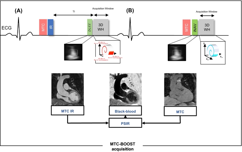
Purpose: Free-breathing Magnetization Transfer Contrast Bright blOOd phase SensiTive (MTC-BOOST) is a prototype balanced-Steady-State Free Precession sequence for 3D whole-heart imaging, that employs the endogenous magnetisation transfer contrast mechanism. This achieves reduction of flow and off-resonance artefacts, that often arise with the clinical T2prepared balanced-Steady-State Free Precession sequence, enabling high quality, contrast-agent free imaging of the thoracic cardiovascular anatomy. Fully-sampled MTC-BOOST acquisition requires long scan times (~10-24 min) and therefore acceleration is needed to permit its clinical incorporation. The aim of this study is to enable and clinically validate the 5-fold accelerated MTC-BOOST acquisition with joint Multi-Scale Variational Neural Network (jMS-VNN) reconstruction.

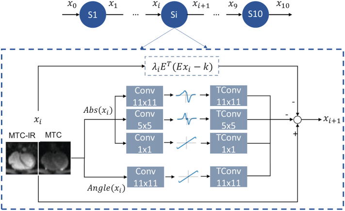
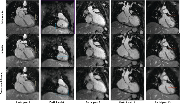
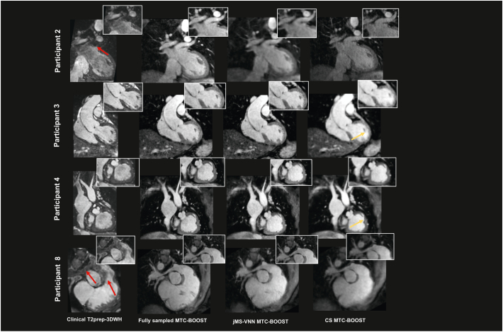
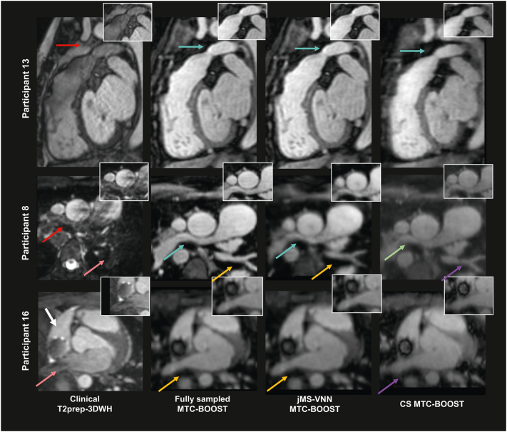
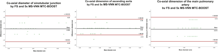
Methods: Thirty-six patients underwent free-breathing, 3D whole-heart imaging with the MTC-BOOST sequence, which is combined with variable density spiral-like Cartesian sampling and 2D image navigators for translational motion estimation. This sequence acquires two differently weighted bright-blood volumes in an interleaved fashion, which are then joined in a phase sensitive inversion recovery reconstruction to obtain a complementary fully co-registered black-blood volume. Data from eighteen patients were used for training, whereas data from the remaining eighteen patients were used for testing/evaluation. The proposed deep-learning based approach adopts a supervised multi-scale variational neural network for joint reconstruction of the two differently weighted bright-blood volumes acquired with the 5-fold accelerated MTC-BOOST. The two contrast images are stacked as different channels in the network to exploit the shared information. The proposed approach is compared to the fully-sampled MTC-BOOST and 5-fold undersampled MTC-BOOST acquisition with Compressed Sensing (CS) reconstruction in terms of scan/reconstruction time and bright-blood image quality. Comparison against conventional 2-fold undersampled T2-prepared 3D bright-blood whole-heart clinical sequence (T2prep-3DWH) is also included.

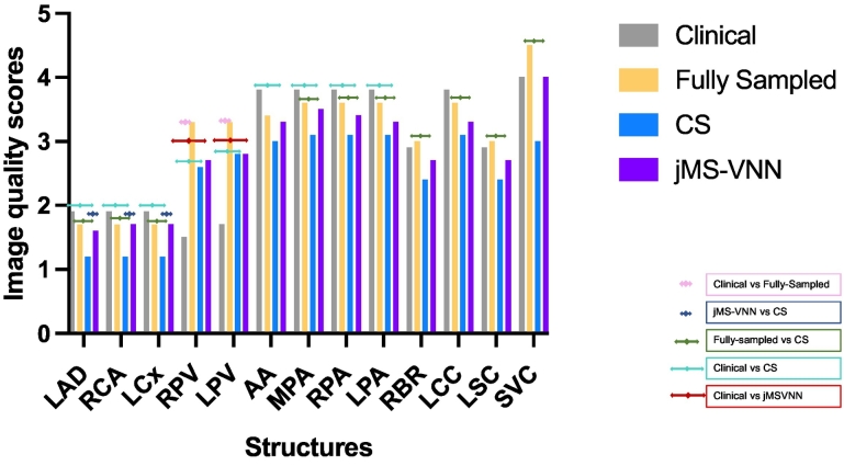
Results: Acquisition time was 3.0 ± 1.0 min for the 5-fold accelerated MTC-BOOST versus 9.0 ± 1.1 min for the fully-sampled MTC-BOOST and 11.1 ± 2.6 min for the T2prep-3DWH (p < 0.001 and p < 0.001, respectively). Reconstruction time was significantly lower with the jMS-VNN method compared to CS (10 ± 0.5 min vs 20 ± 2 s, p < 0.001). Image quality was higher for the proposed 5-fold undersampled jMS-VNN versus conventional CS, comparable or higher to the corresponding T2prep-3DWH dataset and similar to the fully-sampled MTC-BOOST.

Conclusion: The proposed 5-fold accelerated jMS-VNN MTC-BOOST framework provides efficient 3D whole-heart bright-blood imaging in fast acquisition and reconstruction time with concomitant reduction of flow and off-resonance artefacts, that are frequently encountered with the clinical sequence. Image quality of the cardiac anatomy and thoracic vasculature is comparable or superior to the clinical scan and 5-fold CS reconstruction in faster reconstruction time, promising potential clinical adoption.

Keywords: 3D whole-heart imaging; Cardiac MRI; Free-breathing; Neural network.


Conflict of interest statement

All the authors declare that they do not have competing interests.

Figures

Fig. 1
Fig. 1
MTC-BOOST acquisition framework. Two magnetization prepared bright-blood volumes are acquired in odd and even heartbeats. Magnetization transfer in combination with an inversion pulse is used in odd heartbeats (A), whereas magnetization transfer solely is exploited in even heartbeats (B). In odd heartbeats, a short inversion time (TI), Inversion Recovery approach is used to suppress the signal from epicardial fat, whereas frequency-selective pre-saturation is used in even heartbeats. Data acquisition is performed using a 3D Cartesian trajectory with spiral profile order and segmented over multiple heartbeats (green, red, blue). A low-resolution 2D iNAV is acquired in each heartbeat by spatially encoding the ramp-up pulses of the bSSFP sequences. The bright-blood MTC-IR BOOST and MTC BOOST volumes are translational motion corrected at the end-expiratory level and, subsequently, combined in a PSIR-like reconstruction to generate a complementary black-blood volume. Abbreviations: bSSFP: balanced Steady-State Free Precession, MTC: Magnetization Transfer Contrast, IR: Inversion recovery pulse, PSIR: Phase sensitive inversion recovery. (For interpretation of the references to colour in this figure legend, the reader is referred to the web version of this article.)
Fig. 2
Fig. 2
Joint multi-scale variational neural network architecture. Network architecture of the joint multi-scale VNN (jMS-VNN), which consists of 10 stages. In each stage, the first path enforces data consistency, whereas the second path is the regularization operator that applies convolutional filters (Conv), learnable activation functions and corresponding transposed convolutional filters (TConv) respectively to the magnitude and phase of the complex-valued MTC-IR and MTC images. For the magnitude image, a multi-scale approach is applied with three parallel filter sets with size of 11 × 11, 5 × 5 and 1 × 1. For the phase image, one set of 11 × 11 filter kernels is applied. The MTC-IR and MTC images are stacked as different channels for the network input to exploit their shared information. Abbreviations: jMS-VNN: joint multi-scale variational neural network, MTC: Magnetization transfer contrast, IR: Inversion recovery pulse, TConv: transposed convolutional filters.
Fig. 3
Fig. 3
Fully sampled MTC-BOOST versus 5-fold jMS-VNN and 5-fold CS. Bright-blood images in coronal view for five representative participants. Acquisitions were performed with the fully-sampled MTC-BOOST sequence and 5-fold prospectively undersampled MTC-BOOST sequence. Accelerated MTC-BOOST reconstructed with Compressed Sensing (CS) introduced mild blurring that is mitigated with the jMS-VNN reconstruction (participant 2,4,15). Left ventricular wall and papillary muscles (yellow box), right ventricular trabeculations (blue box) and left ventricular wall and papillary muscles (red box) were sharper delineated with the jMS-VNN reconstruction in comparison to CS (participant 2,4,15 respectively). jMS-VNN showed similar image quality to the fully-sampled scan. Abbreviations: CHD: Congenital Heart Disease, CS: Compressed Sensing, jMS-VNN: Joint Multi Scale Variational Neural Network, MTC-BOOST: Magnetisation Transfer Contrast Bright and black blOOd phase SensiTive. (For interpretation of the references to colour in this figure legend, the reader is referred to the web version of this article.)
Fig. 4
Fig. 4
Clinical dataset versus fully sampled MTC-BOOST, 5-fold jMS-VNN and 5-fold CS. Bright blood images for four participants. Acquisitions were performed with the clinical sequence (T2 prepared 3D Whole-Heart), the fully-sampled MTC-BOOST sequence and 5× prospectively undersampled MTC-BOOST sequence. Accelerated MTC-BOOST was reconstructed with Compressed Sensing (CS) and joint Multi-Scale Variational Neural Network (jMS-VNN). Close-up views are shown for each reconstruction. MTC BOOST introduced uniform signal of the cardiac chambers and vessels in comparison to the clinical sequence, suppressing flow and off-resonance artefacts (participant 2 and 8, red arrows), that was preserved with both reconstruction methods, albeit with less noise artefacts in the jMS-VNN reconstruction. Residual blurring in the left and right ventricular wall (yellow arrow) was observed with the CS reconstruction that was reduced with the jMS-VNN. Abbreviations: CHD: Congenital Heart Disease, CS: Compressed Sensing, jMS-VNN: Joint Multi Scale Variational Neural Network, MTC-BOOST: Magnetisation Transfer Contrast Bright and black blOOd phase SensiTive, T2prep-3DWH: T2 prepared 3D Whole-Heart. (For interpretation of the references to colour in this figure legend, the reader is referred to the web version of this article.)
Fig. 5
Fig. 5
Clinical datasets with T2-preparation related MRI artefacts versus the corresponding fully sampled MTC-BOOST, 5-fold jMS-VNN and 5-fold CS. Significant flow related artefacts (red arrow) in the right ventricular outflow tract and main pulmonary artery in participant 13 and in the right pulmonary artery (red arrow) in participant 8, rendering the structures almost indistinguishable with the T2prep-3DWH sequence. Those were mitigated with the fully-sampled MTC-BOOST, 5-fold jMS-VNN and 5-fold CS (light blue and green arrows). Residual blurring was noted in the right pulmonary artery with the CS reconstruction in participant 8. Off-resonance artefacts in the left lower pulmonary veins and right upper pulmonary vein degraded significantly the image quality in the clinical sequence (participant 8 and 16 respectively, pink arrow). Those were attenuated with the fully-sampled MTC-BOOST and 5-fold jMS-VNN sequence (orange arrows). 5× CS introduced mild blurring (purple arrows). Artefact from the stent in the SVC was present in the clinical (white arrow) and the MTC-BOOST acquisition, however the vascular luminal signal and the pulmonary venous return was better appreciated in the fully-sampled MTC-BOOST sequence as well as in the 5-fold jMS-VNN and 5-fold CS. Abbreviations: CHD: Congenital Heart Disease, CS: Compressed Sensing, jMS-VNN: Joint Multi Scale Variational Neural Network, MTC-BOOST: Magnetisation Transfer Contrast Bright and black blOOd phase SensiTive, T2prep-3DWH: T2 prepared 3D Whole-Heart. (For interpretation of the references to colour in this figure legend, the reader is referred to the web version of this article.)
Fig. 6
Fig. 6
Image quality scores for the proposed 5-fold jMS-VNN MTC-BOOST in comparison to the clinical sequence, fully-sampled MTC-BOOST and 5-fold MTC-BOOST with CS reconstruction. Image quality scores for the proposed 5-fold jMS-VNN MTC-BOOST in comparison to the clinical sequence, fully-sampled MTC-BOOST and 5-fold MTC-BOOST with CS reconstruction for the two reviewers. Results have been averaged between the two reviewers. Abbreviations: AA: ascending aorta, CS: Compressed Sensing, jMS-VNN: Joint Multi Scale Variational Neural Network, LAD: left anterior descending coronary artery, LCC: left common carotid, LCx: left circumflex coronary artery, LPA: left pulmonary artery, LPV: left pulmonary vein, LSC: left subclavian, MTC-BOOST: Magnetisation Transfer Contrast Bright and black blOOd phase SensiTive, MPA: main pulmonary artery, RBR: right brachiocephalic, RCA: right coronary artery, RPA: right pulmonary artery, RPV: right pulmonary vein, SVC: superior vena cava.
Fig. 7
Fig. 7
Bland-Altman plots comparing vascular dimensions between the fully-sampled MTC-BOOST and 5-fold jMS-VNN. Bland–Altman analysis comparing the aortic dimensions at the level of sinotubular junction, ascending aorta and main pulmonary artery. Measurements were performed in the 5-fold accelerated acquisition with joint Multi Scale Variational Neural Network (jMS-VNN) reconstruction and Fully Sampled MTC BOOST. The black line indicates the mean bias of the diameter measurements whereas the red lines represent the 95% confidence interval. Values are given in cm. A: co-axial aortic dimensions at the level of sinotubular junction demonstrate excellent agreement with a mean difference of −0.004 cm (95% confidence interval − 0.22. to 0.23, p value 0.7). B: co-axial dimensions of mid ascending aorta result demonstrate excellent agreement with a mean difference of 0.02 cm (95% confidence interval − 0.12 to 0.16, p value 0.09). C: co-axial dimensions of main pulmonary artery diameter demonstrate excellent agreement with a mean difference of −0.014 cm (95% confidence interval − 0.16 to 0.13, p value: 0.3). Abbreviations: FS: Fully-sampled, jMS-VNN: joint Multi Scale Variational Neural Network, MTC-BOOST: Magnetisation Transfer Contrast Bright and black blOOd phase SensiTive. (For interpretation of the references to colour in this figure legend, the reader is referred to the web version of this article.)

References

    1. Liu Y., Chen S., Zühlke L., et al. Global birth prevalence of congenital heart defects 1970–2017: updated systematic review and meta-analysis of 260 studies. Int J Epidemiol. 2019;48(2):455–463. - PMC - PubMed
    1. Buber J., Valente A.M. Predicting survival in adults with congenital heart disease: what are the odds? Heart (British Cardiac Society) 2018;104(20):1643–1644. - PubMed
    1. Fratz S., Chung T., Greil G.F., et al. Guidelines and protocols for cardiovascular magnetic resonance in children and adults with congenital heart disease: SCMR expert consensus group on congenital heart disease. J Cardiovasc Magn Reson. 2013;15(1):51. doi: 10.1186/1532-429X-15-51. - DOI - PMC - PubMed
    1. Rajiah P., Kanne J.P. Cardiac MRI: part 1, cardiovascular shunts. Am J Roentgenol. 2011;197(4):W603–W620. doi: 10.2214/AJR.10.7257. (1976) - DOI - PubMed
    1. Sorensen T.S., Korperich H., Greil G.F., et al. Operator-independent isotropic three-dimensional magnetic resonance imaging for morphology in congenital heart disease: a validation study. Circulation. 2004;110(2):163–169. - PubMed

Publication types

MeSH terms