. 2022 Aug:204:105370.
doi: 10.1016/j.antiviral.2022.105370. Epub 2022 Jun 27.

A circular mRNA vaccine prototype producing VFLIP-X spike confers a broad neutralization of SARS-CoV-2 variants by mouse sera

Affiliations

A circular mRNA vaccine prototype producing VFLIP-X spike confers a broad neutralization of SARS-CoV-2 variants by mouse sera

Chotiwat Seephetdee et al. Antiviral Res. 2022 Aug.

Abstract

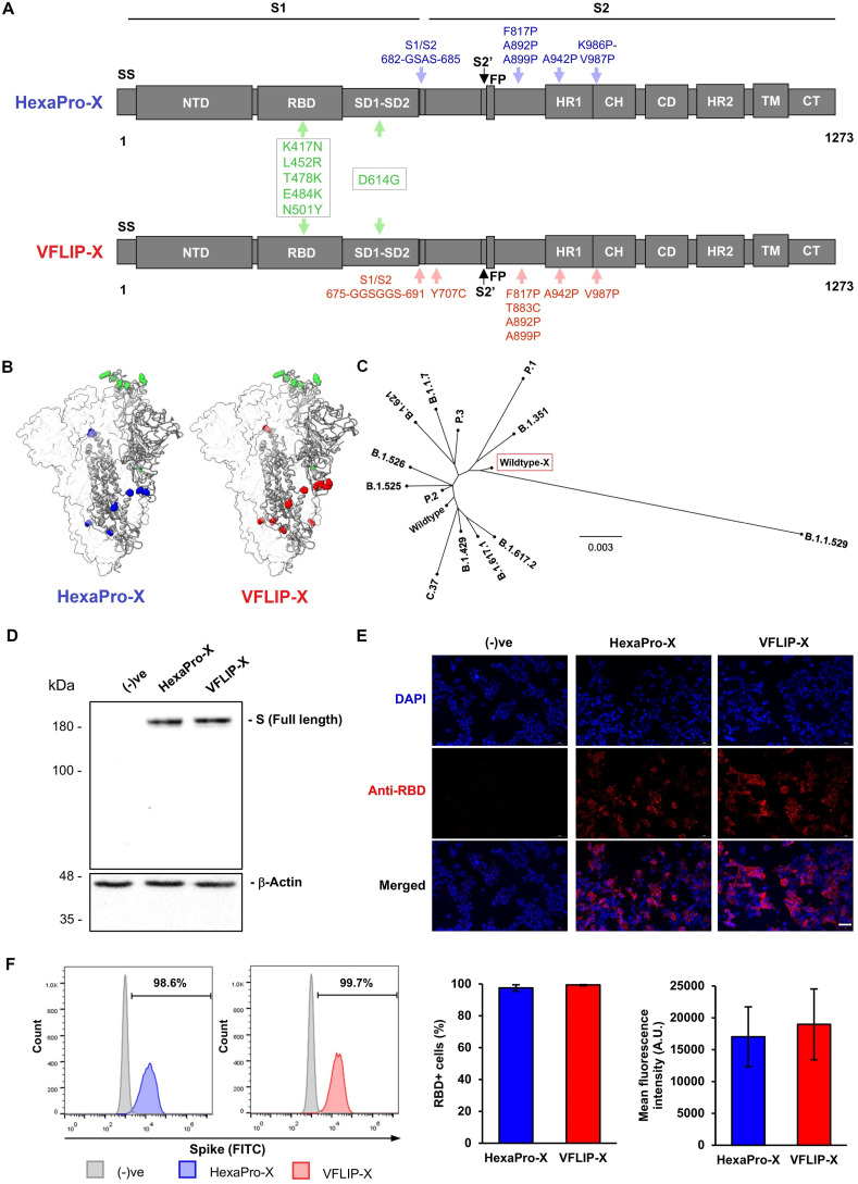
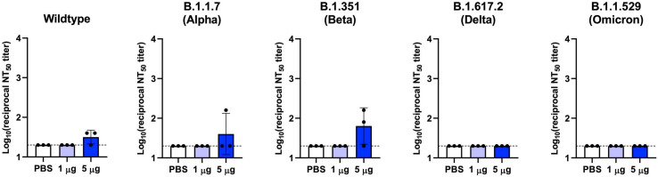
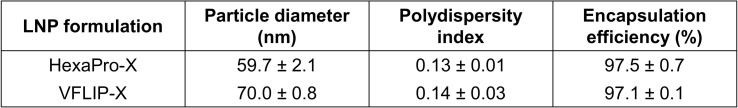
Next-generation COVID-19 vaccines are critical due to the ongoing evolution of SARS-CoV-2 virus and rapid waning duration of the neutralizing antibody response against current vaccines. The mRNA vaccines mRNA-1273 and BNT162b2 were developed using linear transcripts encoding the prefusion-stabilized trimers (S-2P) of the wildtype spike, which have shown a reduced neutralizing activity against the variants of concern B.1.617.2 and B.1.1.529. Recently, a new version of spike trimer, termed VFLIP (five (V) prolines, Flexibly-Linked, Inter-Protomer disulfide) was developed. Based on the original amino acid sequence of the wildtype spike, VFLIP was genetically engineered by using five proline substitutions, a flexible cleavage site amino acid linker, and an inter-protomer disulfide bond. It has been suggested to possess native-like glycosylation, and greater pre-fusion trimeric stability as opposed to S-2P. Here, we report that the spike protein VFLIP-X, containing six rationally substituted amino acids to reflect emerging variants (K417N, L452R, T478K, E484K, N501Y and D614G), offers a promising candidate for a next-generation SARS-CoV-2 vaccine. Mice immunized by a circular mRNA (circRNA) vaccine prototype producing VFLIP-X had detectable neutralizing antibody titers for up to 7 weeks post-boost against SARS-CoV-2 variants of concern (VOCs) and variants of interest (VOIs). In addition, a balance in TH1 and TH2 responses was achieved by immunization with VFLIP-X. Our results indicate that the VFLIP-X delivered by circRNA induces humoral and cellular immune responses, as well as broad neutralizing activity against SARS-CoV-2 variants.

Keywords: SARS-CoV-2; Spike; VFLIP; Vaccine.


Conflict of interest statement

We wish to confirm that there are no known conflicts of interest associated with this publication and there has been no significant financial support for this work that could have influenced its outcome.

Figures

Fig. 1
Fig. 1
Design strategy and expression of SARS-CoV-2 spike proteins harboring six rationally substituted amino acids. (A) Schematic representation of HexaPro-X and VFLIP-X showing the S1 and S2 subunits. Amino acid positions described in the original HexaPro (Hsieh et al., 2020) and VFLIP (Olmedillas et al., 2021) spikes are shown in blue and red colors, respectively. The positions of six rationally substituted amino acids (K417N, L452R, T478K, E484K, N501Y and D614G) are indicated by green color. (B) Molecular models of HexaPro-X and VFLIP-X spike trimers. The RBD-up protomer is shown in ribbons colored corresponding to (A). The structures were prepared in SWISS-MODEL. (C) An unrooted phylogenetic tree comparing amino acid sequences derived from the wildtype spike containing six rationally substituted amino acids (wildtype-X) and spike sequences derived from SARS-CoV-2 VOCs and VOIs. (D) Western blot analysis of full-length SARS-CoV-2 spike expression in HEK293T cells transfected with circRNAs encoding HexaPro-X and VFLIP-X. The proteins were visualized by an anti-RBD antibody. (E) Immunofluorescence analysis of HexaPro-X and VFLIP-X in HEK293T cells immunostained by an anti-RBD antibody. Scale bar, 50 μm. (F) Flow cytometry analysis of HEK293T cells expressing HexaPro-X and VFLIP-X spikes upon delivery by circRNA-LNP. The proteins were visualized by an anti-RBD antibody. Percentage of RBD-positive cells and mean fluorescence intensity from biological duplicates are shown. The error bars indicate the ±SD.
Fig. 2
Fig. 2
Mice immunized with VFLIP-X full length spike produce neutralizing antibodies against B.1.1.529. BALB/c mice (n = 3) were immunized at weeks 0 and 3 with 5 μg of circRNA-LNP encoding HexaPro-X (blue) or VFLIP-X (red). Control mice were administered with PBS (white). (A) IgG levels of two weeks post-boost sera were assessed by enzyme-linked immunosorbent assay (ELISA) using recombinant Omicron (B.1.1.529) spike protein. (B) Neutralization activity of two weeks post-boost sera were determined using B.1.1.529 pseudotyped virus. Data are presented as GMT ± geometric SD. Horizontal dotted lines represent assay limits of detection. Immunized groups were compared by student's t-test (parametric, two-tailed unpaired). ** p < 0.01.
Fig. 3
Fig. 3
VFLIP-X elicits cross-neutralizing antibodies against SARS-CoV-2 VOCs and VOIs. BALB/c mice (n = 3) were immunized at weeks 0 and 3 with 1 μg (pink) or 5 μg (red) of circRNA-LNP encoding VFLIP-X. Control mice were administered with PBS (white). (A) Infectious virus 50% neutralization titers (NT50) of seven weeks post-boost sera were determined against the indicated infectious SARS-CoV-2 variant isolates. (B) Lentivirus-based pseudovirus 50% neutralization titers (pVNT50) of seven weeks post-boost sera were determined against the indicated SARS-CoV-2 variants. Data are presented as GMT ± geometric SD. Horizontal dotted lines represent assay limits of detection. Control group was compared with 5 μg immunized group by student's t-test (parametric, two-tailed unpaired). * p < 0.05, ** p < 0.01, *** p < 0.001, **** p < 0.0001.
Fig. 4
Fig. 4
Humoral and cellular immune responses against B.1.1.529 were raised by VFLIP-X. BALB/c mice (n = 3) were immunized at weeks 0 and 3 with 1 μg (pink) or 5 μg (red) of circRNA-LNP encoding VFLIP-X. Control mice were administered with PBS (white). Sera samples were collected at weeks 2, 5, 8, and 10. Splenocytes isolated from mice were collected at week 10. (A) SARS-CoV-2 Omicron (B.1.1.529) S-specific IgG levels of sera were assessed by ELISA. (B) Lentivirus-based pseudovirus expressing SARS-CoV-2 B.1.1.529 S 50% neutralization titers (pVNT50) of sera were determined. (C) IFN-γ (left) and IL-4 (right) ELISpot of seven weeks post-boost splenocytes restimulated with SARS-CoV-2 B.1.1.529 S peptide pool. (D) SARS-CoV-2 Omicron (B.1.1.529) S-specific IgG2a (left) and IgG1 (right) levels of seven weeks post-boost sera were determined by ELISA. In A, B, and D, data are presented as GMT ± geometric SD. Horizontal dotted lines represent assay limits of detection. In C, data are presented as mean ± SD.
Fig. S1
Fig. S1
In vivo expression of circular RNA in mice. BALB/c mice were inoculated with 1 μg of firefly luciferase-encoding circular mRNA-LNP via intramuscular injection and subjected to IVIS Spectrum imaging at the indicated times after administration.
Fig. S2
Fig. S2
Neutralization of infectious SARS-CoV-2 variants by serum samples from HexaPro-X immunized mice. BALB/c mice (n = 3) were immunized at weeks 0 and 3 with 1 μg (light blue) or 5 μg (blue) of circRNA-LNP encoding HexaPro-X. Control mice were administered with PBS (white). Live virus 50% neutralization titers (NT50) of seven weeks post-boost sera were determined against the indicated infectious SARS-CoV-2 variants. Data are presented as GMT ± geometric SD. Horizontal dotted lines represent assay limits of detection.
Fig. S3
Fig. S3
Neutralization of SARS-CoV-2 variants in convalescent serum samples collected in 2020. (A) Infectious virus 50% neutralization titers (NT50) of convalescent sera were determined against the indicated infectious SARS-CoV-2 variant isolates. (B) Lentivirus-based pseudovirus 50% neutralization titers (pVNT50) of convalescent sera were determined against the indicated SARS-CoV-2 variants. Horizontal dotted lines represent assay limits of detection.
Table S1
Table S1
Physicochemical properties of LNP-RNAs (data are presented as mean ± SD, n = 3).

Similar articles

Cited by

References

    1. Álvarez-Díaz D.A., et al. Low neutralizing antibody titers against the Mu variant of SARS-CoV-2 in 31 BNT162b2 vaccinated individuals in Colombia. Vaccines. 2022;10 doi: 10.3390/vaccines10020180. - DOI - PMC - PubMed
    1. Bowen J.E., et al. SARS-CoV-2 spike conformation determines plasma neutralizing activity. bioRxiv. 2021 doi: 10.1101/2021.12.19.473391. 2012.2019, 2021. - DOI - PMC - PubMed
    1. Cameroni E., et al. Broadly neutralizing antibodies overcome SARS-CoV-2 Omicron antigenic shift. Nature. 2022;602:664–670. doi: 10.1038/s41586-021-04386-2. - DOI - PMC - PubMed
    1. Carnell George, W. et al. SARS-CoV-2 spike protein stabilized in the closed state induces potent neutralizing responses. J. Virol. 95, e00203-00221, doi:10.1128/JVI.00203-21. - PMC - PubMed
    1. Cerutti G., et al. Structural basis for accommodation of emerging B.1.351 and B.1.1.7 variants by two potent SARS-CoV-2 neutralizing antibodies. Structure. 2021;29:655–663. doi: 10.1016/j.str.2021.05.014. e654. - DOI - PMC - PubMed

Publication types

Supplementary concepts